. 2012;8(11):e1002992.
doi: 10.1371/journal.ppat.1002992. Epub 2012 Nov 8.

The polymorphic pseudokinase ROP5 controls virulence in Toxoplasma gondii by regulating the active kinase ROP18

Affiliations

The polymorphic pseudokinase ROP5 controls virulence in Toxoplasma gondii by regulating the active kinase ROP18

Michael S Behnke et al. PLoS Pathog. 2012.

Abstract

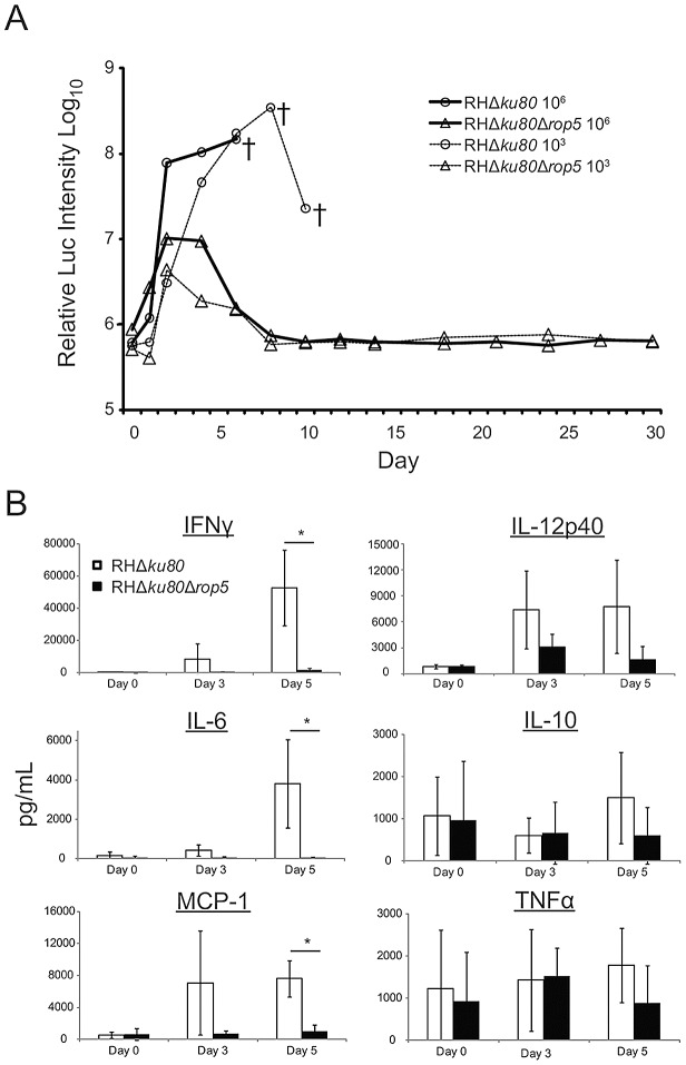
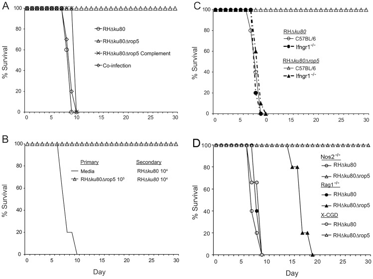
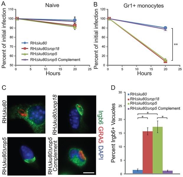
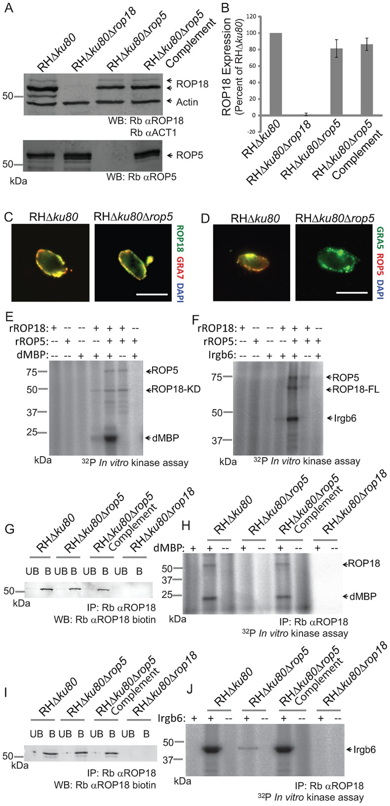
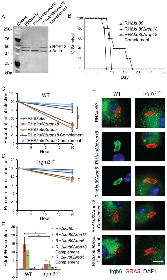
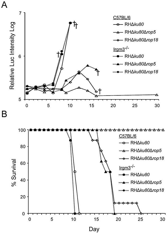
Secretory polymorphic serine/threonine kinases control pathogenesis of Toxoplasma gondii in the mouse. Genetic studies show that the pseudokinase ROP5 is essential for acute virulence, but do not reveal its mechanism of action. Here we demonstrate that ROP5 controls virulence by blocking IFN-γ mediated clearance in activated macrophages. ROP5 was required for the catalytic activity of the active S/T kinase ROP18, which phosphorylates host immunity related GTPases (IRGs) and protects the parasite from clearance. ROP5 directly regulated activity of ROP18 in vitro, and both proteins were necessary to avoid IRG recruitment and clearance in macrophages. Clearance of both the Δrop5 and Δrop18 mutants was reversed in macrophages lacking Irgm3, which is required for IRG function, and the virulence defect was fully restored in Irgm3(-/-) mice. Our findings establish that the pseudokinase ROP5 controls the activity of ROP18, thereby blocking IRG mediated clearance in macrophages. Additionally, ROP5 has other functions that are also Irgm3 and IFN-γ dependent, indicting it plays a general role in governing virulence factors that block immunity.


Conflict of interest statement

The authors have declared that no competing interests exist.

Figures

Figure 1
Figure 1. ROP5 deficient parasites are controlled in wild type mice and trigger a cytokine response proportional to the parasite load.
(A) In vivo parasite growth was monitored using luciferase expressing wild type (RHΔku80) or ROP5 deficient (RHΔku80Δrop5) parasites. CD-1 mice were i.p. injected with either 106 or 103 parasites and imaged on indicated days. † denotes one or more deaths. Mean values shown per group (n = 5). Representative of 2 experiments with similar outcomes. (B) Analysis of cytokines in serum of infected mice used in (A) (106 inoculum) on days 0 (uninfected), 3, and 5. The levels of IFN-γ, IL-6, MCP-1, IL-10, and TNF-α were determined using a Cytometric Bead Array analyzed on a FACS Canto. IL-12p40 was measured by ELISA. Mean ± S.D., n = 3 animals per group. Student's t test * P<0.01. Representative of 2 experiments with similar outcomes.
Figure 2
Figure 2. ROP5 deficient parasites vaccinate against lethal challenge and IFN-γ signaling is required for control of Δrop5 parasites.
(A) CD-1 mice were separately injected i.p. with 103 wild type (RHΔku80), ROP5 deficient (RHΔku80Δrop5) or ROP5 complemented (RHΔku80Δrop5Complement) parasites, or co-infected with 103 each of wild type (RHΔku80) and ROP5 deficient (RHΔku80Δrop5) parasites. Survival was followed for 30 days. Mean values shown per group (n = 5). Representative of 2 experiments with similar outcomes. (B) CD-1 mice were inoculated with either media alone or 103 ROP5 deficient (RHΔku80Δrop5) parasites. After 30 days, mice were challenged with 104 wild type (RHΔku80) parasites and survival followed for 30 days. Mean values shown per group (n = 5). Representative of 2 experiments with similar outcomes. (C) C57BL/6 control or Ifngr1−/− mice were i.p. injected with 103 wild type (RHΔku80) or ROP5 deficient (RHΔku80Δrop5) parasites and survival was followed for 30 days. Mean values shown per group (n = 5). Representative of 3 experiments with similar outcomes. (D) Nos2−/−, Rag1−/−, or X-CGD mice were injected i.p. with 103 wild type (RHΔku80) or ROP5 deficient (RHΔku80Δrop5) parasites and survival was followed for 30 days. Mean values shown per group (n = 5), except X-CGD mice (RHΔku80 n = 3 and RHΔku80Δrop5 n = 4). Representative of 2 experiments with similar outcomes.
Figure 3
Figure 3. Gr1+ F4/80+ inflammatory monocytes are recruited to the peritoneum after infection with wild type or ROP5 deficient parasites.
(A) CD-1 mice were infected with 103 wild type (RHΔku80), ROP5 deficient (RHΔku80Δrop5), or ROP5 complemented (RHΔku80Δrop5Complement) parasites and the number of Gr1+ F4/80+ cells was determined by cell surface staining and FACS. Mean ± S.E.M., n = 3 animals per group. (B) FACS plots of Gr1 and F4/80 gated cells from representative animals at day 3 postinfection in (A). Top left gate represents neutrophils (Gr1+ F4/80), middle gate represents inflammatory monocytes (Gr1+ F4/80+), bottom right gate represents resident macrophages (Gr1 F4/80+). Data are derived from a single experiment, n = 3 mice/group. Plots for representative animals are shown.
Figure 4
Figure 4. ROP5 and ROP18 are required for avoidance of clearance and IRG recruitment.
In vitro clearance of parasites in naïve peritoneal macrophages (A) or Gr1+ inflammatory monocytes (B) was measured by immunofluorescence microscopy. Cells were stained at 0.5 and 20 h post infection for host cell surface markers (see methods) and the parasite surface marker SAG1. Infection rates at 20 h post infection were normalized to initial infection rates. Means ± S.E.M., n = 3 samples each, from 3 combined experiments. Student's t test, **P<0.0005. (C) Immunofluorescence localization of Irgb6 on the parasitophorous vacuole membrane in Gr1+ inflammatory monocytes infected for 0.5 h in vitro. The vacuole membrane was visualized by staining with mAb Tg 17-113 for GRA5 (secondary: Alexa Fluor 594, red). Irgb6 was visualized with rabbit anti-Irgb6 (secondary: Alexa Fluor 488, green). Scale = 5 microns. (D) Quantification of Irgb6 localization to the vacuolar membrane in Gr1+ monocytes. Mean ± SEM, n = 3 samples each, from 3 combined experiments. Student's t test, *P<0.005.
Figure 5
Figure 5. ROP5 regulates the kinase activity of ROP18.
(A) Expression of ROP18 and ROP5 detected by western blotting of parasite lysates with rabbit anti-ROP18 (Rb α-ROP18), rabbit anti-ROP5 (Rb αROP5), and rabbit anti-actin (Rb αACT1) as a loading control. Representative of 3 experiments with similar outcomes. (B) Quantification of ROP18 expression by phosphorimager analysis of western blots, normalized for loading by actin staining. Means ± S.E.M. n = 3 experiments. (C) Immunofluorescence localization of ROP18 on the parasitophorous vacuole membrane in wild type (RHΔku80) and ROP5 deficient (RHΔKu80Δrop5) parasites infecting HFF cells in vitro. ROP18 was localized based on the C-terminal Ty-1 epitope described previously using mAb BB2 (directly conjugated to Alexa Fluor 488, green). The vacuole membrane was stained with polyclonal rabbit anti-GRA7, described previously , (secondary: Alexa Fluor 594, red). Scale = 5 microns. (D) Immunofluorescence localization of ROP5 on the parasitophorous vacuole membrane in wild type (RHΔku80) and ROP5 deficient (RHΔku80Δrop5) parasites infecting HFF cells in vitro. The vacuole membrane was labeled with mAb Tg 17-113 for GRA5 (secondary: Alexa Fluor 594, red). ROP5 was labeled with polyclonal Ab for ROP5 (secondary: Alexa Fluor 488, green). Scale = 5 microns. (E) In vitro kinase reaction using the kinase domain of ROP18 (ROP18-KD, 100 ng) and the heterologous substrate dMBP ± recombinant ROP5 (rROP5, 200 ng). (F) In vitro kinase reaction using full length ROP18 (ROP18-FL, 25 ng) and the natural substrate Irgb6 ± recombinant ROP5 (rROP5, 50 ng). Irgb6 was immunoprecipitated (∼10–20 ng/reaction) from IFN-γ activated RAW cells with polyclonal rabbit anti-Irgb6. (G) Immunoprecipitation of ROP18 (∼5 ng/reaction) from parasite lysates with polyclonal rabbit anti-ROP18 (Rb anti-ROP18). Bound (denoted as B) and unbound (denoted as UB) samples were resolved by SDS-PAGE and blotted for ROP18 (Rb anti-ROP18 biotin). (H) In vitro kinase reaction of ROP18 using the heterologous substrate dMBP. ROP18 immunoprecipitations from (G) were incubated with or without substrate in the presence of 32P ATP. Reactions were resolved by SDS-PAGE and subjected to phosphorimager analysis. (I) Immunoprecipitation of ROP18 (∼5 ng/reaction) from parasite lysates with rabbit anti-ROP18 as in (G). (J) In vitro kinase reaction of ROP18 and a natural substrate Irgb6. ROP18 immunoprecipitations from (I) were incubated with or without substrate in the presence of 32P ATP. Irgb6 was immunoprecipitated from IFN-γ activated RAW cells with polyclonal rabbit anti-Irgb6. Reactions in E, F, H, and J were carried out in the presence of 32P ATP, resolved by SDS-PAGE and subjected to phosphorimager analysis. E–J are representative of 3 or more experiments with similar outcomes.
Figure 6
Figure 6. Irgm3 deficiency reverts the IRG recruitment and clearance defects of both Δrop18 and Δrop5 parasites in vitro.
(A) Western blot analysis of wild type (RHΔku80), ROP18 deficient (RHΔku80Δrop18) and ROP18 complemented (RHΔku80Δrop18Complement) parasites. Western blot probed with rabbit anti-ROP18 and rabbit anti-actin flowed by goat anti-rabbit conjugated to HRP and detected by ECL. (B) Survival of female outbred CD1 mice challenged with 100 parasites by i.p. inoculation. n = 5 animals per group from a single experiment. In vitro clearance of parasites in wild type C57BL/6 (WT) (C) or Irgm3 deficient (Irgm3−/−) (D) bone marrow derived macrophages following IFN-γ activation (50 units/mL). Means ± S.D. n = 6 samples from 2 combined experiments. For statistical analysis, parasite survival in Irgm3 deficient cells was compared to survival in wild-type cells. Student's t test, **P<0.001. Quantification (E) and immunofluorescence localization (F) of Irgb6 localization to the vacuolar membrane in WT or Irgm3−/− IFN-γ-activated macrophages. (E) Means ± S.D. n = 6 samples from 2 combined experiments. Student's t test, *P<0.005. (F) The vacuole membrane was visualized by staining with mAb Tg 17-113 for GRA5 (secondary: Alexa Fluor 594, red). Irgb6 was visualized with rabbit-Irgb6 (secondary: Alexa Fluor 488, green). Scale = 5 microns.
Figure 7
Figure 7. Irgm3 deficient mice revert the phenotypes of Δrop18 and Δrop5 parasites.
(A) Wild type (C57BL/6) (n = 4 per parasite strain) or Irgm3 deficient (Irgm3−/−) (n = 4 per parasite strain) mice were infected with 102 luciferase expressing wild type (RHΔku80), ROP5 deficient (RHΔku80Δrop5), or ROP18 deficient (RHΔku80Δrop18) parasites by s.c. injection and imaged on indicated days. † denotes one or more death per group. Representative experiment. (B) Survival curves for mice challenged with T. gondii strains as shown. Combination of two experiments (n = 8 animals per group).

Similar articles

Cited by

References

    1. Dubey JP (2010) Toxoplasmosis of animals and humans. Boca Raton: CRC Press. 313 p.
    1. Howe DK, Sibley LD (1995) Toxoplasma gondii comprises three clonal lineages: correlation of parasite genotype with human disease. J Infect Dis 172: 1561–1566. - PubMed
    1. Khan A, Dubey JP, Su C, Ajioka JW, Rosenthal BM, et al. (2011) Genetic analyses of atypical Toxoplasma gondii strains reveals a fourth clonal lineage in North America. Int J Parasitol 41: 645–655. - PMC - PubMed
    1. Khan A, Taylor S, Su C, Mackey AJ, Boyle J, et al. (2005) Composite genome map and recombination parameters derived from three archetypal lineages of Toxoplasma gondii . Nuc Acids Res 33: 2980–2992. - PMC - PubMed
    1. Su C, Howe DK, Dubey JP, Ajioka JW, Sibley LD (2002) Identification of quantitative trait loci controlling acute virulence in Toxoplasma gondii . Proc Natl Acad Sci U S A 99: 10753–10758. - PMC - PubMed

Publication types

MeSH terms

Substances

Associated data