. 2012;8(11):e1003026.
doi: 10.1371/journal.pgen.1003026. Epub 2012 Nov 8.

Microhomology directs diverse DNA break repair pathways and chromosomal translocations

Affiliations

Microhomology directs diverse DNA break repair pathways and chromosomal translocations

Diana D Villarreal et al. PLoS Genet. 2012.

Abstract

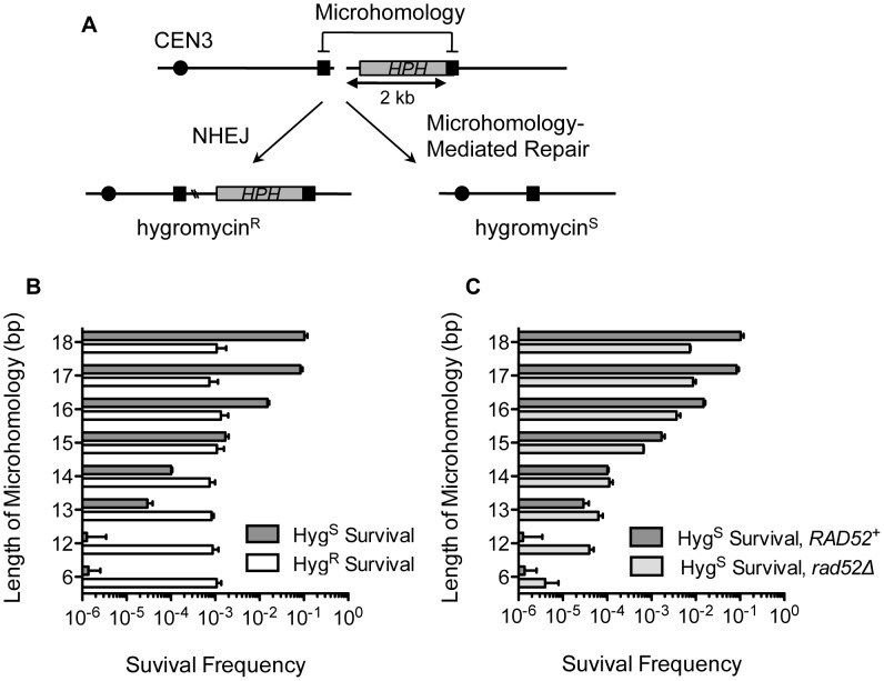
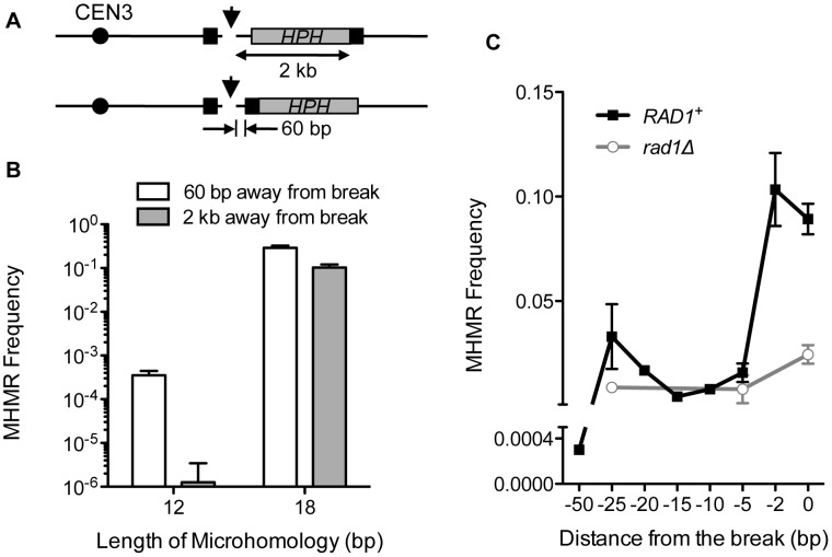
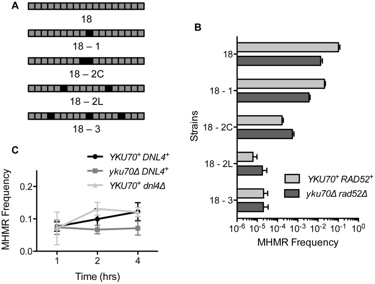
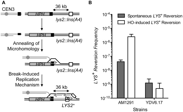
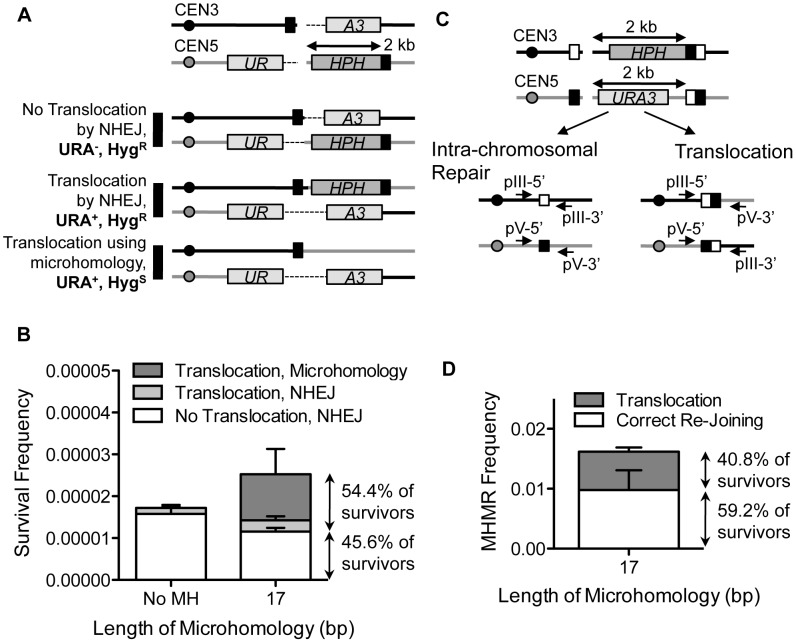
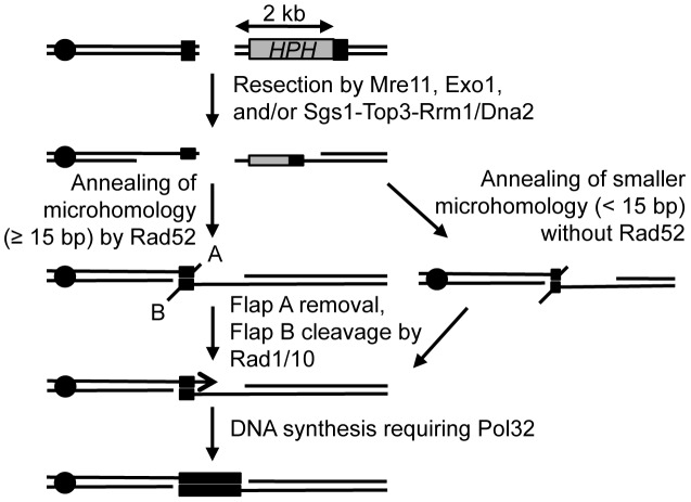
Chromosomal structural change triggers carcinogenesis and the formation of other genetic diseases. The breakpoint junctions of these rearrangements often contain small overlapping sequences called "microhomology," yet the genetic pathway(s) responsible have yet to be defined. We report a simple genetic system to detect microhomology-mediated repair (MHMR) events after a DNA double-strand break (DSB) in budding yeast cells. MHMR using >15 bp operates as a single-strand annealing variant, requiring the non-essential DNA polymerase subunit Pol32. MHMR is inhibited by sequence mismatches, but independent of extensive DNA synthesis like break-induced replication. However, MHMR using less than 14 bp is genetically distinct from that using longer microhomology and far less efficient for the repair of distant DSBs. MHMR catalyzes chromosomal translocation almost as efficiently as intra-chromosomal repair. The results suggest that the intrinsic annealing propensity between microhomology sequences efficiently leads to chromosomal rearrangements.


Conflict of interest statement

The authors have declared that no competing interests exist.

Figures

Figure 1
Figure 1. MHMR frequency increases 10-fold per nucleotide length from 12 bp to 17 bp.
(A) A diagram of the genetic system used to distinguish microhomology-mediated repair from NHEJ-mediated repair. HPH represents the hygromycin B phosphotransferase gene that confers resistance to hygromycin treatment. The locations of HO cut site, microhomology, and centromere are shown. Two repair outcomes are distinguishable based on the sensitivity to hygromycin. Repair by NHEJ is shown by double crossing lines at the break site. (B) Graph showing survival frequency ± S.D. using the microhomology (Hygs) and by NHEJ (HygR). Survival frequency was calculated by dividing the number of colonies surviving on the YEP-galactose plates by the number of colonies surviving on the YEPD plates. The results are the average of three independent experiments. (C) Survival frequency using the microhomology (Hygs) is shown in wild type and rad52Δ. Survival frequency was calculated as shown in Figure 1B. The results are the average of three independent experiments ± S.D.
Figure 2
Figure 2. The distance from the DSB inversely affects MHMR frequency.
(A) A diagram showing strains with microhomology inserted 2 kb and 60 bp from the DSB. HPH (gray box) represents the hygromycin B phosphotransferase gene. The position of the microhomology (black box) and the distance to the HO cleavage site are shown. (B) Survival using the microhomology for strains with 12 or 18 bp of microhomology located at 60 bp or 2 kb from the DSB. Survival frequency was calculated as shown in Figure 1B. For MHMR using microhomology at 60 bp from the DSB, the repair events were distinguished by sequencing of the repair junctions. The results are the average of three independent experiments ± S.D. (C) MHMR frequency using the 18 bp microhomology at various locations from the DSB in wild type and rad1Δ. Survival frequency was calculated as shown in Figure 1B. The results are the average of three independent experiments ± S.D.
Figure 3
Figure 3. Mismatched microhomology inhibits repair efficiency, but NHEJ does not inhibit MHMR.
(A) Illustration of strains with mismatched sequences. Black boxes indicate mismatched nucleotides. (B) MHMR frequency using 18 bp mismatched microhomology 2 kb from the DSB in wild type and yku70Δ rad52Δ. Survival frequency was calculated as shown in Figure 1B. The results are the average of three independent experiments ± S.D. (C) Graph showing MHMR frequency using the 17 bp of microhomology located 2 kb from the DSB in wild type, yku70Δ and dnl4Δ mutants. Survival frequency was calculated as shown in Figure 1B. The results are the average of three independent experiments ± S.D.
Figure 4
Figure 4. MHMR does not depend on long-range repair synthesis.
(A) A diagram illustrating how microhomology repairs the break using a BIR mechanism. BIR has been shown to increase mutational frequency of lys2::Ins(A4) to LYS2+ by frameshift mutation, which is shown as a white box . The position of the centromere (gray circle), HPH (gray boxes) and the microhomology (black boxes) are shown. (B) Spontaneous and HO-induced LYS+ reversion frequency was calculated using the average median value of five strains in three independent experiments in AM1291 that uses BIR to repair a DSB and YDV6.17 that carries 17 bp of microhomology. The results are the average of the median values ± S.D.
Figure 5
Figure 5. MHMR stimulates HO break-induced chromosomal translocations.
(A) Diagram of the strain with one HO recognition sequence on chromosome III and one HO recognition sequence on chromosome V. 17 bp of microhomology (shown in black box) found on the centromeric side of the DSB on chromosome III is also found on the telomeric side of the DSB on chromosome V, 2 kb from the break site. In this strain, one part of the URA3 gene (UR) is found on the centromeric side of chromosome V, and the other part of the URA3 gene (A3) is found on the telomeric side of chromosome III. Since artificial introns (the dotted lines) are inserted between the gene and the HO cut-site, the URA3 gene is expressed in the event of reciprocal translocation. Three possible repair outcomes are shown at the bottom. (B) The frequencies of the repair outcomes from the yeast strain with breaks on two different chromosomes are shown. Survival frequency was calculated as shown in Figure 1B. The results are the average of three independent experiments ± S.D. (C) Diagram illustrating the strain that allows for competition between intra-chromosomal and inter-chromosomal MHMR. The positions of microhomology (white and black boxes), and HPH and URA3 markers are shown. The location of sequences homologous to two sets of primers used to check the types of repair events are shown in arrows. (D) Survival frequency of intra- and inter-chromosomal MHMR. Survival frequency was calculated as shown in Figure 1B. The results are the average of three independent experiments ± S.D. 100 colonies from each survival experiment were assessed by PCR to detect intra-chromosomal or inter-chromosomal repair products.
Figure 6
Figure 6. Proposed mechanism for repair of a DSB using microhomology.
After a break, ends are resected to single-stranded DNA, and microhomology flanking the break (shown in black boxes) are brought together and annealed. Rad52 facilitates repair using microhomology that is 15 bp or longer, and microhomology less than 15 bp is inhibited by Rad52. A 2 bp 3′ flap (A) is removed, likely through the proof-reading activity of Polymerase δ, and a 2 kb 3′ flap (B) is removed by the Rad1/Rad10 heterodimer endonuclease. Pol32 stabilizes the annealed microhomology and the break is healed, deleting the intervening DNA and one of the microhomology sequences, similar to SSA. The location of the centromere (black circle) is shown.

Similar articles

Cited by

References

    1. Hastings PJ, Lupski JR, Rosenberg SM, Ira G (2009) Mechanisms of change in gene copy number. Nat Rev Genet 10: 551–564. - PMC - PubMed
    1. Kidd JM, Graves T, Newman TL, Fulton R, Hayden HS, et al. (2010) A human genome structural variation sequencing resource reveals insights into mutational mechanisms. Cell 143: 837–847. - PMC - PubMed
    1. Conrad DF, Bird C, Blackburne B, Lindsay S, Mamanova L, et al. (2010) Mutation spectrum revealed by breakpoint sequencing of human germline CNVs. Nat Genet 42: 385–391. - PMC - PubMed
    1. Aplan PD (2006) Causes of oncogenic chromosomal translocation. Trends Genet 22: 46–55. - PMC - PubMed
    1. Elliott B, Jasin M (2002) Double-strand breaks and translocations in cancer. Cell Mol Life Sci 59: 373–385. - PMC - PubMed

Publication types

MeSH terms

Substances