. 2012 Jul;7(4):461-472.
doi: 10.1586/eem.12.34.

Calcium regulation of keratinocyte differentiation

Affiliations

Calcium regulation of keratinocyte differentiation

Daniel D Bikle et al. Expert Rev Endocrinol Metab. 2012 Jul.

Abstract

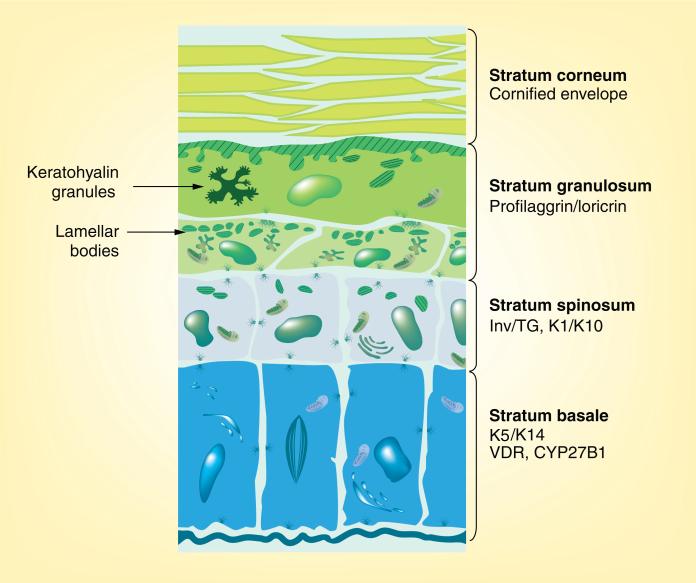
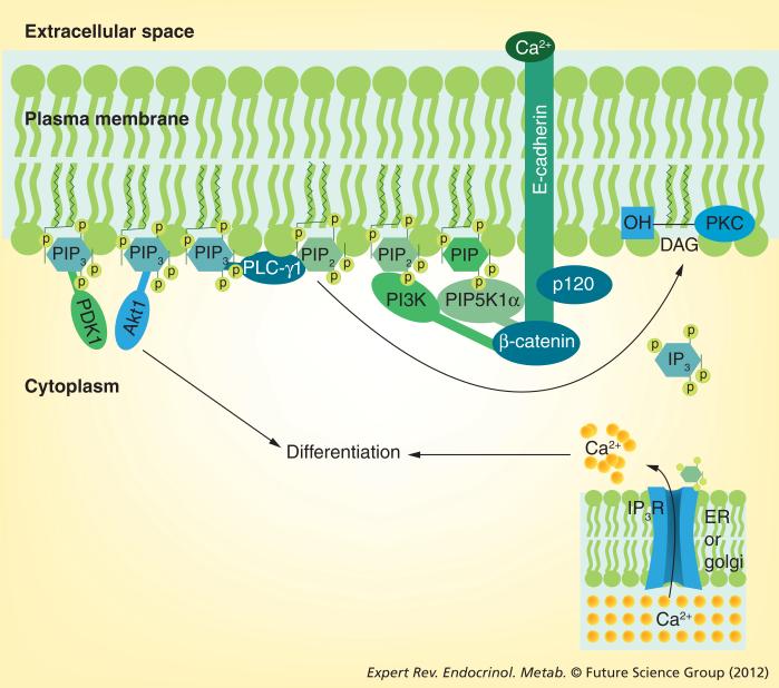
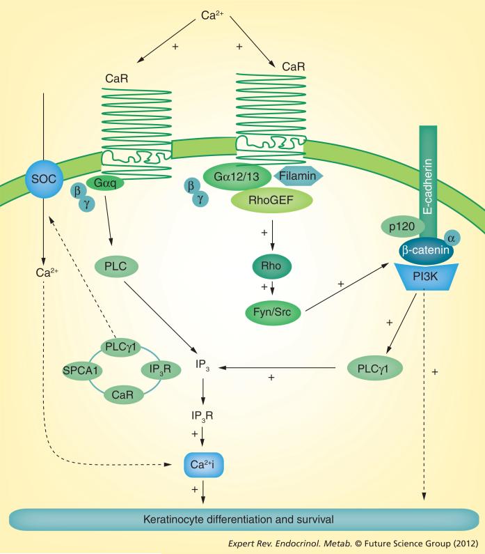
Calcium is the major regulator of keratinocyte differentiation in vivo and in vitro. A calcium gradient within the epidermis promotes the sequential differentiation of keratinocytes as they traverse the different layers of the epidermis to form the permeability barrier of the stratum corneum. Calcium promotes differentiation by both outside-in and inside-out signaling. A number of signaling pathways involved with differentiation are regulated by calcium, including the formation of desmosomes, adherens junctions and tight junctions, which maintain cell-cell adhesion and play an important intracellular signaling role through their activation of various kinases and phospholipases that produce second messengers that regulate intracellular free calcium and PKC activity, critical for the differentiation process. The calcium receptor plays a central role by initiating the intracellular signaling events that drive differentiation in response to extracellular calcium. This review will discuss these mechanisms.


Figures

Figure 1
Figure 1. Microanatomy of the epidermis
The epidermis is composed of four functionally different layers that may vary in thickness depending on location and species. The stratum basale rests on the basal lamina and contains the stem cells. This layer is distinguished by its production of keratins 5 and 14. The VDR and the 25OHD-1α CYP27B1 responsible for producing the active metabolite of vitamin D (1,25[OH]2D) are found in the highest concentration in this layer. Differentiation is initiated as the cells move from the stratum basale to the stratum spinosum, where involucrin, transglutaminase-I and the keratins K1 and K10 are expressed. In the next layer, the stratum granulosum, profilaggrin and loricrin are produced and packaged in keratohyalin granules. This layer is also where lipids for the waterproofing of the permeability barrier are produced and packaged into lamellar bodies. The stratum corneum is the enucleated layer critical for barrier function containing the cornified envelope within and the lipid matrix without the cells. VDR: Vitamin D receptor. Adapted with permission from [133].
Figure 2
Figure 2. Signaling by the E-cadherin–catenin complex
Following the calcium switch, the E-cadherin–catenin complex forms in the plasma membrane. Extracellular calcium serves to link the extracellular domains of E-cadherin together, forming the adherens junctions (outside–in signaling). Within the cell, p120- and β-catenins bind to the juxtamembrane and cytoplasmic tail, respectively, of E-cadherin. γ-catenin (not shown) competes with β-catenin for this site, but in human keratinocytes it does not appear to have a major role in calcium-induced keratinocyte differentiation. α-catenin (not shown) attaches to β-catenin and links the complex to the cytoskeleton. β-catenin also serves as the binding site for PIP5K1α and PI3K, enzymes that phosphorylate PIP to PIP2 and PIP2 to PIP3, respectively. PIP3 activates PLC-γ1, which hydrolyzes PIP2 to IP3 and DAG, leading to the release of calcium from intracellular stores and activation of PKC, respectively. PIP3 also serves as a binding site for Akt and PDK1, which also contribute to the differentiation process. DAG: Diacylglycerol; ER: Endoplasmic reticulum; IP3: Inositol trisphosphate; IP3R: Inositol trisphosphate receptor; P: Phosphorylation; PIP: Phosphatidylinositol phosphate; PIP2: Phosphatidylinositol bisphosphate; PIP3: Phosphatidylinositol trisphosphate; PIP5K1α: Phosphatidylinositol 4 phosphate 5 kinase 1α.
Figure 3
Figure 3. The role of the calcium receptor in calcium-induced differentiation
Calcium stimulates intracellular signaling (inside–out signaling) through its binding to CaR. In one pathway, CaR activates the G protein Gαq, leading to PLC activation and generation of IP3 with the release of calcium from intracellular stores. This is the predominant mechanism for the initial spike in Ca2+i concentration following the calcium switch. CaR also activates Rho that, in turn, activates the src kinases Src and Fyn to tyrosine phosphorylate the catenins, enabling their binding to the E-cadherin–catenin complex. As noted in the legend of Figure 2, this results in PLC-γ1 activation, leading to a sustained increase in Ca2+ i concentration both by release of calcium from intracellular stores and by stimulating the influx of calcium through store-operated channels. The latter role may be played by PLC-γ1 as part of the complex in Golgi with the Golgi calcium pump SPCA1, CaR and the IP3 receptor. Ca2+i: Intracellular calcium; CaR: Calcium receptor; IP3: Inositol trisphosphate; IP3R: Inositol trisphosphate receptor; PLC: Phospholipase C; SOC: Store-operated channel.

References

    1. Green KJ, Simpson CL. Desmosomes: new perspectives on a classic. J. Invest. Dermatol. 2007;127(11):2499–2515. - PubMed
    1. Yuki T, Haratake A, Koishikawa H, Morita K, Miyachi Y, Inoue S. Tight junction proteins in keratinocytes: localization and contribution to barrier function. Exp. Dermatol. 2007;16(4):324–330. - PubMed
    1. Marchisio PC, Bondanza S, Cremona O, Cancedda R, De Luca M. Polarized expression of integrin receptors (α 6 β 4, α 2 β 1, α 3 β 1, and α v β 5) and their relationship with the cytoskeleton and basement membrane matrix in cultured human keratinocytes. J. Cell Biol. 1991;112(4):761–773. - PMC - PubMed
    1. Peltonen J, Larjava H, Jaakkola S, et al. Localization of integrin receptors for fibronectin, collagen, and laminin in human skin. Variable expression in basal and squamous cell carcinomas. J. Clin. Invest. 1989;84(6):1916–1923. - PMC - PubMed
    1. Guo M, Kim LT, Akiyama SK, Gralnick HR, Yamada KM, Grinnell F. Altered processing of integrin receptors during keratinocyte activation. Exp. Cell Res. 1991;195(2):315–322. - PubMed