. 2012;7(10):e48313.
doi: 10.1371/journal.pone.0048313. Epub 2012 Oct 29.

Effects of the oral administration of viable and heat-killed Streptococcus bovis HC5 cells to pre-sensitized BALB/c mice

Affiliations

Effects of the oral administration of viable and heat-killed Streptococcus bovis HC5 cells to pre-sensitized BALB/c mice

Aline D Paiva et al. PLoS One. 2012.

Abstract

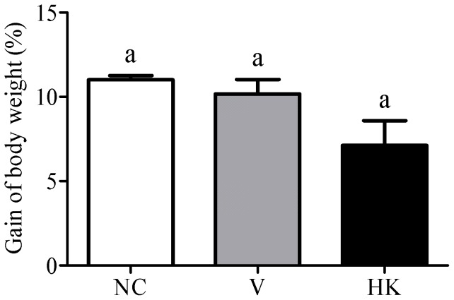
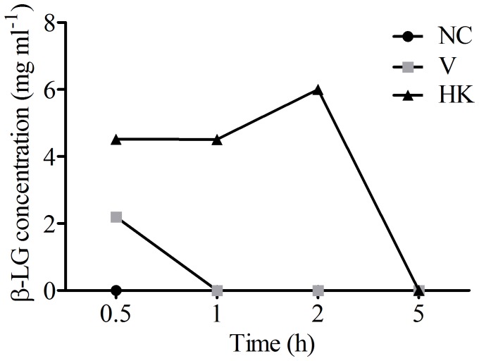
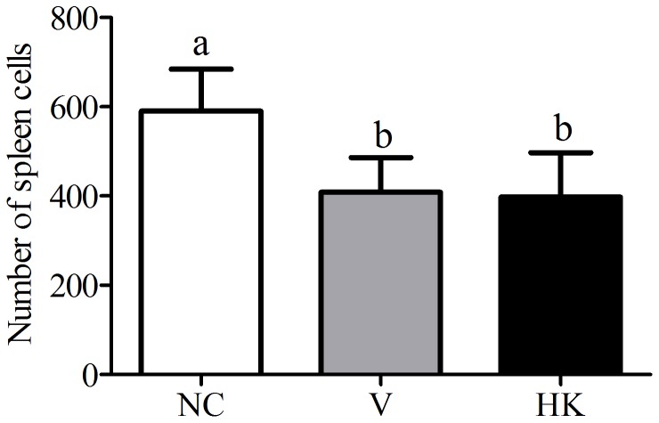
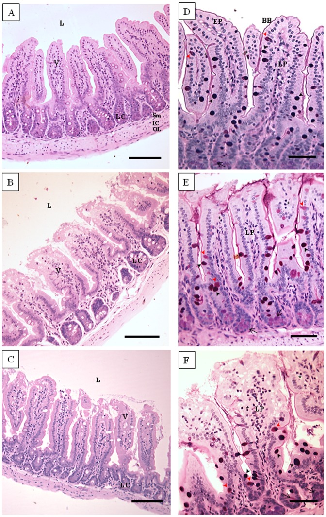
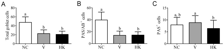
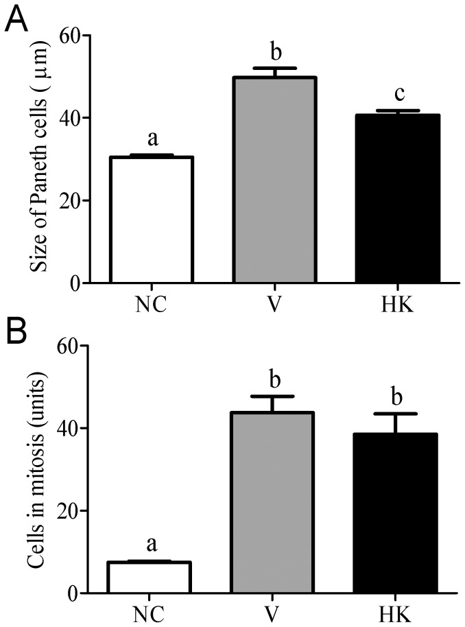
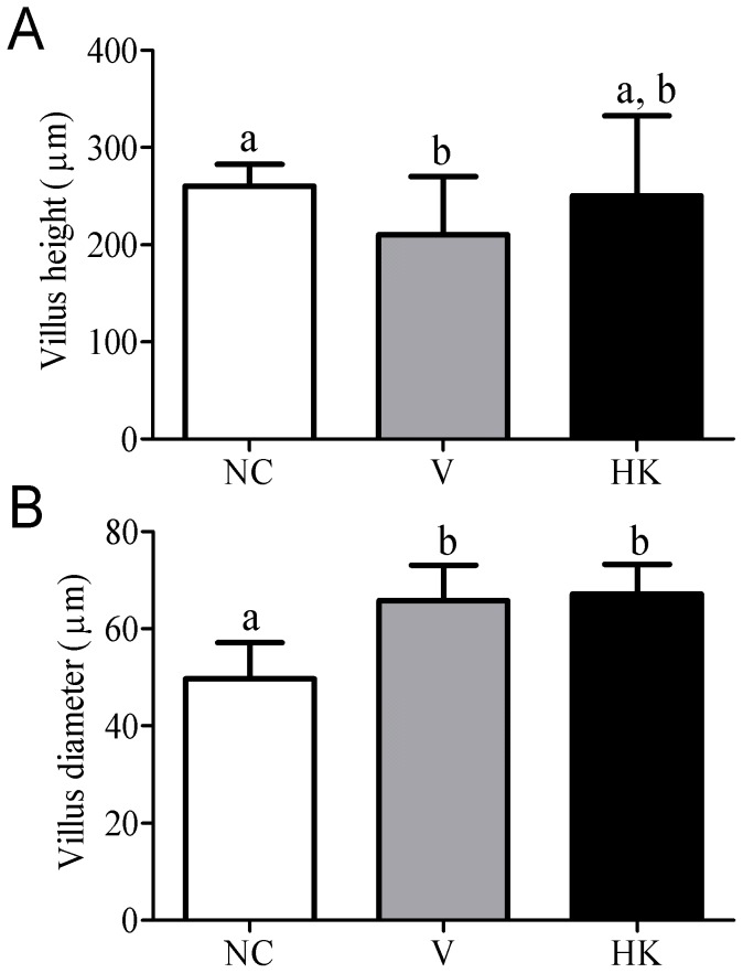
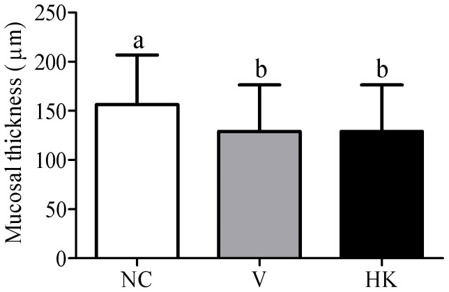
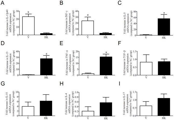
Antimicrobial peptides have been suggested as an alternative to classical antibiotics in livestock production and bacteriocin-producing bacteria could be added to animal feeds to deliver bacteriocins in the gastrointestinal (GI) tract of ruminant and monogastric animals. In this study, viable (V) and heat-killed (HK) Streptococcus bovis HC5 cells were orally administered to pre-sensitized mice in order to assess the effects of a bacteriocin-producing bacteria on histological parameters and the immune response of the GI tract of monogastric animals. The administration of V and HK S. bovis HC5 cells during 58 days to BALB/c mice did not affect weight gain, but an increase in gut permeability was detected in animals receiving the HK cells. Viable and heat killed cells caused similar morphological alterations in the GI tract of the animals, but the most prominent effects were detected in the small intestine. The oral administration of S. bovis HC5 also influenced cytokine production in the small intestine, and the immune-mediated activity differed between V and HK cells. The relative expression of IL-12 and INF-γ was significantly higher in the small intestine of mice treated with V cells, while an increase in IL-5, IL-13 and TNF-α expression was only detected in mice treated with HK cells. Considering that even under a condition of severe challenge (pre-sensitization followed by daily exposure to the same bacterial immunogen) the general health of the animals was maintained, it appears that oral administration of S. bovis HC5 cells could be a useful route to deliver bacteriocin in the GI tract of livestock animals.


Conflict of interest statement

Competing Interests: The authors have declared that no competing interests exist.

Figures

Figure 1
Figure 1. Effect of the oral administration of S. bovis HC5 on percent weight gain of BALB/c mice.
The weight of the animals is visualized as percentage of the animals' weight, which was calculated comparing the weight at the end of the experiment (day 58) to the weight at the day of the first sensitization (day 0). Each bar represents the mean value from six determinations with the standard error of the mean (SEM). Treatments with identical lowercase letters were considered not statistically significant by the Dunn's multiple comparison test (p>0.05). (NC) negative control group; (V) mice treated with viable S. bovis HC5 cells; (HK) mice treated with heat-killed S. bovis HC5 cells.
Figure 2
Figure 2. β-lactoglobulin levels in animal sera from the treatment groups.
An intragastrically dose of β-LG (20 mg) was administered as a bystander protein to the negative control group and the groups that received S. bovis HC5 (viable and heat-killed cells). At the indicated time points following β-LG administration, the levels of β-LG in mice sera were determined by FPLC. The results show an average of the β-LG level detected in a pool of animal's sera from each group. β-LG was not detected in all serum samples from negative control group. (NC) negative control group; (V) mice treated with viable S. bovis HC5 cells; (HK) mice treated with heat-killed S. bovis HC5 cells (HK).
Figure 3
Figure 3. Number of total spleen cells among the experimental groups.
Data represent the average±SEM. Differences among treatments were indicated by different lowercase letters and were considered statistically significant by the Dunn's multiple comparison test (p<0.05). (NC) negative control group; (V) mice treated with viable S. bovis HC5 cells; (HK) mice treated with heat-killed S. bovis HC5 cells.
Figure 4
Figure 4. Photomicrographs of histological sections of small intestine of the animal groups studied.
Jejunum segments were collected and processed for optical microscopy analysis at the end of the experiment. (NC), negative control group, figures A and D; (V) mice treated with viable S. bovis HC5 cells, figures B and E; (HK) mice treated with heat-killed S. bovis HC5 cells, figures C and F. The sections were stained with hematoxylin and eosin (HE; right panel) or PAS/Alcian Blue (left panel). Abbreviations: L: lumen; EP: simple cuboidal epithelium; BB: brush border; V: villum; LP: lamina propria; LC: Lieberkühn crypt; E: edema; V: blood vessel; Sm: submucosa; IC: inner circular muscle layer; OL: outer longitudinal muscle layer. The asterisks indicate intraepithelial lymphocytes; simple arrow indicates Paneth cells. Black arrow head indicates goblet cells PAS/AB+; red arrow head indicates PAS+ cells. Right panel – Scale bar: 100 µm; Left panel – Scale bar: 50 µm.
Figure 5
Figure 5. Comparison of the number of total goblet cells and mucin production among the experimental groups.
(A) total number of cells; (B) PAS/AB+ cells; (C) PAS+ cells. Data were shown as average±SEM. Differences among treatments were indicated by different lowercase letters and were considered statistically significant by the Dunn's multiple comparison test (p<0.05). (NC) negative control group; (V) mice treated with viable S. bovis HC5 cells; (HK) mice treated with heat-killed S. bovis HC5 cells (HK).
Figure 6
Figure 6. Size of Paneth cells (A) and number of cells in mitosis (B) at the small intestinal crypts of animals from different experimental groups.
Data were shown as average±SEM. Differences among treatments were indicated by different lowercase letters and were considered statistically significant by the Dunn's multiple comparison test (p<0.05). (NC) negative control group; (V) mice treated with viable S. bovis HC5 cells; (HK) mice treated with heat-killed S. bovis HC5 cells (HK).
Figure 7
Figure 7. Height (A) and diameter (B) of the small intestinal villi of animals from different experimental groups.
Data were shown as average±SEM. Differences among treatments were indicated by different lowercase letters and were considered statistically significant by the Dunn's multiple comparison test (p<0.05). (NC) negative control group; (V) mice treated with viable S. bovis HC5 cells; (HK) mice treated with heat-killed S. bovis HC5 cells (HK).
Figure 8
Figure 8. Mucosal thickness of the large intestine of the mice at the experimental groups.
Data were shown as average±SEM. Differences among treatments were indicated by different lowercase letters and were considered statistically significant by the Dunn's multiple comparison test (p<0.05). (NC) negative control group; (V) mice treated with viable S. bovis HC5 cells; (HK) mice treated with heat-killed S. bovis HC5 cells (HK).
Figure 9
Figure 9. Cytokine production in small intestine of five-week old female BALB/c mice that received S. bovis HC5 cells.
Segments of jejunum were collected on day 58 of the experiment and mRNA was extracted. The relative expression of the interleukin genes determined by real time-PCR was calculated in reference to the β-actin in each sample. (A) IL-12p40; (B) INF-γ; (C) IL-5; (D) IL-13; (E) TNF-α; (F) TGF-β; (G) IL-10; (H) IL-4; (I) IL-17. Results are shown as the mean value±SEM of duplicate samples from three independent mice within the V and HK groups, relative to the NC group. Differences between the relative cytokine expression in small intestine from mice treated with viable S. bovis HC5 cells (V) and treated with heat-killed S. bovis HC5 cells (HK) were indicated by (*) and were considered statistically significant by the t test (p<0.05).

References

    1. Gill HS, Rutherfurd KJ (2001) Immune enhancement conferred by oral delivery of Lactobacillus rhamnosus HN001 in different milk-based substrates. J. Dairy Res. 68: 611–616. - PubMed
    1. Nagafuchi S, Takahashi T, Yajima T, Kuwata T, Hirayama K, et al. (1999) Strain dependency of the immunopotentiating activity of Lactobacillus delbrueckii subsp. bulgaricus. Biosci. Biotech. Biochem. 63: 474–479. - PubMed
    1. Chiang YJ, Kolw HK, Brown K, Naramura M, Fukuhara S, et al. (2000) CbI-b regulates the CD28 dependence of T-cell activation. Nature 403: 216–220. - PubMed
    1. Parra MD, Martinez de Moretin BE, Cobo JM, Mateos A, Martinez JA (2004) Daily ingestion of fermented milk containing Lactobacillus casei DN114001 improves innate-defense capacity in healthy middle-aged people. J. Physiol. Biochem. 60: 85–91. - PubMed
    1. Valeur N, Engel P, Carbajal N, Connolly E, Ladefoged K (2004) Colonization and immunomodulation by Lactobacillus reuteri ATCC 55730 in the human gastrointestinal tract. Appl. Environ. Microbiol. 70: 1176–1181. - PMC - PubMed

Publication types

MeSH terms