. 2012;7(10):e48475.
doi: 10.1371/journal.pone.0048475. Epub 2012 Oct 29.

Urea amidolyase (DUR1,2) contributes to virulence and kidney pathogenesis of Candida albicans

Affiliations

Urea amidolyase (DUR1,2) contributes to virulence and kidney pathogenesis of Candida albicans

Dhammika H M L P Navarathna et al. PLoS One. 2012.

Abstract

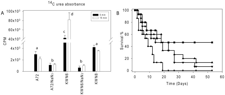
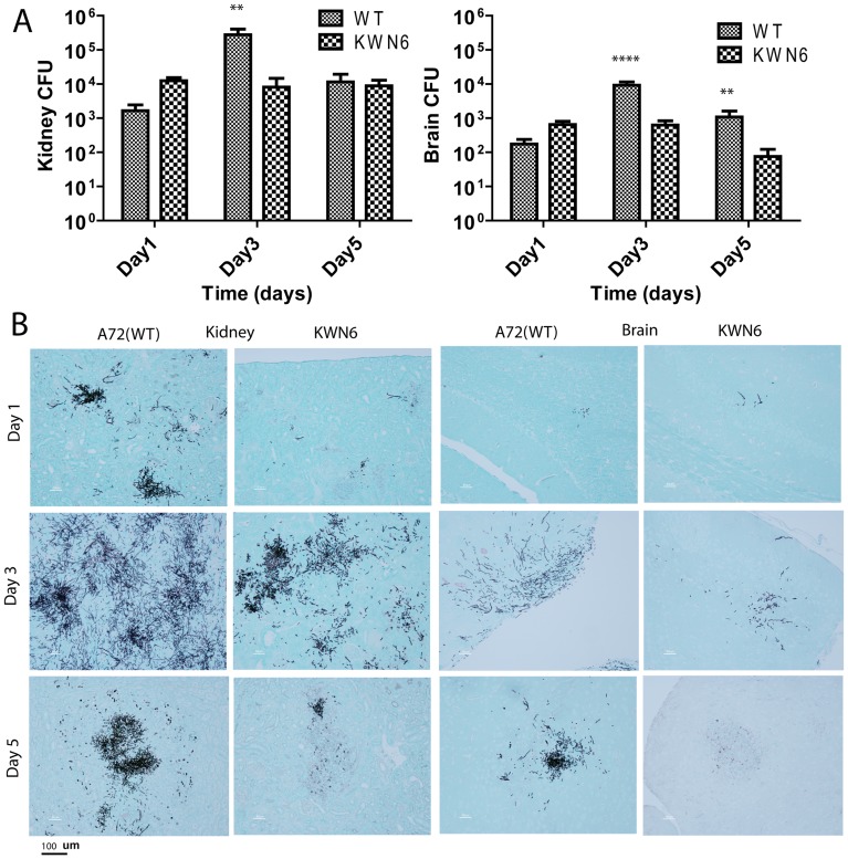
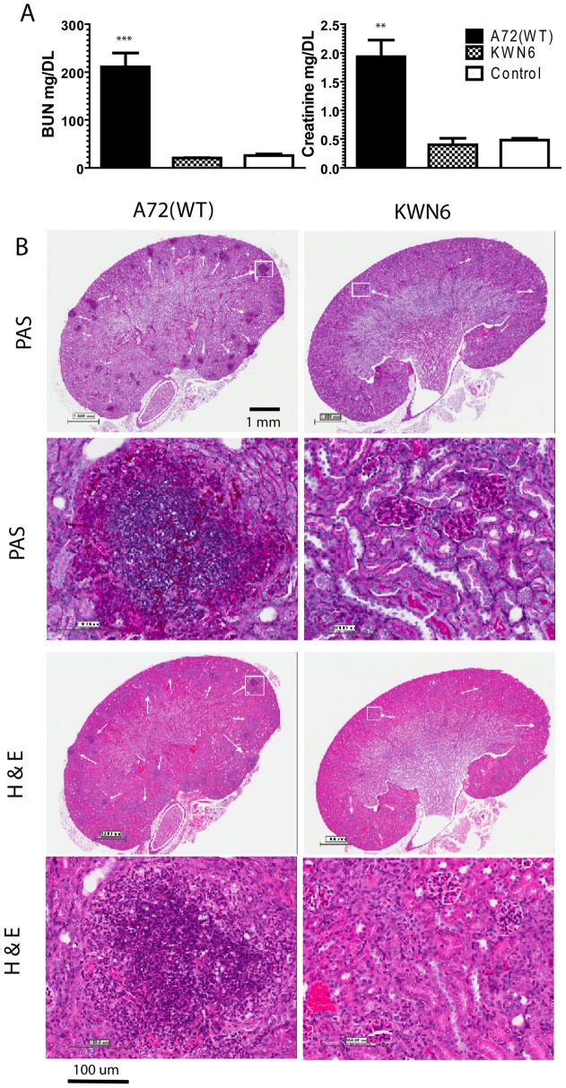
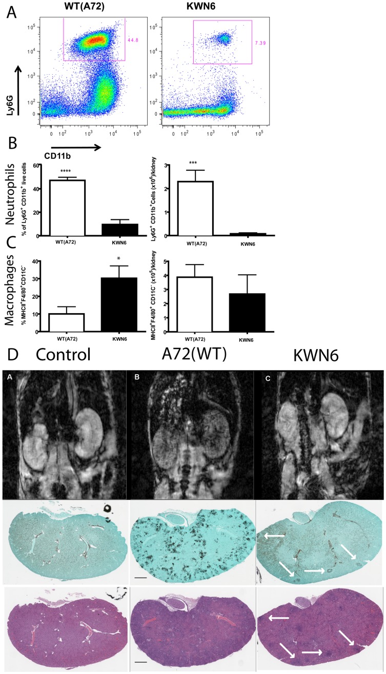
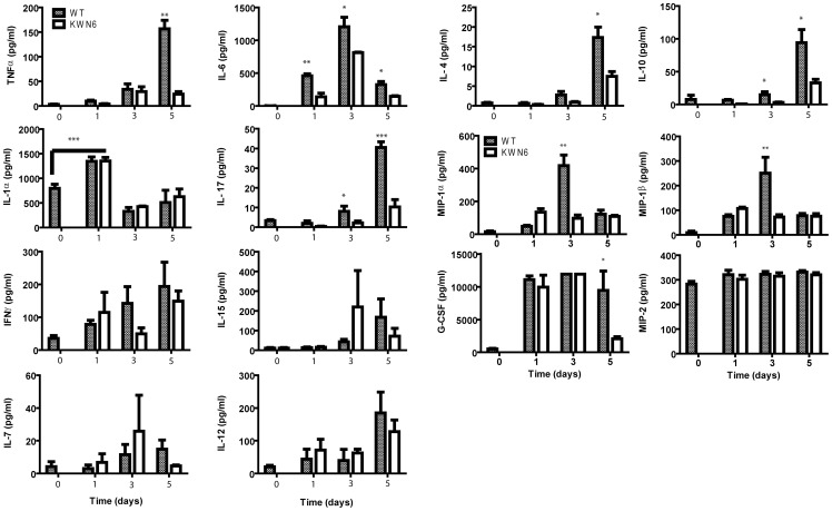
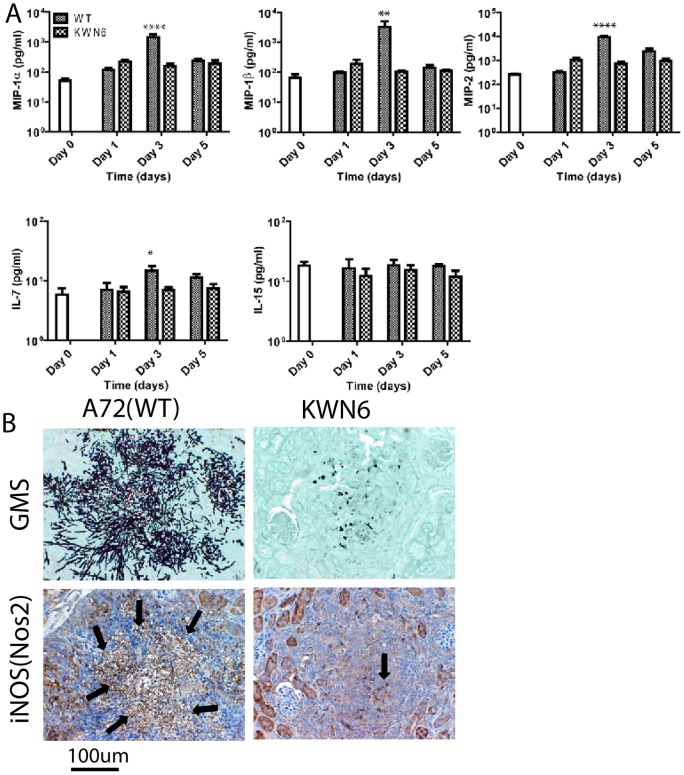
The intracellular enzyme urea amidolyase (Dur1,2p) enables C. albicans to utilize urea as a sole nitrogen source. Because deletion of the DUR1,2 gene reduces survival of C. albicans co-cultured with a murine macrophage cell line, we investigated the role of Dur1,2p in pathogenesis using a mouse model of disseminated candidiasis. A dur1,2Δ/dur1,2Δ strain was significantly less virulent than the wild-type strain, showing significantly higher survival rate, better renal function, and decreased and less sustained fungal colonization in kidney and brain. Complementation of the mutant restored virulence. DUR1,2 deletion resulted in a milder host inflammatory reaction. Immunohistochemistry, flow cytometry, and magnetic resonance imaging showed decreased phagocytic infiltration into infected kidneys. Systemic cytokine levels of wild-type mice infected with the dur1,2 mutant showed a more balanced systemic pro-inflammatory cytokine response. Host gene expression and protein analysis in infected kidneys revealed parallel changes in the local immune response. Significant differences were observed in the kidney IL-1 inflammatory pathway, IL-15 signaling, MAP kinase signaling, and the alternative complement pathway. We conclude that Dur1,2p is important for kidney colonization during disseminated candidiasis and contributes to an unbalanced host inflammatory response and subsequent renal failure. Therefore, this Candida-specific enzyme may represent a useful drug target to protect the host from kidney damage associated with disseminated candidiasis.


Conflict of interest statement

Competing Interests: The authors have declared that no competing interests exist.

Figures

Figure 1
Figure 1. Dur1,2p function in urea metabolism and virulence.
(A). Urea uptake by WT (A72), dur1,2Δ/dur1,2Δ mutant and reconstituted C. albicans strain dur1,2Δ::DUR1,2/dur1,2Δ::DUR1,2. Radioactive uptake into 4−5×10⁶ cells is presented after exposure to 14C urea for 5 and 15 min in the absence or presence of sodium azide. Values shown are the average of triplicate experiments ± SE. Closed bars indicate 5 min uptake, and open bars indicate 15 min uptake of radioactive urea. (B). Effect of DUR1,2 deletion on mouse mortality following intravenous infection. Survival of mice injected with WT C. albicans A72 (•), the null mutant KWN6 (▪), single copy reconstituted KWN7 (▾), fully reconstituted KWN8 (▴), and an uninfected control group of 5 mice (not shown) was assessed daily. Each infected group contained 15 mice.
Figure 2
Figure 2. Dur1,2p expression promotes kidney and brain colonization.
(A). Kidney and brain fungal burdens of mice infected with parental A72 and mutant KWN6 strains. Gray bars represent mean CFU of WT infected left kidney homogenates determined by CFU on BiGGY agar from six representative serial dilutions from each kidney, representing three mice per time point infected. Checkered bars represent similar mean values for mice administered with KWN6. (B) GMS-stained mouse kidney and brain tissues to determine C. albicans colonization. Representative kidney and brain sections harvested day 1, 3 and 5 PI from mice infected with WT or KWN6 are shown. Scale bar indicates 100 μm.
Figure 3
Figure 3. Dur1,2p expression impairs kidney function and increases inflammation.
A. Kidney function tests of mice infected with A72 (WT) vs KWN6 at day 3 PI. Open bars represent serum levels of BUN and creatinine in non-infected control mice. Checkered bars represent KWN6 infected mice, and closed bars represent A72 (WT) infected mice. Note the levels of BUN and creatinine are significantly higher in WT infected sera compared with KWN6 infected mice. B. PAS and H&E-stained mouse kidney tissues to assess inflammatory responses and tissue necrosis caused by C. albicans colonization. Representative sections of infected mouse kidneys (group of 3 mice) harvested at day 3 PI are shown. Scale bars indicate magnification. Arrows indicate regions of PMN and C. albicans accumulation in PAS stains and PMN inflammatory foci in H&E stains. Demarcated regions by white squares are shown under higher magnifications in the lower panels.
Figure 4
Figure 4. Dur1,2p expression increases inflammatory cell infiltration in infected kidneys. A.
Representative FACS plots of kidney neutrophils at day 3 PI with WT and KWN6. B Neutrophils expressed as percent of CD45+ cells (left panel) and as absolute numbers per kidney (right). C. Macrophages expressed as percent of CD45+ cells (left panel) and as absolute numbers per kidney (right). D. Top panels show transverse MR images of 8 week old BALB/c mice 24 h post injection of USPIO agent. Images are representative of at least three non-infected mice per group (left panels), Candida infected (middle panels), and infected with KWN6 strain (right panels). Small localized candida colonization by KWN6 is indicated by arrowheads. Massive phagocytic infiltration in kidneys infected with WT is demonstrated by T2* spoiled signal in the kidney cortex and medulla. Lower panels show GMS and H&E stained sections indicating colonization and inflammatory reactions in the respective kidneys imaged in the top panels.
Figure 5
Figure 5. Effects of Dur1,2p expression in C. albicans on host serum cytokines and chemokines.
Serum levels of the indicated proteins were assessed at 1 to 5 days PI for mice infected iv with WT (gray bars) or KWN6 (open bars). Values at time 0 are mean values determined for sera from 5 control mice, and other data are mean ± standard deviation for 3 mice at each time point. *  =  p<0.05; **  =  p<0.01; ***  = p<0.001.
Figure 6
Figure 6. Effects of Dur1,2p expression in local inflammatory markers in infected kidneys.
(A) Concentrations of cytokines and chemokines in kidney extracts from C. albicans-infected mice at 1 to 5 days PI. Values at time 0 are mean values determined for sera from 5 control mice, and other data are mean ± standard deviation for 5 mice at each time point. Mice were infected iv using the wild type strain A72 or the dur1,2 mutant strain KWN6. Asterisks above an individual bar indicate that the serum value for that protein was significantly greater in mice infected with WT C. albicans than in mice infected with KWN6, *  =  p<0.05; **  =  p<0.01; **** = p<0.0001. (B) Kidney iNOS expression in cellular infiltrates. Left panels represent WT infected kidneys, and right panels represent KWN6 infected kidneys. Upper panels show Candida colonization detected using GMS stain. Lower two panels show micrographs from the same area of an adjacent section stained using an antibody. Arrows indicate regions positive for iNOS (brown stain). Scale bar  = 100 µm.

Similar articles

Cited by

References

    1. Vincent JL, Norrenberg M (2009) Intensive care unit-acquired weakness: framing the topic. Crit Care Med 37: S296–298. - PubMed
    1. Trick WE, Fridkin SK, Edwards JR, Hajjeh RA, Gaynes RP (2002) Secular trend of hospital-acquired candidemia among intensive care unit patients in the United States during 1989–1999. Clin Infect Dis 35: 627–630. - PubMed
    1. Calderone AR, Clancy JC, editors (2012) Candida and candidiasis. 2 ed: Amer Society for Microbiology. 534 p.
    1. Odds FC (1988) Candida and candidiasis. London: Bailliere Tindall.
    1. Navarathna DH, Hornby JM, Krishnan N, Parkhurst A, Duhamel GE, et al. (2007) Effect of farnesol on a mouse model of systemic candidiasis, determined by use of a DPP3 knockout mutant of Candida albicans. Infect Immun 75: 1609–1618. - PMC - PubMed

Publication types

MeSH terms