. 2012;7(11):e44826.
doi: 10.1371/journal.pone.0044826. Epub 2012 Nov 5.

Relevance of brain lesion location to cognition in relapsing multiple sclerosis

Affiliations

Relevance of brain lesion location to cognition in relapsing multiple sclerosis

Francesca Rossi et al. PLoS One. 2012.

Abstract

Objective: To assess the relationship between cognition and brain white matter (WM) lesion distribution and frequency in patients with relapsing-remitting multiple sclerosis (RR MS).

Methods: MRI-based T2 lesion probability map (LPM) was used to assess the relevance of brain lesion location for cognitive impairment in a group of 142 consecutive patients with RRMS. Significance of voxelwise analyses was p<0.05, cluster-corrected for multiple comparisons. The Rao Brief Repeatable Battery was administered at the time of brain MRI to categorize the MS population into cognitively preserved (CP) and cognitively impaired (CI).

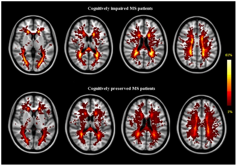
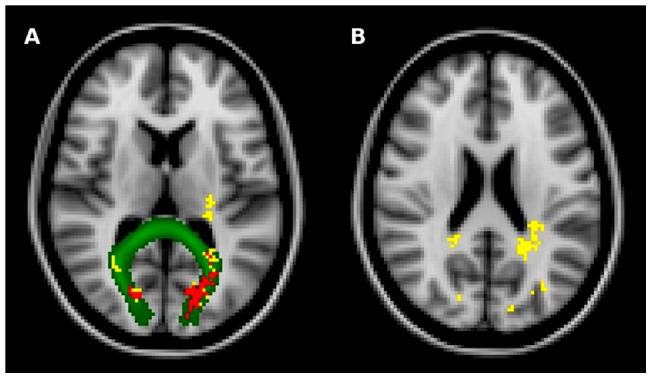
Results: Out of 142 RRMS, 106 were classified as CP and 36 as CI. Although the CI group had greater WM lesion volume than the CP group (p = 0.001), T2 lesions tended to be less widespread across the WM. The peak of lesion frequency was almost twice higher in CI (61% in the forceps major) than in CP patients (37% in the posterior corona radiata). The voxelwise analysis confirmed that lesion frequency was higher in CI than in CP patients with significant bilateral clusters in the forceps major and in the splenium of the corpus callosum (p<0.05, corrected). Low scores of the Symbol Digit Modalities Test correlated with higher lesion frequency in these WM regions.

Conclusions: Overall these results suggest that in MS patients, areas relevant for cognition lie mostly in the commissural fiber tracts. This supports the notion of a functional (multiple) disconnection between grey matter structures, secondary to damage located in specific WM areas, as one of the most important mechanisms leading to cognitive impairment in MS.


Conflict of interest statement

Competing Interests: The authors have read the journal's policy and have the following conflicts: Emilio Portaccio serves on a scientific advisory board for Biogen Idec and receives research support from Merck Serono, Biogen Idec, Bayer Schering Pharma, and Sanofi- Aventis. Benedetta Goretti serves on a scientific advisory board for Biogen Idec; has received speaker honoraria from Biogen Idec and Novartis; and receives research support from Biogen Idec, Merck Serono, and Novartis. Maria Pia Amato serves on scientific advisory boards for and has received speaker honoraria and research support from Biogen Idec, Merck Serono, Bayer Schering Pharma, and Sanofi-Aventis and serves on the editorial board of BMC Neurology. Nicola De Stefano serves on a scientific advisory board for Merck Serono; has received funding for travel from Teva Pharmaceutical Industries Ltd. and Merck Serono; has received speaker honoraria from Teva Pharmaceutical Industries Ltd., BioMS Medical, Biogen-Dompé AG, Bayer Schering Pharma, and Merck Serono; and receives research support from the Italian MS Society. Francesca Rossi, Antonio Giorgio, Marco Battaglini, and Maria Laura Stromillo have nothing to disclose. This does not alter the authors' adherence to all the PLOS ONE policies on sharing data and materials.

Figures

Figure 1
Figure 1. LPM in cognitively impaired and cognitively preserved patients with MS.
T2-weighted lesion probability maps in cognitively impaired (upper panel, n = 36) and cognitively preserved (lower panel, n = 106) patients with MS. The color overlay created on top of the Montreal Neurological Institute standard brain shows the probability of each voxel containing a lesion in each patient group. The color bar denotes the probability range. The maximum local probability for lesions was higher in cognitively impaired patients (61% peak probability in the forceps major) than in cognitively preserved patients (37% peak probability in the posterior corona radiata). Images are shown in radiological convention.
Figure 2
Figure 2. Clusters of high lesion frequency in cognitively impaired MS patients.
Yellow shows the clusters of voxels where lesions, after controlling for age and sex, were more frequent (p<0.05, corrected) in cognitively impaired (n = 36) than in cognitively preserved (n = 106) patients with MS (forceps major in A and splenium of the corpus callosum in B). Red shows the clusters of voxels that also survived correction for T2-LV (forceps major, in A). Green represents the WM fiber tract (forceps major, in A) obtained from the FSL probabilistic tractography atlas. Background image is the MNI152 standard space image. Images are shown in radiological convention.

Similar articles

Cited by

References

    1. Ferreira ML (2010) Cognitive deficits in multiple sclerosis: a systematic review. Arq Neuropsiquiatr 68: 632–641. - PubMed
    1. Chiaravalloti ND, DeLuca J (2008) Cognitive impairment in multiple sclerosis. Lancet Neurol 7: 1139–1151. - PubMed
    1. DeLuca J, Chelune GJ, Tulsky DS, Lengenfelder J, Chiaravalloti ND (2004) Is speed of processing or working memory the primary information processing deficit in multiple sclerosis? J Clin Exp Neuropsychol 26: 550–562. - PubMed
    1. Filippi M, Rocca MA, Benedict RH, DeLuca J, Geurts JJ, et al. (2010) The contribution of MRI in assessing cognitive impairment in multiple sclerosis. Neurology 75: 2121–2128. - PMC - PubMed
    1. Amato MP, Portaccio E, Stromillo ML, Goretti B, Zipoli V, et al. (2008) Cognitive assessment and quantitative magnetic resonance metrics can help to identify benign multiple sclerosis. Neurology 71: 632–638. - PubMed

Publication types