. 2012;7(11):e48470.
doi: 10.1371/journal.pone.0048470. Epub 2012 Nov 7.

Neurons of the dentate molecular layer in the rabbit hippocampus

Affiliations

Neurons of the dentate molecular layer in the rabbit hippocampus

Francisco J Sancho-Bielsa et al. PLoS One. 2012.

Abstract

The molecular layer of the dentate gyrus appears as the main entrance gate for information into the hippocampus, i.e., where the perforant path axons from the entorhinal cortex synapse onto the spines and dendrites of granule cells. A few dispersed neuronal somata appear intermingled in between and probably control the flow of information in this area. In rabbits, the number of neurons in the molecular layer increases in the first week of postnatal life and then stabilizes to appear permanent and heterogeneous over the individuals' life span, including old animals. By means of Golgi impregnations, NADPH histochemistry, immunocytochemical stainings and intracellular labelings (lucifer yellow and biocytin injections), eight neuronal morphological types have been detected in the molecular layer of developing adult and old rabbits. Six of them appear as interneurons displaying smooth dendrites and GABA immunoreactivity: those here called as globoid, vertical, small horizontal, large horizontal, inverted pyramidal and polymorphic. Additionally there are two GABA negative types: the sarmentous and ectopic granular neurons. The distribution of the somata and dendritic trees of these neurons shows preferences for a definite sublayer of the molecular layer: small horizontal, sarmentous and inverted pyramidal neurons are preferably found in the outer third of the molecular layer; vertical, globoid and polymorph neurons locate the intermediate third, while large horizontal and ectopic granular neurons occupy the inner third or the juxtagranular molecular layer. Our results reveal substantial differences in the morphology and electrophysiological behaviour between each neuronal archetype in the dentate molecular layer, allowing us to propose a new classification for this neural population.


Conflict of interest statement

Competing Interests: The authors have declared that no competing interests exist.

Figures

Figure 1
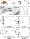
Figure 1. Hippocampal structure, nissl staining and cell counts along development.
A, lateral view of the left hemisphere of a rabbit (P30), vertical lines show the levels of the transversal sections studied. B, Drawing of the silhouettes; profiles of the transversal sections, from anterior to posterior, dark gray marks the hippocampus. C, Drawing of transparent sections with the exception of the hippocampus. D, Drawing as only an opaque molecular layer. E, Nissl staining in a transversal section of a P12 rabbit showing the extent of the molecular layer. F, Enlarged view of the molecular layer with some neuronal somata and abundant glial cells. G, Histogram showing the estimated mean numbers of the neurons in the molecular layer of one hemisphere for postnatal, young and adult rabbits (vertical bars mean deviation). H, Plots of the estimated numbers of neurons in the molecular layer of one hemisphere after the stereological study. I, Enlarged view of the first two months of life. J, Plots of the numbers of glia. K, Enlarged view of the first two months of life.
Figure 2
Figure 2. Stainings of globoid and vertical neurons.
A, Calbindine immunostaining of a globoid soma with very thin dendrites, B, Golgi-impregnated globoid neuron, C, NADPH diaphorase histochemical staining of a vertical neuron, D, Calretinin immunostaining of a vertical neuron. Scale bars, A–D: 25 µm.
Figure 3
Figure 3. Electron micrographs of hippocampal molecular layer neurons.
Different cellular archetypes were firstly injected with Lucifer Yellow and then filled with biocytin (see methods). In these stained neurons, GABA was detected using a post-embedding immunogold (immunoparticles 10 nm) method (arrows). A1: Biocytin-stained globoid neuron soma. A2, Soma magnification showing immunoparticles for GABA mainly detected in the soma. Asterisk shows a glial cell close to the injected neuron. B1, B2: Sarmentous neuron soma and magnification without immunoparticles. C1, C2: Vertical neuron and amplification of immunogold particles detected in the soma (arrows) and presynaptic contacts (crossed arrows). D1, D2: Ectopic granular neuron. Immunogold particles were detected in presynaptic contacts (crossed arrows) localized along somatic and dendritic plasma membrane. E, F, G and H micrographs show, respectively, inverted pyramidal, polymorphic, large horizontal and small horizontal neurons with immunoparticles magnification mainly located in the soma. Scale bars, A–H: 10 µm.
Figure 4
Figure 4. Camera lucida drawings and electrophysiological recording of globoid and vertical neurons.
A, Globoid neuron with axon (ax) in the hippocampal fissure (hp) and dendrites crossing the granular layer (gr) reaching the hilus (hi). B, drawings of different globoid neurons. C, electrophysiological recording of action potentials in a globoid neuron elicited by a stimulus of 300 ms. D, E, drawings of vertical neurons. Scale bars, A–E: 25 µm.
Figure 5
Figure 5. Stainings of ectopic granular and small horizontal neurons.
A, NADPH diaphorase histochemical staining of an ectopic granular neuron. B, ABC-diaminobenzidine-nickel staining of biocytin injected into a granular ectopic neuron with an axon (arrow head) crossing the granular layer. C, NADPH diaphorase histochemical staining of a small horizontal neuron. D, idem, parvalbumin immunostaining. E, idem, calretinin immunostaining. F, Golgi impregnated small horizontal neuron (arrow head) and two granule cells (arrows). Scale bars, A–F: 25 µm.
Figure 6
Figure 6. Camera lucida drawings and electrophysiological recordings of granular ectopic and small horizontal neurons.
A, B, drawings of ectopic granular neurons in A, the axon crossing the granular layer, bifurcates and arborization in the hilus (hi). C, electrophysiological record from an ectopic granular neuron. D, E, drawings of the small horizontal neurons. Observed in D, the profuse axonal arborization close to the hippocampal fissure (hp). F, electrophysiological recording of a small horizontal neuron. Scale bars, A–F: 25 µm.
Figure 7
Figure 7. Stainings of inverted pyramidal and large horizontal neurons.
A, NADPH diaphorase histochemical staining of an inverted pyramidal neuron. B, ABC-DAB-nickel staining of biocytin injected into an inverted pyramidal neuron. C, NADPH diaphorase histochemical staining of a large horizontal neuron in the inferior blade of the dentate gyrus. D, Golgi impregnation of a large horizontal neuron. E, parvalbumin immunostaining of a large horizontal neuron in the superior blade of the dentate gyrus; note the counterstained granular layer. Scale bars, A–E, 25 µm.
Figure 8
Figure 8. Camera lucida drawings and electrophysiological recordings of inverted pyramidal and large horizontal neurons.
A, B, Drawings of inverted pyramidal neurons. The neuron in A is that shown in Fig. 6B; note the place of the axonal hillock marked by triangles. C, electrophysiological recording of an inverted pyramidal neuron. D, E, drawings of large horizontal neurons. F, electrophysiological recording of a large horizontal neuron. Scale bars, A–E: 25 µm.
Figure 9
Figure 9. Stainings of sarmentous and polymorphic neurons.
A, NADPH diaphorase histochemical staining of a sarmentous neuron (arrow) (inverted pyramidal on the left and globoid on the right). B, Golgi impregnation of a sarmentous neuron (arrow). C, ABC-DAB-nickel staining of biocytin injected into a polymorphic neuron with two dendrites crossing the granular layer (gr). D, Golgi impregnation of a polymorphic neuron. E, parvalbumin immunostaining of a polymorphic neuron, toluidine blue counterstaining. Scale bars, A–E: 25 µm.
Figure 10
Figure 10. Camera lucida drawings and electrophysiological recordings of sarmetous and polymorphic neurons.
A, B, Drawings of sarmentous neurons. C, electrophysiological recording of a sarmentous neuron. D, E, drawings of polymorphic neurons; note the dense axonal arborization in D distributed by the outer and middle molecular layers. F, elecrophysiological record of a polymorphic neuron. Scale bars, A–E: 25 µm.
Figure 11
Figure 11. Morphometry and distribution of the neuronal somata in the molecular layer of the dentate gyrus.
A, Profiles of neuronal somata from different neurons taken as prototypes; the length of the two principal axes and the perimeter length were the main parameters analyzed, (v- vertical neuron, g- globular neuron, e- ectopic granular neuron, sh- small horizontal neuron, lh- large horizontal neuron, s- sarmentous neuron, P- polymorphic neuron). B, Frequency of location of neuronal types in the inner, middle and outer molecular layer strata (from a pool of 120 neurons from a 30-day-old rabbit; 15 neurons/type). C, Plots of the minor axis value against the major axis value; observe how specific neurons segregate in distinct populations. D, Idem, plots of the surface value of the profile (soma area) (ordinate) against the axes ratio (minor/major) and some types also segregate. E, Plots of the minor axis values (abscise) in front of the major axis values (ordinate) of the neurons which are more frequent in the outer molecular: sarmentous, inverted pyramidals and small horizontals. F, Idem, for the more frequent neurons in the middle molecular: verticals, globoid and polymorphic. G, Idem, for those more frequently located in the inner molecular: ectopic granular and large horizontal. Scale bars, A–G: 25 µm.
Figure 12
Figure 12. Diagram of distribution and putative role of the eight identified archetypes in the hippocampal circuitry, and their temporal evolution in the early postnatal period.
A, Drawing composed of individual camera lucida reconstructions located according to the archetype preferred position in the layer. From left to right: vertical, polymorphic, large horizontal, sarmentous, inverted pyramidal, globoid, ectopic granular and small horizontal neuron. In addition, this drawing shows the main afferent and efferent projections of each neuronal which suggest reveal their putative roles in the hippocampal circuitry. The inhibitory or excitatory nature of each neuronal archetype is represented by (−) and (+) symbols respectively. Scale bars, 25 µm. B, Relative frequency distributions at P0, P3, P6, P10, P14 and P34 which show a moderate increase of globoid neurons and a simultaneous decrease of small horizontals.
Figure 13
Figure 13. Retrogradely labeled neurons in the molecular layer of the rabbit gyrus dentatus.
A, Injection place of DiL; note the red track of the microsyringe in the subiculum. B, Red fluorescent labeling in a cell soma located in the molecular layer (40×). C, Impalement of cell somata and the injection of Lucifer Yellow; note the bright signal from the soma and from some thin projections. D, Different depth focus plane of the impaled soma and surroundings (double illumination with transmitted light plus DiL fluorescence); note the presence of several cell somata closely attached to the labeled soma. E, Microglia cell surrounding a toluidine blue stained neuronal soma in the molecular layer of an 8-year-old rabbit; (Timm-Danscher staining for ionic zinc). F, Confocal microscopic image of a retrogradelly labeled soma and associated glial cells; observe the very thin cell processes from an oligodendrocyte attached to neuronal somata whose processes appear less intensively labeled. G, Confocal planes pictures of the cell shown in F (3); observe the close attachment of up to 6 cell somata with different histological appearances, one of which is clearly identified as oligodendroglia (2). H, Confocal plane pictures from a retrogradely labeled neuronal soma in the molecular layer, also displaying attached glial cells; here one of the glial cells (3) is an astrocyte with a vascular end foot process. Scale bars, A: 100 µm; B–H: 10 µm.

Similar articles

Cited by

References

    1. Amaral DG, Witter MP (1989) The three-dimensional organization of the hippocampal formation: a review of anatomical data. Neuroscience 31 571–591: 0306–4522(89)90424-7. - PubMed
    1. Ramón y Cajal S (1883) Estructura del Asta de Amon. Anal Soc Esp Hist Nat 53–114.
    1. Lorente de Nó R (1934) Studies on the structure of the cerebral cortex. II. Continuation of the study of the Ammonic System. J Psychol Neurol 46: 113–177.
    1. Stensaas LJ (1967) The development of hippocampal and dorsolateral pallial regions of the cerebral hemisphere in fetal rabbits. III. Twenty-nine millimeter stage, marginal lamina. J Comp Neurol 130: 149–162. - PubMed
    1. Stensaas LJ (1967) The development of hippocampal and dorsolateral pallial region of the cerebral hemisphere in fetal rabbits. V. Sixty millimeter stage, glial cell morphology. J Comp Neurol 131: 423–436. - PubMed

Publication types