. 2012;7(11):e48775.
doi: 10.1371/journal.pone.0048775. Epub 2012 Nov 7.

Endogenous thrombospondin-1 regulates leukocyte recruitment and activation and accelerates death from systemic candidiasis

Affiliations

Endogenous thrombospondin-1 regulates leukocyte recruitment and activation and accelerates death from systemic candidiasis

Gema Martin-Manso et al. PLoS One. 2012.

Abstract

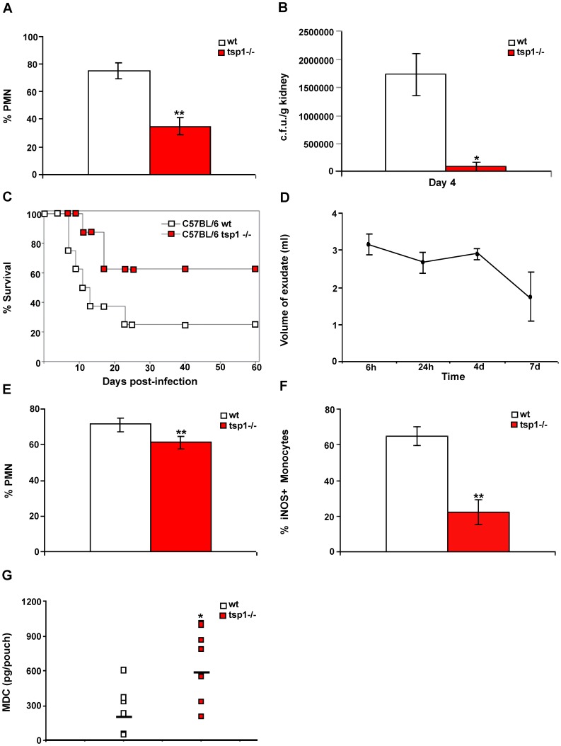
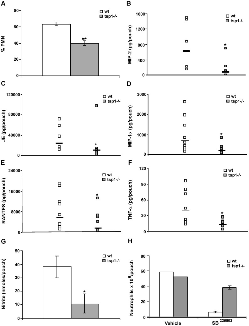
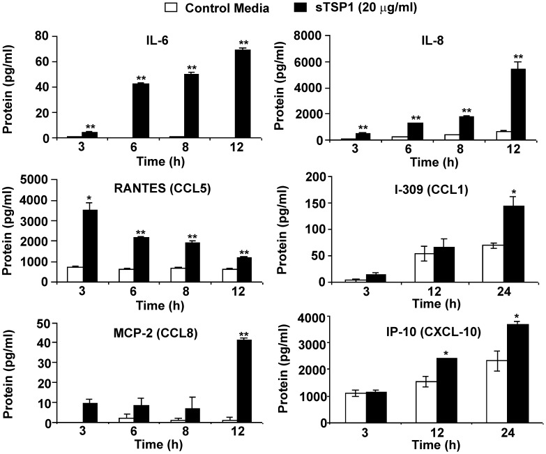
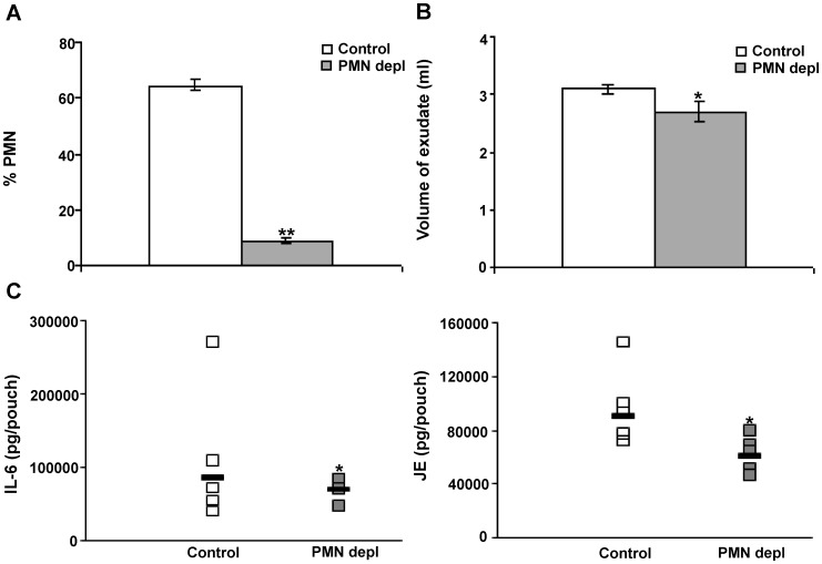
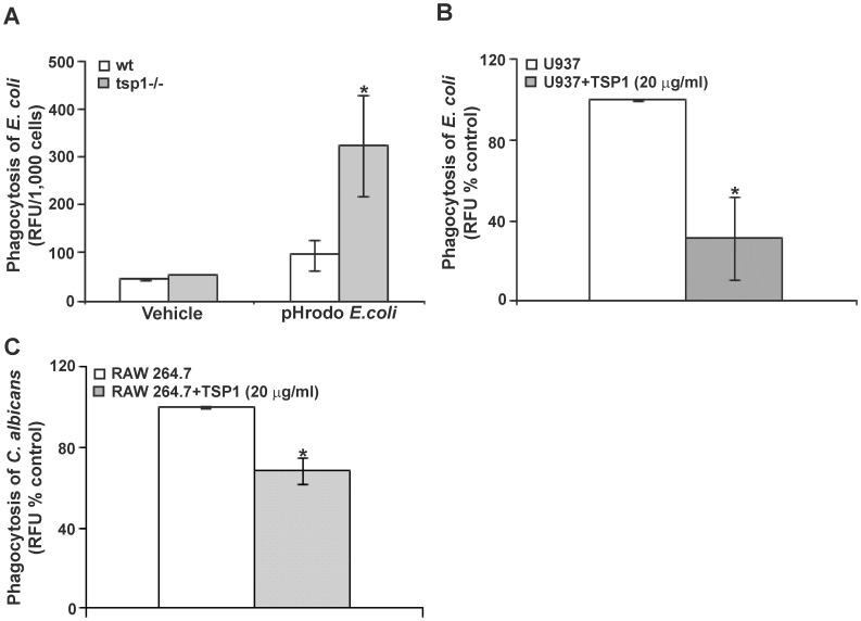
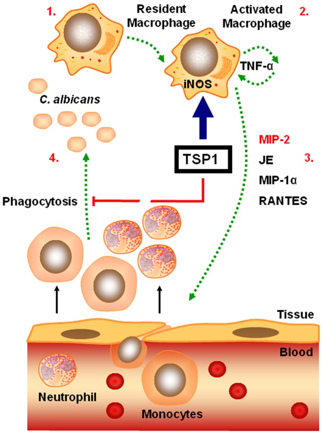
Disseminated Candida albicans infection results in high morbidity and mortality despite treatment with existing antifungal drugs. Recent studies suggest that modulating the host immune response can improve survival, but specific host targets for accomplishing this goal remain to be identified. The extracellular matrix protein thrombospondin-1 is released at sites of tissue injury and modulates several immune functions, but its role in C. albicans pathogenesis has not been investigated. Here, we show that mice lacking thrombospondin-1 have an advantage in surviving disseminated candidiasis and more efficiently clear the initial colonization from kidneys despite exhibiting fewer infiltrating leukocytes. By examining local and systemic cytokine responses to C. albicans and other standard inflammatory stimuli, we identify a crucial function of phagocytes in this enhanced resistance. Subcutaneous air pouch and systemic candidiasis models demonstrated that endogenous thrombospondin-1 enhances the early innate immune response against C. albicans and promotes activation of inflammatory macrophages (inducible nitric oxide synthase⁺, IL-6(high), TNF-α(high), IL-10(low)), release of the chemokines MIP-2, JE, MIP-1α, and RANTES, and CXCR2-driven polymorphonuclear leukocytes recruitment. However, thrombospondin-1 inhibited the phagocytic capacity of inflammatory leukocytes in vivo and in vitro, resulting in increased fungal burden in the kidney and increased mortality in wild type mice. Thus, thrombospondin-1 enhances the pathogenesis of disseminated candidiasis by creating an imbalance in the host immune response that ultimately leads to reduced phagocytic function, impaired fungal clearance, and increased mortality. Conversely, inhibitors of thrombospondin-1 may be useful drugs to improve patient recovery from disseminated candidiasis.


Conflict of interest statement

Competing Interests: David D. Roberts, Ph.D. is an Academic Editor. This does not alter the authors’ adherence to all the PLOS ONE policies on sharing data and materials.

Figures

Figure 1
Figure 1. Endogenous TSP1 contributes to the pathogenesis of disseminated C. albicans infection.
(A) Tissue sections cut from infected kidneys harvested and paraffin-embedded 48 h post-infection were stained with H&E. Quantitative analysis of PMN infiltration into the specimens was performed by a pathologist evaluating the number of cells in 10 different 400× fields with inflammatory infiltrate. Bars, mean ± SD, n = 4 mice/group. (B) Kidney fungal burden. Bars, mean ± SEM, n = 4 mice/group. (C) The probability of survival as a function of time was determined by the Kaplan-Meier method. Data are representative of two independent experiments (n = 8 mice/group). Hazard ratio estimates of 4.2 and 95% confidence interval (1.131–16.74) indicated that the wt mice infected with C. albicans had higher lethality. (D-G) Six day-old air pouches received an inoculum of 108 heat-inactivated C. albicans. (D) Mice were sacrificed at the indicated time points, pouches were washed with saline, and exudates were collected. Results represent mean volume of exudate ± SD, n = 3 wt mice/time point. (E–G) wt and tsp1−/− mice were sacrificed 24 h after injection of heat-inactivated C. albicans. Quantitative analysis of leukocyte infiltration was performed by a pathologist evaluating the percentage of PMN (E) and iNOS+ monocytes (F) in 10 different 400× fields with inflammatory infiltrate. Data are pooled from two independent experiments. Bars, mean ± SD, n = 8 mice/group. (G) The levels of mouse MDC in the air pouch lavage were determined using a multiplexed ELISA array (Quansys Biosciences), as described in Materials and Methods. Data are pooled from two independent experiments and represent geometric mean, n = 8 mice/group. *indicates p<0.05; **indicates p<0.001.
Figure 2
Figure 2. TSP1 enhances inflammation-mediated activation of macrophages in vivo.
(A–F) Six day-old air pouches received 1 ml of 1% λ-carrageenan. (A) Mice were sacrificed at the indicated time points before (time 0 h) or after λ-carrageenan injection. For the tissue control, mice without air pouches were subcutaneously injected in the back with 1 ml of saline and the lavage fluid was collected. The levels of mouse TSP1 in the air pouch exudates were determined by immunoassay. Data are pooled from two independent experiments. Bars, mean ± SD, n = 4 wt mice/group. One-way ANOVA (p = 0.0097). (B–F) wt and tsp1−/− mice were sacrificed 6 h after injection of 1% λ-carrageenan. Quantitative analysis of leukocyte infiltration was performed by a pathologist evaluating the percentage of PMN and MN (B) and iNOS+ monocytes (C) in 10 different 400× fields with inflammatory infiltrate. Bars, mean ± SEM, n = 8−10 mice/group. (D–F) IL-6, TNF-α, and IL-10 were determined in the air pouch lavage using a multiplexed ELISA array. Data represent geometric mean, n = 8−10 mice/group. *indicates p<0.05; **indicates p<0.001.
Figure 3
Figure 3. TSP1 increases PMN recruitment to sites of acute inflammation.
Six day-old air pouches received 1 ml of 1% λ-carrageenan, and the mice were sacrificed 24 h after injection. (A) The percentage of infiltrating PMN was quantified in 10 different 400× fields with inflammatory infiltrate. Bars, mean ± SEM, n = 5−6 mice/group. (B–F) The levels of mouse MIP-2, JE, MIP-1α and RANTES, and TNF-α in the air pouch lavage were determined using a MIP-2 Quantikine® ELISA or a multiplexed ELISA array (Quansys Biosciences), as described in Materials and Methods. Data are pooled from two independent experiments and represent geometric mean, n = 5−11 mice/group. (G) 50 µl of exudates were used for NO2 detection using a Griess Reagent System. All samples were run in triplicate, as described in Materials and Methods. Bars, mean ± SEM, n = 4 mice/group. (H) The absolute number of neutrophils was determined in the presence or absence of SB225002 (50 µM) or vehicle (saline) by FACS, as described in Materials and Methods. Bars, mean ± SEM, n = 3 wt and tsp1−/− mice. *indicates p<0.05; **indicates p<0.001.
Figure 4
Figure 4. TSP1 enhances cytokine and chemokine production by human IFN-γ-differentiated macrophages in vitro.
1×10⁶ IFN-γ-differentiated U937 cells/condition were incubated in the absence or the presence of soluble TSP1. At the indicated times the supernatants were harvested, and total IL-6, IL-8, RANTES, I-309, MCP-2 and IP-10 were determined using a Multiplexed ELISA array. Bars, mean ± SD, are representative of at least four different experiments. *indicates p<0.05; **indicates p<0.001.
Figure 5
Figure 5. Neutrophil depletion leads to a reduction of the inflammatory response.
wt mice with 6-day-old air pouches received an intraperitoneal injection of a LEAF™ purified anti-mouse Ly-6G/Ly-6C (Gr-1) antibody 18 h before carrageenan challenge. On day 7, 1 ml of 1% λ-carrageenan was injected into the pouch cavity. Mice were sacrificed 4 h after λ-carrageenan injection. (A) The percentage of PMN was quantified in 10 different 400× fields with inflammatory infiltrate. (B) The volume of exudate collected was quantified. Bars, mean ± SEM, n = 5−7 mice/group. (C) The levels of mouse IL-6 and JE in the air pouch lavage were determined using a multiplexed ELISA array. Data represent geometric mean, n = 4−5 mice/group. *indicates p<0.05; **indicates p<0.001.
Figure 6
Figure 6. TSP1 inhibits phagocytosis in vivo and in vitro.
(A) wt and tsp1−/− mice received 1 ml of 1% λ-carrageenan (intra-pouch) and 23 hours later were injected with pHrodo-labeled E. coli Bioparticles® or vehicle control (HBSS/20 mM HEPES pH 7.4). After 1 h incubation, cells were harvested, counted and placed in a fluorometer. The results are presented as the ratio relative fluorescence units (RFU)/1,000 cells. Data are pooled from two independent experiments and represent the mean ± SEM, n = 6−8 mice/group. (B) 1×10⁵ IFN-γ-differentiated U937 cells/condition were incubated with FITC-labeled E. coli in the absence or the presence of soluble TSP1 for 2 h. The fluorescence intensity (mean ± SD) is presented as a % of control RFU and is representative of four independent experiments. (C) 2×10⁵ RAW 264.7 cells/condition were incubated with FITC-labeled C. albicans (2 yeast: 1 macrophage) in the absence or the presence of soluble TSP1 for 45 min. The results are presented as a % of control RFU. Data are pooled from three independent experiments (mean ± SEM). *indicates p<0.05.
Figure 7
Figure 7. Model for the role of TSP1 in the pathogenesis of disseminated candidiasis.
TSP1 is rapidly released at sites of acute infection and inflammation and promotes activation of inflammatory macrophages (iNOS+, IL-6high, TNF-αhigh, IL-10low) (1–2). TNF-α stimulates PMN recruitment by up-regulating the expression of specific chemokines such as MIP-2, JE, and MIP-1α (2–3). This initial influx of PMN precedes the extravasation of inflammatory monocytes that amplify the inflammatory response. In addition, TSP1 inhibits the phagocytic function of inflammatory leukocytes (3–4). Thus, TSP1 promotes a sustained recruitment and activation of leukocytes that leads to tissue damage while reducing phagocytic function and fungal clearance.

Similar articles

Cited by

References

    1. Raugi GJ, Olerud JE, Gown AM (1987) Thrombospondin in early human wound tissue. J Invest Dermatol 89: 551–554. - PubMed
    1. DiPietro LA, Nissen NN, Gamelli RL, Koch AE, Pyle JM, et al. (1996) Thrombospondin 1 synthesis and function in wound repair. Am J Pathol 148: 1851–1860. - PMC - PubMed
    1. Gotis-Graham I, Hogg PJ, McNeil HP (1997) Significant correlation between thrombospondin 1 and serine proteinase expression in rheumatoid synovium. Arthritis Rheum 40: 1780–1787. - PubMed
    1. Narizhneva NV, Razorenova OV, Podrez EA, Chen J, Chandrasekharan UM, et al. (2005) Thrombospondin-1 up-regulates expression of cell adhesion molecules and promotes monocyte binding to endothelium. Faseb J 19: 1158–1160. - PMC - PubMed
    1. Mansfield PJ, Suchard SJ (1994) Thrombospondin promotes chemotaxis and haptotaxis of human peripheral blood monocytes. J Immunol 153: 4219–4229. - PubMed

Publication types

MeSH terms

Supplementary concepts