. 2012;7(11):e48797.
doi: 10.1371/journal.pone.0048797. Epub 2012 Nov 7.

Protein tyrosine phosphatase receptor type z negatively regulates oligodendrocyte differentiation and myelination

Affiliations

Protein tyrosine phosphatase receptor type z negatively regulates oligodendrocyte differentiation and myelination

Kazuya Kuboyama et al. PLoS One. 2012.

Abstract

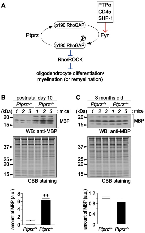
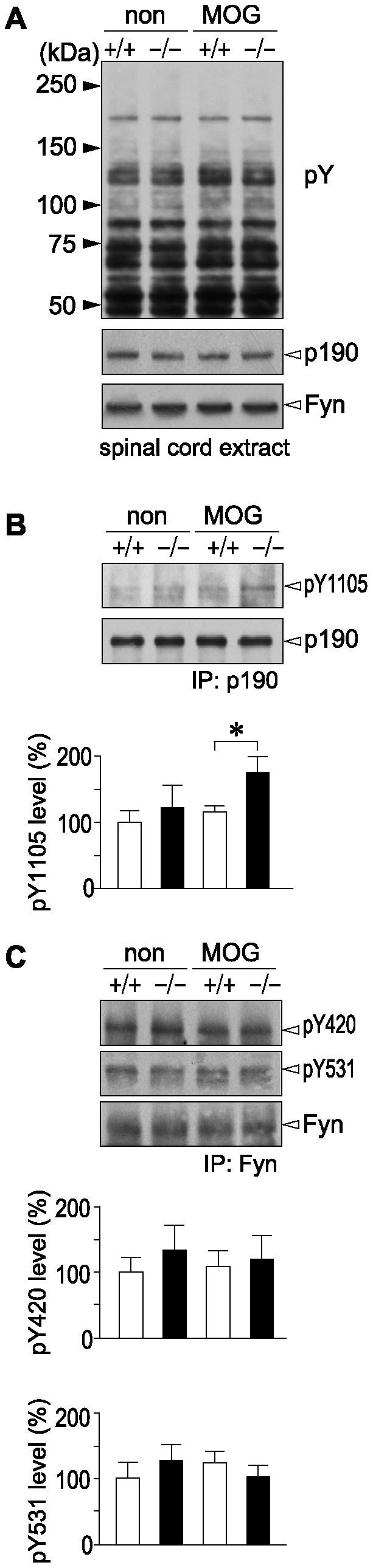
Background: Fyn tyrosine kinase-mediated down-regulation of Rho activity through activation of p190RhoGAP is crucial for oligodendrocyte differentiation and myelination. Therefore, the loss of function of its counterpart protein tyrosine phosphatase (PTP) may enhance myelination during development and remyelination in demyelinating diseases. To test this hypothesis, we investigated whether Ptprz, a receptor-like PTP (RPTP) expressed abuntantly in oligodendrocyte lineage cells, is involved in this process, because we recently revealed that p190RhoGAP is a physiological substrate for Ptprz.

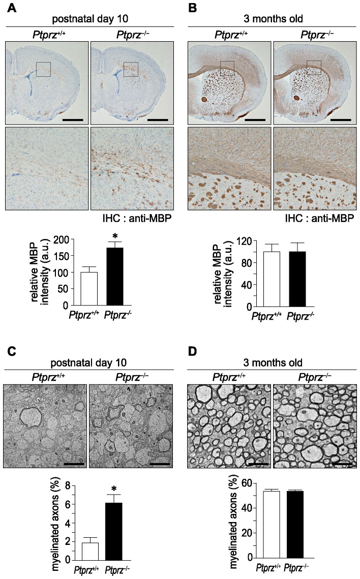
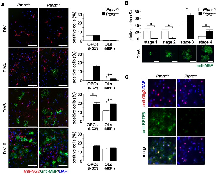
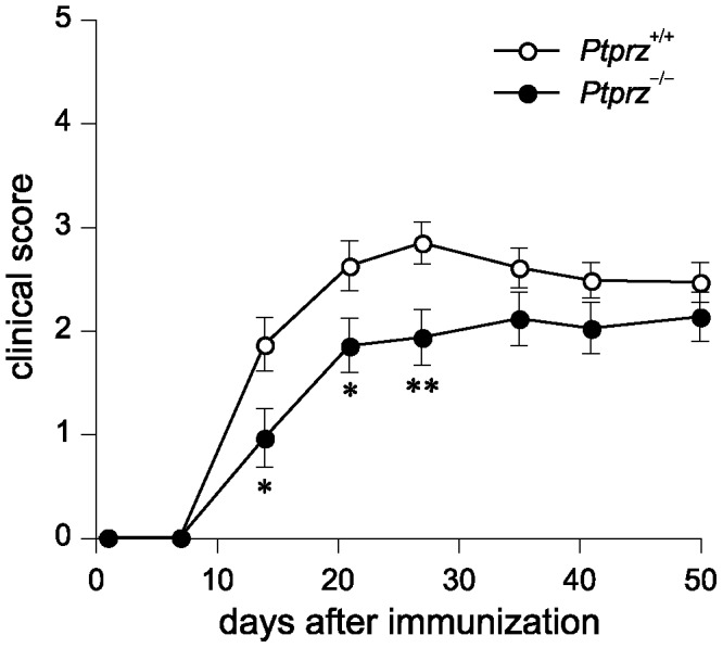
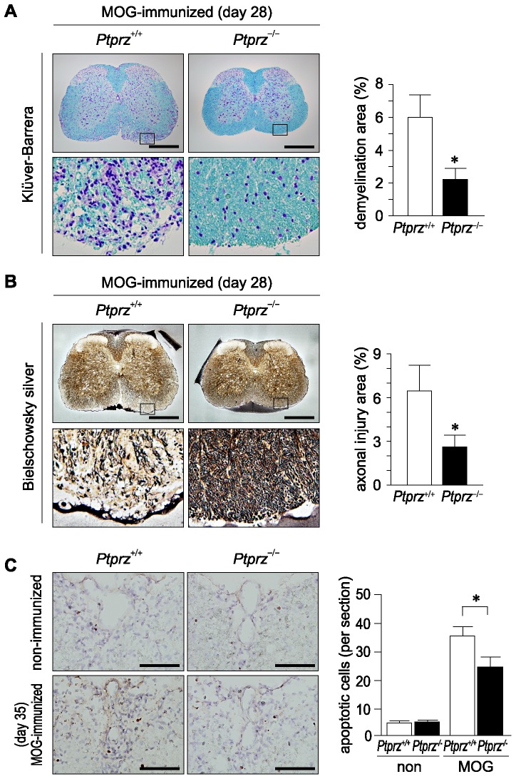
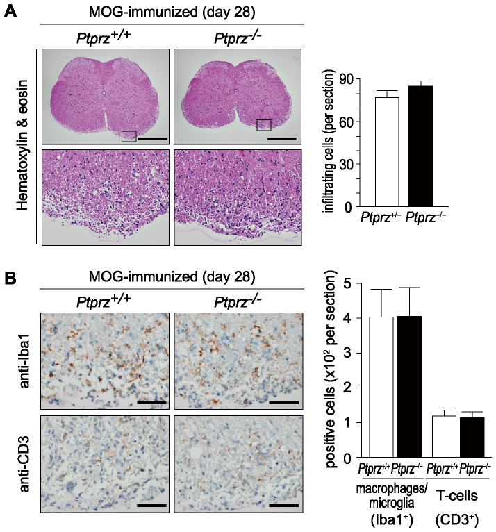
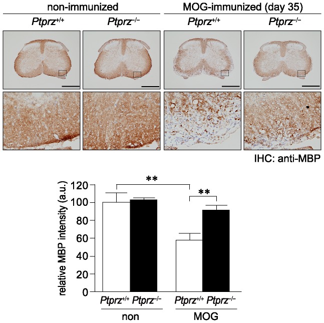
Methodology/principal findings: We found an early onset of the expression of myelin basic protein (MBP), a major protein of the myelin sheath, and early initiation of myelination in vivo during development of the Ptprz-deficient mouse, as compared with the wild-type. In addition, oligodendrocytes appeared earlier in primary cultures from Ptprz-deficient mice than wild-type mice. Furthermore, adult Ptprz-deficient mice were less susceptible to experimental autoimmune encephalomyelitis (EAE) induced by active immunization with myelin/oligodendrocyte glycoprotein (MOG) peptide than were wild-type mice. After EAE was induced, the tyrosine phosphorylation of p190RhoGAP increased significantly, and the EAE-induced loss of MBP was markedly suppressed in the white matter of the spinal cord in Ptprz-deficient mice. Here, the number of T-cells and macrophages/microglia infiltrating into the spinal cord did not differ between the two genotypes after MOG immunization. All these findings strongly support the validity of our hypothesis.

Conclusions/significance: Ptprz plays a negative role in oligodendrocyte differentiation in early central nervous system (CNS) development and remyelination in demyelinating CNS diseases, through the dephosphorylation of substrates such as p190RhoGAP.


Conflict of interest statement

Competing Interests: M. Masumura is employed by Asubio Pharma Co. Ltd. There are no patents, products in development or marketed products to declare. This does not alter the authors' adherence to all the PLOS ONE policies on sharing data and materials.

Figures

Figure 1
Figure 1. Early onset of MBP expression in the brain of Ptprz-deficient mice.
A, Schematic drawing of postulated signaling mechanisms of Ptprz and Fyn in oligodendrocyte differentiation and myelination. Fyn and Ptprz may also act on yet unidentified substrates other than p190RhoGAP to regulate the differentiation. The red arrow shows activation, whereas the blunt blue arrows represent inhibition. B, C, Western blot analyses of MBP expression in the cerebral cortex of mice at postnatal day 10 (B), and 3 months old (C). Applied protein amounts were verified by Coomassie Brilliant Blue (CBB) staining. The amounts of MBP are presented as densitometric units normalized to the value for respective wild-type controls, and are shown at the lower position of each panel. Data are the mean ± SEM (n = 6 for each group). **p<0.01 (Student's t-test). a.u., arbitrary unit.
Figure 2
Figure 2. Early initiation of myelination in Ptprz-deficient mice.
A, B, Immunohistochemical analyses of MBP expression in mouse brains at postnatal day 10 (A), and 3 months old (B). Scale bars, 1 mm. The results of the densitometric analysis of MBP signals are normalized to the value for respective wild-type controls, and shown at the lower position of each panel. Data are the mean ± SEM (n = 3 for each group). *p<0.05 (Student's t-test). a.u., arbitrary unit. C, D, Electron micrographs of transverse sections at the corpus callosum from mice at postnatal day 10 (C), and 3 months old (D). Scale bars, 2 µm. Percentages of myelinated axons in total axons are shown at the lower position of each panel. Data are the mean ± SEM (n = 4 for each group). *p<0.05 (Student's t-test).
Figure 3
Figure 3. Early onset of oligodendrocyte differentiation in Ptprz-deficient mice.
A, Immunohistochemistry of cultured oligodendrocyte precursor cells (OPCs, NG2-postive cells, red) and oligodendrocytes (OLs, MBP-postive cells, green) from Ptprz-deficient mice and wild-type mice. Scale bars, 100 µm. The percentages of OPCs and OLs among total cells (DAPI-positive nuclei, blue) are shown at the right of each panel. Data are the mean ± SEM from five independent experiments. *p<0.05 and **p<0.01 (Student's t-test). B, Morphological assessment of cultured OLs at DIV6. The MBP-postive cells were classified into four categories. Representative images are shown in lower panels; Stage 1, three or less primary processes longer than a cell body with minimal development of secondary and tertiary processes; Stage 2, three or more primary processess with moderate secondary and tertiary processes; Stage 3, five or more primary processes with extensive secondry and teriary processes; Stage 4, extending myelin-like membrane structures and branched processes. Scale bars, 50 µm. Data are the mean ± SEM from three independent experiments. *p<0.05 (Mann-Whitney U-test). C, Ptprz expression in oligodendrocyte lineage cells. Cultured cells at DIV10 were triple stained with anti-RPTPβ (specific for Ptprz receptor isoforms, green), anti-Olig2 (red), and DAPI (blue). Scale bars, 50 µm.
Figure 4
Figure 4. Expression of Ptprz in the spinal cord of adult mice.
Immunohistochemical staining of the spinal cord with anti-Ptprz-S (left panels) and anti-RPTPβ (right panels). The lower images are enlargements of the areas enclosed by squares in the upper images. Scale bars, 500 µm.
Figure 5
Figure 5. Reduced clinical severity of EAE in Ptprz-deficient mice.
Clinical scores in wild-type mice and Ptprz-deficient mice after the MOG peptide injection. Clinical scores are 0, no disease; 1, limp tail; 2, ataxia and/or paresis of hindlimbs; 3, paralysis of hindlimbs and/or paresis of forelimbs; 4, tetraparalysis; 5, moribund or death. Data are the mean ± SEM (Ptprz +/+, n = 25; Ptprz −/−, n = 23). The comparison of clinical scores between the two groups at each time point was performed with Mann-Whitney's U-test, *p<0.05, **p<0.01.
Figure 6
Figure 6. Reduced tissue damage and increased oligodendrocyte survival in Ptprz-deficient mice with EAE.
A, B, Klüver-Barrera (A) and Bielschowsky silver staining (B) of the spinal cord obtained from wild-type and Ptprz-deficient mice 28 days after MOG immunization. The lower images are enlargements of the areas enclosed by squares in the upper images, respectively. The extent of demyelination and axon injury determined by Klüver-Barrera staining and Bielschowsky silver staining is shown as the percentage of damaged areas at the right of each panel. Scale bars, 500 µm. Data are the mean ± SEM (n = 10 for each group). *p<0.05 (Student's t-test). C, TUNEL staining of spinal cord sections 35 days after MOG immunization, or non-immunized control animals. Scale bars, 100 µm. TUNEL-positive cells were counted in six sections from each animal, and the numbers of TUNEL-positive cells per section are shown at the right. Data are the mean ± SEM (n = 6 for each group). *p<0.05 (Student's t-test).
Figure 7
Figure 7. No genotypic differences in infiltrating T-cells and macrophages/microglia within the spinal cord after EAE induction.
A, Hematoxylin and eosin staining of the spinal cord 28 days after MOG immunization. The lower images are enlargements of the areas enclosed by squares in the upper images. Scale bars, 500 µm. The numbers of infiltrating cells per section are shown at the right. Data are the mean ± SEM (n = 10 for each group). B, Immunohistochemistry of infiltrating T-cells (detected with anti-CD3) or macrophages/microglia (with anti-Iba1) in the spinal cords 28 days after MOG immunization. Scale bars, 50 µm. The numbers of CD3-positive or Iba1-positive cells are shown at the right. Data are the mean ± SEM (n = 10 for each group). No significant differences were detected between the two genotypes.
Figure 8
Figure 8. Normal proliferative responses in T-cells of Ptprz-deficient mice.
T-cell preparations taken from the axillary and inguinal lymph nodes of mice at 10, 28, or 35 days after MOG immunization (imm.), or non-immunized control (non) mice were cultivated in the presence of the MOG peptide (top), anti-CD3/CD28 antibodies (middle), or vehicle (bottom). BrdU incorporation was measured as an index of cell proliferation, and is expressed as the relative change (fold-increase) compared with the vehicle-treated sample of the non-immunized wild-type control. Data are the mean ± SEM (n = 6 for each group). No significant differences were detected between the two genotypes. a.u., arbitrary unit.
Figure 9
Figure 9. Increased phosphorylation of Tyr 1105 on p190RhoGAP in the spinal cord of Ptprz-deficient mice after EAE induction.
A, Overall tyrosine phosphorylation patterns of total protein and expression of p190RhoGAP and Fyn in the spinal cord. The third to sixth lumbar spinal cord extracts were prepared from wild-type (+/+) and Ptprz-deficient mice (−/−) 35 days after MOG immunization, or non-immunized control animals, and examined by Western blotting using anti-phosphotyrosine PY20 (top), anti-p190RhoGAP (middle), and anti-Fyn (bottom) antibodies, respectively. B, Tyrosine phosphorylation of Tyr 1105 on p190RhoGAP. The spinal cord extracts were immunoprecipitated with anti-p190RhoGAP antibody and immunoblotted with anti-pY1105 p190RhoGAP (upper), or anti-p190RhoGAP (lower). The densitometric data for anti-pY1105 p190RhoGAP signals are presented as a percentage of the non-immunized wild-type control, and shown at the bottom. Data are the mean ± SEM (n = 4 pooled samples from two animals per each group). *p<0.05 (Student's t-test). C, No siginificant differences in tyrosine phosphorylation of Fyn among the four groups. The spinal cord extracts prepared as above were immunoprecipitated with anti-Fyn antibody and immunoblotted with anti-pY420 (top), anti-pY531 (middle), or anti-Fyn (bottom). The densitometric data for anti-pY420 and anti-pY531 signals are presented as a percentage of the non-immunized wild-type control, and shown at the bottom. Data are the mean ± SEM (n = 4 pooled samples from two animals per group). No significant differences were detected between the two genotypes.
Figure 10
Figure 10. Reduced MBP loss in Ptprz-deficient mice after EAE induction.
Anti-MBP staining of the spinal cord sections from wild-type and Ptprz-deficient mice 35 days after MOG immunization, or non-immunized control mice. The lower images are enlargements of the areas enclosed by squares in the upper images. Scale bars, 500 µm. The densitometric data for MBP signals are expressed as the relative change (fold-increase) compared with the non-immunized wild-type mice, and shown at the bottom. Data are the mean ± SEM (n = 6 for each group). **p<0.01 (Student's t-test). a.u., arbitrary unit.

Similar articles

Cited by

References

    1. Frohman EM, Racke MK, Raine CS (2006) Multiple sclerosis - the plaque and its pathogenesis. N Engl J Med 354: 942–955. - PubMed
    1. Krämer-Albers EM, White R (2011) From axon-glial signalling to myelination: the integrating role of oligodendroglial Fyn kinase. Cell Mol Life Sci 68: 2003–2012. - PMC - PubMed
    1. Scarlato M, Beesley J, Pleasure D (2000) Analysis of oligodendroglial differentiation using cDNA arrays. J Neurosci Res 59: 430–435. - PubMed
    1. Umemori H, Kadowaki Y, Hirosawa K, Yoshida Y, Hironaka K, et al. (1999) Stimulation of myelin basic protein gene transcription by Fyn tyrosine kinase for myelination. J Neurosci 19: 1393–1397. - PMC - PubMed
    1. Umemori H, Sato S, Yagi T, Aizawa S, Yamamoto T (1994) Initial events of myelination involve Fyn tyrosine kinase signalling. Nature 367: 572–576. - PubMed

Publication types

MeSH terms

Substances