. 2012;7(11):e49079.
doi: 10.1371/journal.pone.0049079. Epub 2012 Nov 8.

Modulation of pluripotency in the porcine embryo and iPS cells

Affiliations

Modulation of pluripotency in the porcine embryo and iPS cells

Aida Rodríguez et al. PLoS One. 2012.

Abstract

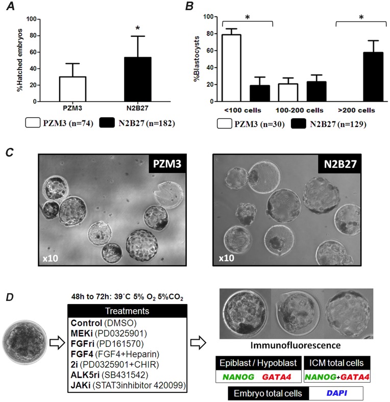
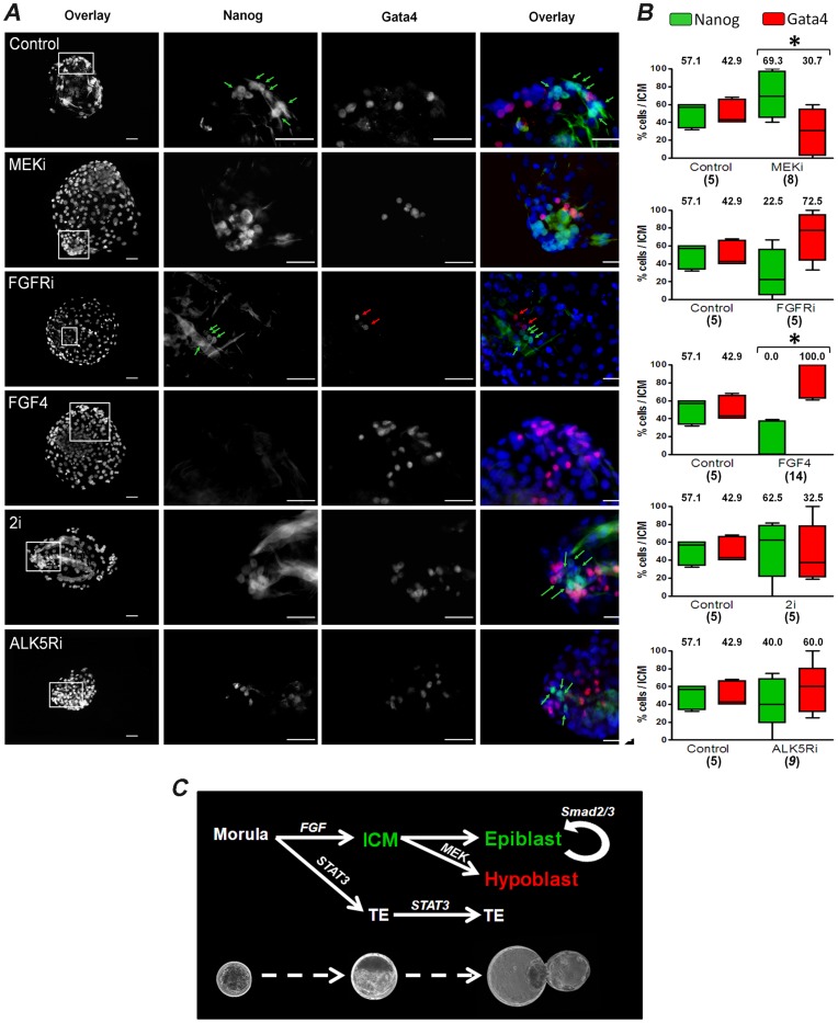
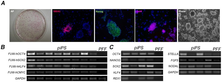
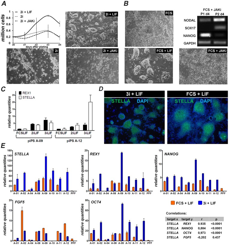
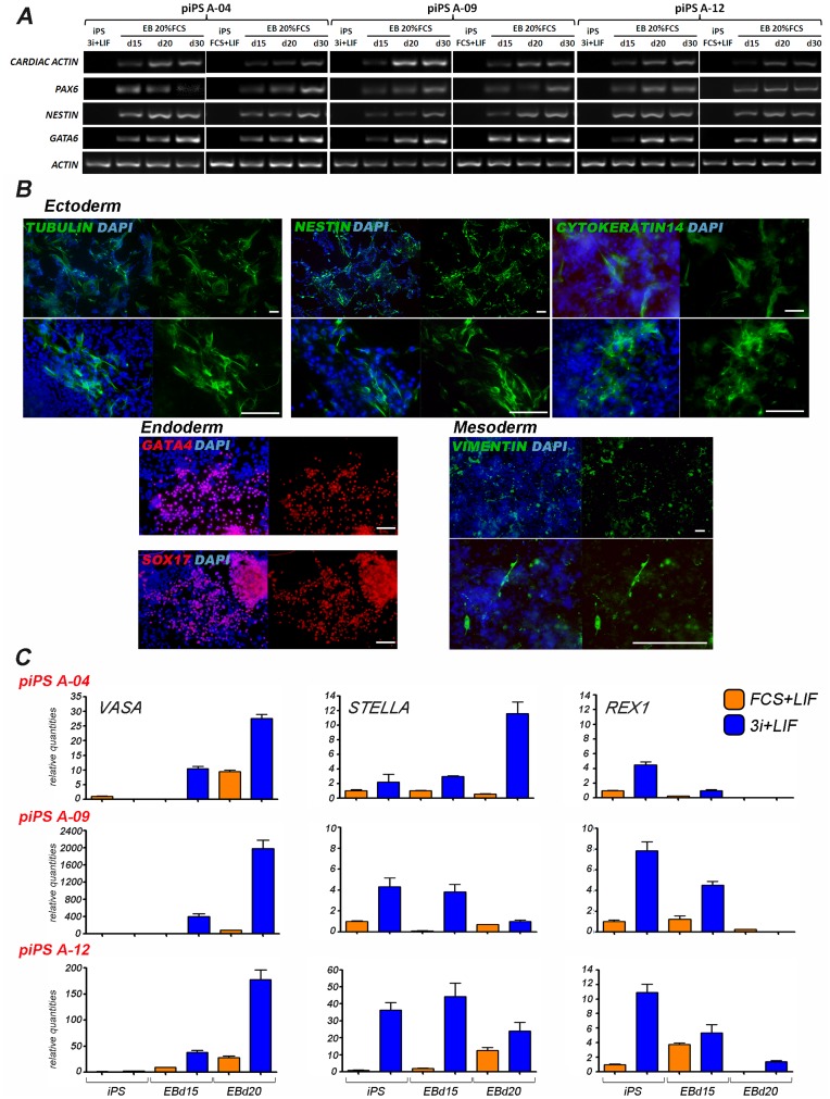
The establishment of the pluripotent ICM during early mammalian development is characterized by the differential expression of the transcription factors NANOG and GATA4/6, indicative of the epiblast and hypoblast, respectively. Differences in the mechanisms regulating the segregation of these lineages have been reported in many species, however little is known about this process in the porcine embryo. The aim of this study was to investigate the signalling pathways participating in the formation of the porcine ICM, and to establish whether their modulation can be used to increase the developmental potential of pluripotent cells. We show that blocking MEK signalling enhances the proportion of NANOG expressing cells in the ICM, but does not prevent the segregation of GATA-4 cells. Interestingly, inhibition of FGF signalling does not alter the segregation of NANOG and GATA-4 cells, but affects the number of ICM cells. This indicates that FGF signalling participates in the formation of the founders of the ICM. Inhibition of MEK signalling combined with GSK3β inhibition and LIF supplementation was used to modulate pluripotency in porcine iPS (piPS) cells. We demonstrate that under these stringent culture conditions piPS cells acquire features of naive pluripotency, characterized by the expression of STELLA and REX1, and increased in vitro germline differentiation capacity. We propose that small molecule inhibitors can be used to increase the homogeneity of induced pluripotent stem cell cultures. These improved culture conditions will pave the way for the generation of germline competent stem cells in this species.


Conflict of interest statement

Competing Interests: The authors have declared that no competing interests exist.

Figures

Figure 1
Figure 1. Development of pig morulae after in vitro culture.
A) Proportion of embryos hatching from the zona pellucida after culture in PZM3 and N2B27. B) Cell count of blastocysts developing after culture in PZM3 and N2B27. C) Images of embryos obtained after incubation of morulae in PZM3 and N2B27. D) Diagram depicting the experimental design used to study signalling pathways involved in the segregation of ICM. * P<0.05.
Figure 2
Figure 2. Effect of signalling inhibition in lineage segregation.
A) Immunofluorescence images of NANOG and GATA-4 of hatched blastocysts after incubation with the indicated molecules from the morula stage. Blue: nuclei stained with DAPI. Scale bar: 50 µm. B) Box-whisker plots depicting the proportion of NANOG (green) and GATA-4 (red) cells of embryos treated with different molecules. Numbers indicate median values for each group. * P<0.05. C) Model depicting the stages of embryo development studied in this report. Inhibition of STAT3 signalling interferes with the cavitation process affecting the development of the ICM and the TE during the transition from early to late blastocyst. In addition, inhibition of FGF receptors at the morula stage prevents the formation of the ICM, whereas inhibition of MEK signalling reduces, but does not totally abolish, the segregation of hypoblast (GATA-4) in late blastocysts. Inhibition of SMAD2/3 does not interfere with the activation of NANOG in the blastocyst, but is needed for the development of the epiblast, as previously shown .
Figure 3
Figure 3. Characterization of piPS cells.
A) Alkaline phosphatase, SSEA1, NANOG and OCT-4 staining in colonies of piPS cells. Phase contrast image shows the morphology of cells under low magnification. B) Expression of exogenous factors was determined by RT-PCR in 8 cell lines. PFF: porcine fetal fibroblasts used for generating the iPS cells. C) RT-PCR analysis of four iPS cell lines shows variables levels of gene expression of endogenous genes.
Figure 4
Figure 4. Induction of naive features in piPS cells cultured with 2/3i + LIF.
A) piPS cells were seeded in the indicated culture conditions and their growth was quantified daily for 4 days. B) piPS cells were seeded in the indicated culture conditions and collected 4 days after seeding at passage 1 and 2. RT-PCR was performed to analyse gene expression. C) qRT-PCR for REX1 and STELLA of two cell lines grown in FCS + LIF or in 2/3i + LIF. Expression was normalized to FCS + LIF piPS cells. D) STELLA immunostaining in piPS colonies seeded in the indicated culture conditions. E) qRT-PCR for STELLA, OCT-4, NANOG, REX1 and FGF5 of 8 iPS cell lines grown in FCS + LIF or in 3i + LIF. Expression was normalized to PFF sample. Pearson’s correlation coefficient analysis was performed to study interactions between genes.
Figure 5
Figure 5. Differentiation potential of iPS cells grown under different conditions.
A) RT-PCR performed on three iPS cell lines (A04, A-09, and A-12) grown under 3i + LIF or FCS + LIF were spontaneously differentiated in medium containing 20% FCS. Gene expression analysis was performed at the indicated time points. B) Immunocytochemistry analysis was carried out in spontaneously differentiated piPS cells. Scale bar: 10 µM. C) qRT-PCR analysis of three iPS cell lines induced to differentiate in culture medium containing 20% FCS over 20 days. Expression levels are relative to iPS cells grown in FCS + LIF.

References

    1. Nichols J, Smith A (2009) Naive and primed pluripotent states. Cell Stem Cell 4: 487–492. - PubMed
    1. Hanna J, Markoulaki S, Mitalipova M, Cheng AW, Cassady JP, et al. (2009) Metastable pluripotent states in NOD-mouse-derived ESCs. Cell Stem Cell 4: 513–524. - PMC - PubMed
    1. Vallier L, Alexander M, Pedersen RA (2005) Activin/Nodal and FGF pathways cooperate to maintain pluripotency of human embryonic stem cells. J Cell Sci 118: 4495–4509. - PubMed
    1. Alberio R, Croxall N, Allegrucci C (2010) Pig epiblast stem cells depend on activin/nodal signaling for pluripotency and self-renewal. Stem Cells Dev 19: 1627–1636. - PMC - PubMed
    1. Tachibana M, Sparman M, Ramsey C, Ma H, Lee HS, et al. (2012) Generation of chimeric rhesus monkeys. Cell 148: 285–295. - PMC - PubMed

Publication types

MeSH terms

Substances