. 2012;7(11):e49150.
doi: 10.1371/journal.pone.0049150. Epub 2012 Nov 7.

Induced tauopathy in a novel 3D-culture model mediates neurodegenerative processes: a real-time study on biochips

Affiliations

Induced tauopathy in a novel 3D-culture model mediates neurodegenerative processes: a real-time study on biochips

Diana Seidel et al. PLoS One. 2012.

Abstract

Tauopathies including Alzheimer's disease represent one of the major health problems of aging population worldwide. Therefore, a better understanding of tau-dependent pathologies and consequently, tau-related intervention strategies is highly demanded. In recent years, several tau-focused therapies have been proposed with the aim to stop disease progression. However, to develop efficient active pharmaceutical ingredients for the broad treatment of Alzheimer's disease patients, further improvements are necessary for understanding the detailed neurodegenerative processes as well as the mechanism and side effects of potential active pharmaceutical ingredients (API) in the neuronal system. In this context, there is a lack of suitable complex in vitro cell culture models recapitulating major aspects of taupathological degenerative processes in sufficient time and reproducible manner.Herewith, we describe a novel 3D SH-SY5Y cell-based, tauopathy model that shows advanced characteristics of matured neurons in comparison to monolayer cultures without the need of artificial differentiation promoting agents. Moreover, the recombinant expression of a novel highly pathologic fourfold mutated human tau variant lead to a fast and emphasized degeneration of neuritic processes. The neurodegenerative effects could be analyzed in real time and with high sensitivity using our unique microcavity array-based impedance spectroscopy measurement system. We were able to quantify a time- and concentration-dependent relative impedance decrease when Alzheimer's disease-like tau pathology was induced in the neuronal 3D cell culture model. In combination with the collected optical information, the degenerative processes within each 3D-culture could be monitored and analyzed. More strikingly, tau-specific regenerative effects caused by tau-focused active pharmaceutical ingredients could be quantitatively monitored by impedance spectroscopy.Bringing together our novel complex 3D cell culture taupathology model and our microcavity array-based impedimetric measurement system, we provide a powerful tool for the label-free investigation of tau-related pathology processes as well as the high content analysis of potential active pharmaceutical ingredient candidates.


Conflict of interest statement

Competing Interests: Till G.A. Mack and Frank Striggow were formerly employed by the the KeyNeurotek Pharmaceuticals AG. This does not alter the authors' adherence to all the PLOS ONE policies on sharing data and materials.

Figures

Figure 1
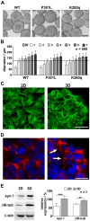
Figure 1. 3D cultivation of tau expressing SH-SY5Y cells leads to enhanced neuronal differentiation. A.
Stably EGFP-tau fusion protein expressing SH-SY5Y cells were cultivated on a gyratory shaker in order to generate spheroids within the first five days in vitro (DIV) (bar = 100 µm). B. Statistical analysis of spheroid diameter revealed comparable size development over time for wildtype (WT), single-mutated (P301L) and poly-mutated (K280q) tau expressing SH-SY5Y cells (mean ± s.d.). C. Live cell imaging of the EGFP-fused wildtype tau (green) and D. immunocytochemical staining of the neuronal marker NF200 (red) ensured the overall existence of matured neural cells and revealed extended neuritic processes (arrows) in five days cultivated spheroids (3D) in comparison to monolayer cultures (2D) (bar = 20 µm, nuclear stain DAPI blue). E. The expression of the neuronal markers synapsin I (syn I)and β III-tubulin (βIII-tub) in wildtype tau expressing 2D and 3D cultures at culture day seven were analyzed via Western blot and quantified relative to the housekeeping protein β-tubulin.(n ≥3, **p<0.01, ***p<0.001).
Figure 2
Figure 2. Analysis of cellular alterations after okadaic acid induced hyperphosphorylation in wildtype and mutated tau expressing SH-SY5Y spheroids.
A. Western blot analysis of five days cultivated WT, P301L and K280q tau expressing SH-SY5Y spheroids after incubation with okadaic acid (OA) for 24 hours. Hyperphosphorylation was proven by determination of the pathological relevant tau epitopes T212 (p-tauT212), S262 (p-tauS262) and S422 (p-tauS422). Cellular degradation was analyzed by the caspase-3 cleavage products tauD421 (cl-tau) and cleaved-PARP (cl-PARP) as well as neurofilament-L degradation (NF-L). B. The phosphorylation of the three tau epitopes was quantified relative to total tau, NF-L expression and cleaved protein amounts were quantified relative to β-tubulin (β-tub) C. Significant changes between the tau variants were analyzed to proof the tau pathology state in the tau mutants. (n = 3, *p<0.05, **p<0.01, ***p<0.001). D. For analysis of tauopathy-related alterations in cellular localization and aggregation state of the pathological tau conformation MC-1 (red) and E. tau phosphorylated at S262 (red), five days cultivated WT and K280q spheroids were incubated with 10 nM OA for 24 hours. Immunocytochemical staining of untreated WT, P301L and K280q tau expressing spheroids showed the sporadic accumulation (arrows) and aggregation (arrowheads) of pathological microtubule-associated tau protein. Incubation with OA led to a spread accumulation of aggregated MC-1 and p-tauS262 tau (arrowheads). Fibrous structures indicate the existence of tau fibrils. (bar = 10 µm, nuclear stain DAPI blue).
Figure 3
Figure 3. Quantification of tau-specific impedimetric and morphologic alterations induced by okadaic acid treatment. A.
SH-SY5Y spheroids expressing WT, P301L and K280q tau were treated with 5 nM, 10 nM, 25 nM and 100 nM okadaic acid (OA) for 24, 48 and 72 hours. Statistical analysis of relative impedance changes normalized to experimental start (100%) and the control revealed tau mutation dependent degeneration of spheroids. B. Analysis of cross section area showed a different mutation dependent pattern. (n = 30, *p<0.05, **p<0.01, ***p<0.001) C. Example microscopic images of WT and K280q tau expressing spheroids after 24 and 72 hours. (bar = 100 µm) D. For analysis of morphological and tau localization alterations, five days cultivated WT and K280q spheroids were incubated with 25 nM OA for 72 hours. Live cell imaging of EGFP-fused untreated WT and K280q tau-expressing spheroids showed the ordered arrangement (arrowheads) of microtubule-associated tau protein especially in the long neuronal processes (arrows). Incubation with OA led to a degeneration of cells and alteration of tau localization (arrows) as well as accumulation (arrowheads) in WT and moreover K280q spheroids. (bar = 50 µm).
Figure 4
Figure 4. Impedimetric quantification of compound efficacy for tau-specific neurodegeneration attenuation and detection of toxic side effects.
A. Scheme of impedimetric detection of tau-specific neurodegeneration by using our WT and K280q expressing SH-SY5Y 3D culture model. The induction of tau pathology by OA reduces the physiological impedance level (100% normalization for the untreated tau variants) in a tau mutant manner leading to the pathologic impedance level for WT and K280q tau. Based on this OA induced tau-specific impedance decrease, compounds can be analyzed for their ability to recover induced pathology. For quantitative comparison, all values are normalized to the pathological level of 25 nM OA treated cultures (PL set to 1.0). The Comparison of WT and K280q recovery values allow quantitative distinction of tau-specific effects (recovery level K280q > WT ≥1), common cellular recovery (recovery level WT ≥ K280q ≥1) and potential toxic side effects (recovery level ≤1). B. Impedimetric analysis of the reference compounds AR-A014418 and hymenialdisine (tau kinase inhibitors), 17-AAG (HSP-90 inhibitor) and methylene blue (aggregation inhibitor). The pathology recovery (x-fold change to the pathology level, PL) of a 24 h and 72 h treatment of spheroids with 25 nM OA and the reference compounds compared in comparison to cultures treated only with 25 nM OA. (n = 20 *p<0.05, **p<0.01, ***p<0.001).
Figure 5
Figure 5. Scheme of tau mutation dependent cellular neurodegeneration mechanism based on observed relative impedance and cross section area alteration monitoring.
Combination of time-dependent relative impedance (dark grey circles) and cross section area (light grey quad) alterations lead to a model of spheroid degradation that is in line with the obtained molecular and microscopic data. Comparing wildtype tau expressing spheroids incubated with 5 nM and 25 nM OA, hyperphosphorylation leads to an extended degeneration. More strikingly, comparison of wildtype tau (WT) and K280q tau (K280q) expressing spheroids reveals the tau-specific neurodegenerative processes.

Comment in

Similar articles

Cited by

References

    1. Rodgers AB (2008) ALZHEIMER’S DISEASE Unraveling the Mystery. In: Aging NIoHNIo, editor. Silver Spring. 79.
    1. Ferri CP, Prince M, Brayne C, Brodaty H, Fratiglioni L, et al. (2005) Global prevalence of dementia: a Delphi consensus study. Lancet 366: 2112–2117. - PMC - PubMed
    1. Brookmeyer R, Johnson E, Ziegler-Graham K, Arrighi HM (2007) Forecasting the global burden of Alzheimer’s disease. Alzheimers Dement 3: 186–191. - PubMed
    1. Ittner LM, Gotz J (2011) Amyloid-beta and tau–a toxic pas de deux in Alzheimer’s disease. Nat Rev Neurosci 12: 65–72. - PubMed
    1. Israel MA, Yuan SH, Bardy C, Reyna SM, Mu Y, et al. (2012) Probing sporadic and familial Alzheimer’s disease using induced pluripotent stem cells. Nature 482: 216–220. - PMC - PubMed

Publication types