AHCYL1 is mediated by estrogen-induced ERK1/2 MAPK cell signaling and microRNA regulation to effect functional aspects of the avian oviduct
- PMID: 23145124
- PMCID: PMC3492294
- DOI: 10.1371/journal.pone.0049204
AHCYL1 is mediated by estrogen-induced ERK1/2 MAPK cell signaling and microRNA regulation to effect functional aspects of the avian oviduct
Abstract
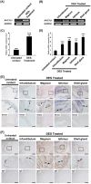
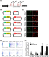
S-adenosylhomocysteine hydrolase-like protein 1 (AHCYL1), also known as IP(3) receptor-binding protein released with IP(3) (IRBIT), regulates IP(3)-induced Ca(2+) release into the cytoplasm of cells. AHCYL1 is a critical regulator of early developmental stages in zebrafish, but little is known about the function of AHCYL1 or hormonal regulation of expression of the AHCYL1 gene in avian species. Therefore, we investigated differential expression profiles of the AHCYL1 gene in various adult organs and in oviducts from estrogen-treated chickens. Chicken AHCYL1 encodes for a protein of 540 amino acids that is highly conserved and has considerable homology to mammalian AHCYL1 proteins (>94% identity). AHCYL1 mRNA was expressed abundantly in various organs of chickens. Further, the synthetic estrogen agonist induced AHCYL1 mRNA and protein predominantly in luminal and glandular epithelial cells of the chick oviduct. In addition, estrogen activated AHCYL1 through the ERK1/2 signal transduction cascade and that activated expression of AHCYL1 regulated genes affecting oviduct development in chicks as well as calcium release in epithelial cells of the oviduct. Also, microRNAs, miR-124a, miR-1669, miR-1710 and miR-1782 influenced AHCYL1 expression in vitro via its 3'-UTR which suggests that post-transcriptional events are involved in the regulation of AHCYL1 expression in the chick oviduct. In conclusion, these results indicate that AHCYL1 is a novel estrogen-stimulated gene expressed in epithelial cells of the chicken oviduct that likely affects growth, development and calcium metabolism of the mature oviduct of hens via an estrogen-mediated ERK1/2 MAPK cell signaling pathway.
Conflict of interest statement
Figures


Similar articles
-
Differential expression of neuregulin 1 (NRG1) and candidate miRNA regulating NRG1 transcription in the chicken oviduct in response to hormonal changes.J Anim Sci. 2017 Sep;95(9):3885-3904. doi: 10.2527/jas2017.1663. J Anim Sci. 2017. PMID: 28992000
-
ERBB receptor feedback inhibitor 1: identification and regulation by estrogen in chickens.Gen Comp Endocrinol. 2012 Jan 1;175(1):194-205. doi: 10.1016/j.ygcen.2011.11.013. Epub 2011 Nov 23. Gen Comp Endocrinol. 2012. PMID: 22137914
-
RAS-related protein 1: an estrogen-responsive gene involved in development and molting-mediated regeneration of the female reproductive tract in chickens.Animal. 2018 Aug;12(8):1594-1601. doi: 10.1017/S1751731117003226. Epub 2017 Dec 4. Animal. 2018. PMID: 29198267
-
Avian WNT4 in the female reproductive tracts: potential role of oviduct development and ovarian carcinogenesis.PLoS One. 2013 Jul 2;8(7):e65935. doi: 10.1371/journal.pone.0065935. Print 2013. PLoS One. 2013. PMID: 23843947 Free PMC article.
-
The role of S-adenosylhomocysteine hydrolase-like 1 in cancer.Biochim Biophys Acta Mol Cell Res. 2024 Oct;1871(7):119819. doi: 10.1016/j.bbamcr.2024.119819. Epub 2024 Aug 17. Biochim Biophys Acta Mol Cell Res. 2024. PMID: 39154900 Review.
Cited by
-
DNA methylome and transcriptome identified Key genes and pathways involved in Speckled Eggshell formation in aged laying hens.BMC Genomics. 2023 Jan 19;24(1):31. doi: 10.1186/s12864-022-09100-8. BMC Genomics. 2023. PMID: 36658492 Free PMC article.
-
IRBIT activates NBCe1-B by releasing the auto-inhibition module from the transmembrane domain.J Physiol. 2021 Feb;599(4):1151-1172. doi: 10.1113/JP280578. Epub 2020 Dec 9. J Physiol. 2021. PMID: 33237573 Free PMC article.
-
IRBITs, signaling molecules of great functional diversity.Pflugers Arch. 2025 Aug;477(8):1007-1036. doi: 10.1007/s00424-025-03095-3. Epub 2025 May 30. Pflugers Arch. 2025. PMID: 40445299 Review.
-
RNA-Sequencing based analysis of bovine endometrium during the maternal recognition of pregnancy.BMC Genomics. 2022 Jul 7;23(1):494. doi: 10.1186/s12864-022-08720-4. BMC Genomics. 2022. PMID: 35799127 Free PMC article.
-
Molecular and cellular mechanism of okadaic acid (OKA)-induced neurotoxicity: a novel tool for Alzheimer's disease therapeutic application.Mol Neurobiol. 2014 Dec;50(3):852-65. doi: 10.1007/s12035-014-8699-4. Epub 2014 Apr 8. Mol Neurobiol. 2014. PMID: 24710687 Review.
References
-
- Cooper BJ, Key B, Carter A, Angel NZ, Hart DN, et al. (2006) Suppression and overexpression of adenosylhomocysteine hydrolase-like protein 1 (AHCYL1) influences zebrafish embryo development: a possible role for AHCYL1 in inositol phospholipid signaling. J Biol Chem 281: 22471–22484. - PubMed
-
- Ando H, Mizutani A, Matsu-ura T, Mikoshiba K (2003) IRBIT, a novel inositol 1,4,5-trisphosphate (IP3) receptor-binding protein, is released from the IP3 receptor upon IP3 binding to the receptor. J Biol Chem 278: 10602–10612. - PubMed
-
- De La Haba G, Cantoni GL (1959) The enzymatic synthesis of S-adenosyl-L-homocysteine from adenosine and homocysteine. J Biol Chem 234: 603–608. - PubMed
-
- Bujnicki JM, Prigge ST, Caridha D, Chiang PK (2003) Structure, evolution, and inhibitor interaction of S-adenosyl-L-homocysteine hydrolase from Plasmodium falciparum. Proteins 52: 624–632. - PubMed
-
- Gomi T, Takusagawa F, Nishizawa M, Agussalim B, Usui I, et al. (2008) Cloning, bacterial expression, and unique structure of adenosylhomocysteine hydrolase-like protein 1, or inositol 1,4,5-triphosphate receptor-binding protein from mouse kidney. Biochim Biophys Acta 1784: 1786–1794. - PubMed
Publication types
MeSH terms
Substances
LinkOut - more resources
Full Text Sources
Molecular Biology Databases
Research Materials
Miscellaneous

