. 2012;7(11):e49402.
doi: 10.1371/journal.pone.0049402. Epub 2012 Nov 8.

Induction of brain microvascular endothelial cell urokinase expression by Cryptococcus neoformans facilitates blood-brain barrier invasion

Affiliations

Induction of brain microvascular endothelial cell urokinase expression by Cryptococcus neoformans facilitates blood-brain barrier invasion

Jamal Stie et al. PLoS One. 2012.

Abstract

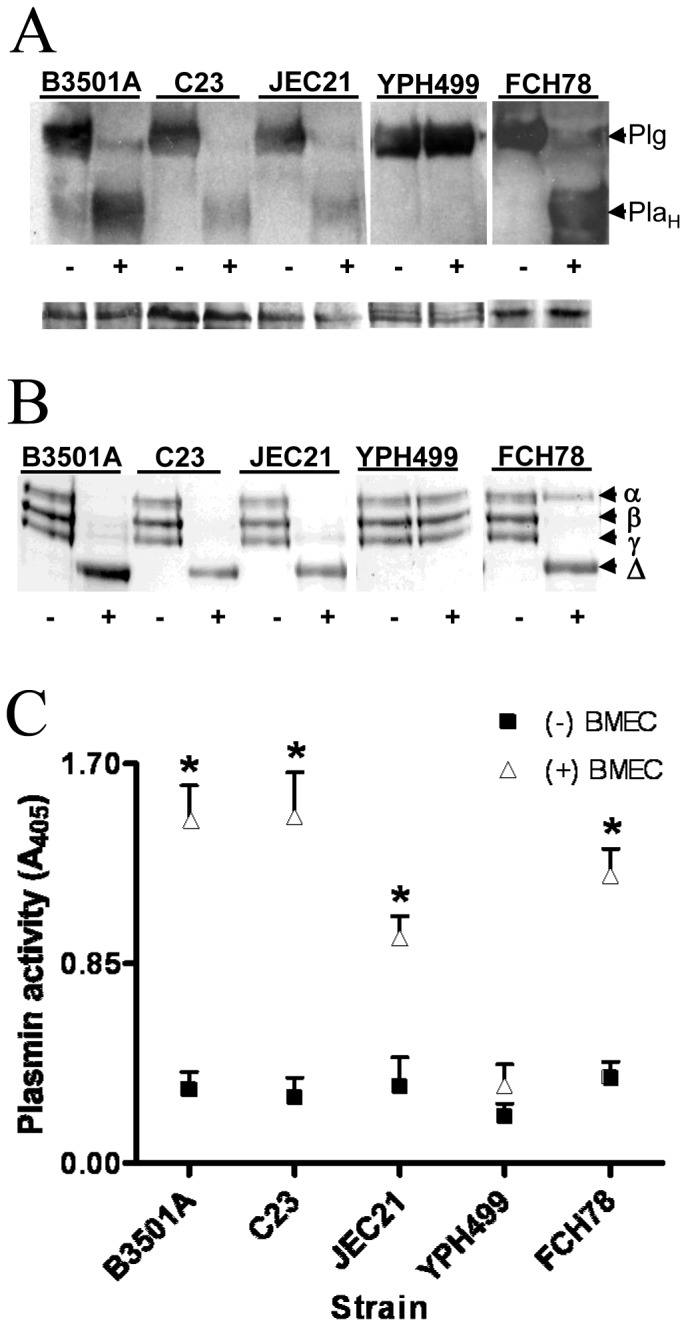
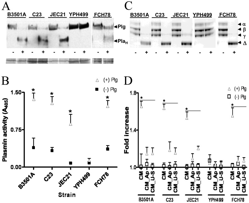
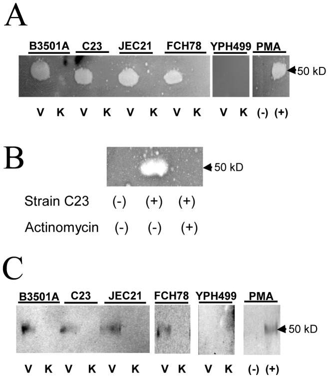
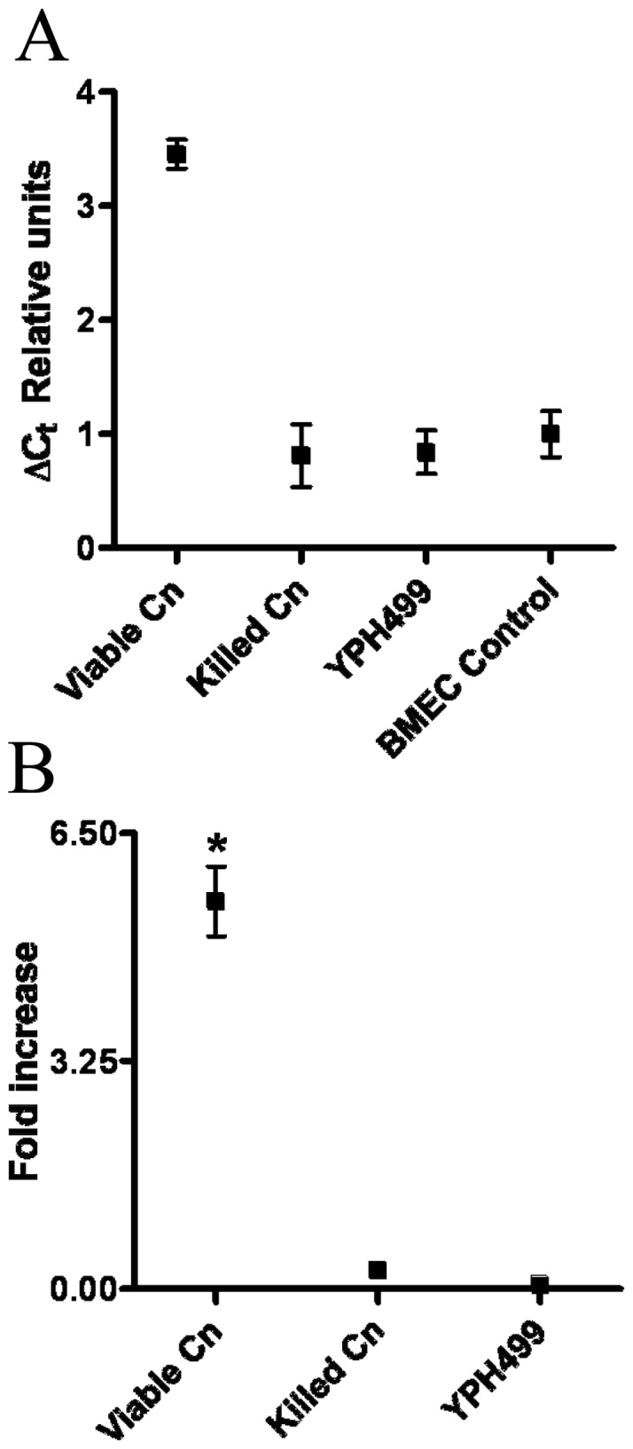
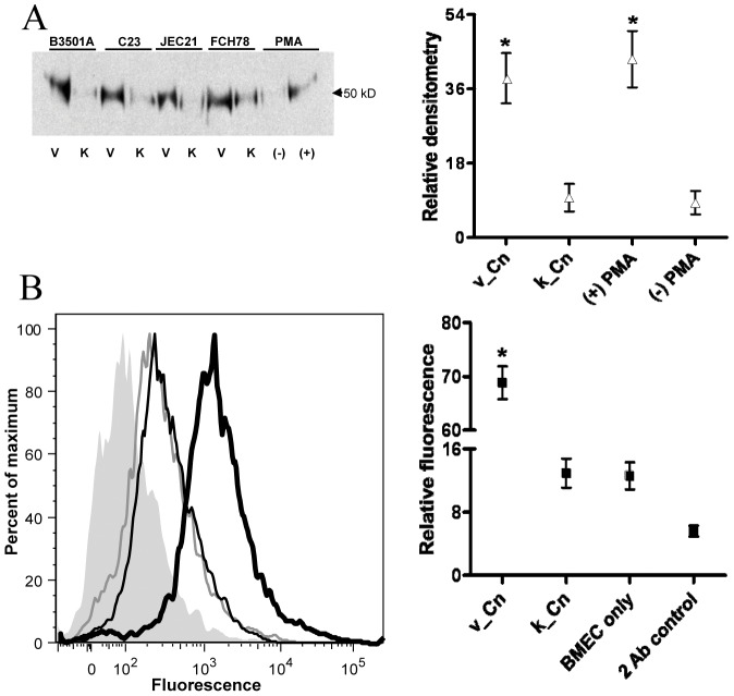
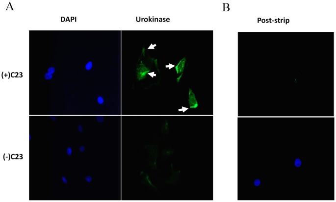
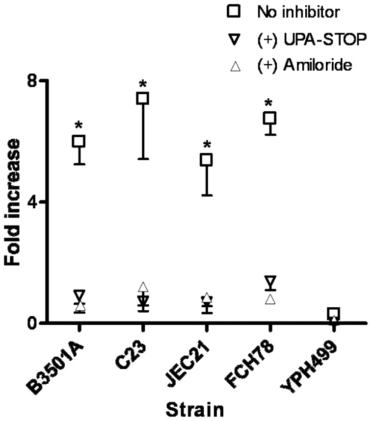
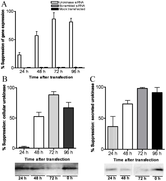
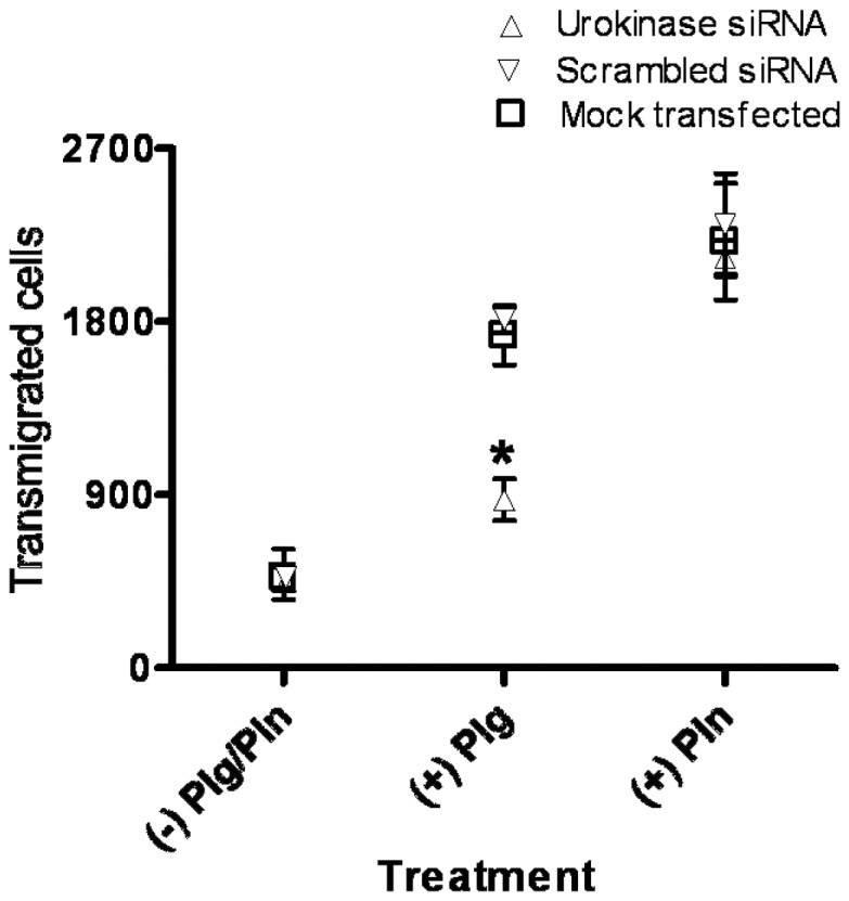
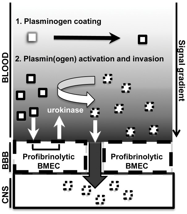
The invasive ability of the blood-borne fungal pathogen Cryptococcus neoformans can be enhanced through interactions with host plasma components, such as plasminogen. Previously we showed by in vitro studies that plasminogen coats the surface of C. neoformans and is converted to the active serine protease, plasmin, by host plasminogen activators. Viable, but not formaldehyde- or sodium azide-killed, cryptococcal strains undergo brain microvascular endothelial cell-dependent plasminogen-to-plasmin activation, which results in enhanced, plasmin-dependent cryptococcal invasion of primary bovine brain microvascular endothelial cells and fungal ability to degrade plasmin substrates. In the present work, brain microvascular endothelial cells cultured with viable, but not killed, cryptococcal strains led to significant increases in both urokinase mRNA transcription and cell-associated urokinase protein expression. Soluble urokinase was also detected in conditioned medium from brain microvascular endothelial cells cultured with viable, but not killed, C. neoformans. Exposure of plasminogen pre-coated viable C. neoformans to conditioned medium from strain-matched brain microvascular endothelial cell-fungal co-cultures resulted in plasminogen-to-plasmin activation and plasmin-dependent cryptococcal invasion. siRNA-mediated silencing of urokinase gene expression or the use of specific inhibitors of urokinase activity abrogated both plasminogen-to-plasmin activation on C. neoformans and cryptococcal-brain microvascular endothelial cell invasion. Our results suggest that pathogen exploitation of the host urokinase-plasmin(ogen) system may contribute to C. neoformans virulence during invasive cryptococcosis.


Conflict of interest statement

Competing Interests: The authors have declared that no competing interests exist.

Figures

Figure 1
Figure 1. Plasminogen-to-plasmin conversion on C. neoformans is facilitated by exposure to BMEC.
(A) Fungi were pre-coated with plasminogen, incubated in the presence (+) or absence (−) of BMEC, and analyzed by Western blot for plasminogen (Plg) or the heavy chain component (PlaH) of plasminogen activator-cleaved plasminogen. 50 µg protein was loaded per lane on SDS-PAGE gels, with protein loading controls shown below each blot. Representative of four experiments. (B–C) Strains were pre-coated (+) or not pre-coated (−) with plasminogen and incubated with BMEC for 12 h. Fungal cells were recovered from co-culture supernatants and analyzed by fibrin overlay zymography (B) or plasmin activity assay (C) against plasmin-specific substrates, fibrinogen (B) and Chromogenix (S-2251) (C). In (B), the α, β and γ bands of fibrinogen are indicated, with the principle degradation product labeled as Δ. Results from three experiments are shown. The Western blots shown in (A) are from separate gels processed in parallel, while the data shown in (B) are from a single gel. *p<0.05 by t-test for same-strain comparisons under the indicated conditions.
Figure 2
Figure 2. Conditioned medium from BMEC-C. neoformans co-cultures mediate cleavage of surface-bound plasminogen and promotes plasmin-dependent invasion of Matrigel.
(A) Western blot showing the conversion of surface-bound plasminogen to its cleavage product (PlaH) after exposure of plasminogen pre-coated strains to conditioned medium (CM), prepared from a previous incubation with the same strain. The lower blot shows the protein load per lane of the upper blot. Representative of 3 experiments. (B–C) Surface plasmin activity against plasmin substrates, Chromogenix (B) and fibrinogen (C), of strains after CM exposure in (A). In (C), the α, β and γ bands of fibrinogen are indicated, with the principle degradation product labeled as Δ. The Western blots shown in (A) are from separate gels processed in parallel, while the data shown in (C) are from a single gel. *p<0.05 by t-test for same-strain comparisons under the indicated conditions. ‘Same-strain comparison’ refers to the comparison of activity detected when individual strains are assayed under the indicated conditions. Results from 3 experiments are shown. (D) The effect of CM on Matrigel invasion by plasminogen pre-coated (+) or non-pre-coated (−) strains. CM was prepared from 12 h cultures of each strain with BMEC or from uninfected control BMEC [CM (control)]. Invasion assays were performed with urokinase (US) or aprotinin (Ap), as indicated. The y-axis shows the number of invading fungal cells. *p<0.05 by ANOVA for the comparisons under each bar. Results from 4 experiments are shown.
Figure 3
Figure 3. Characterization of plasminogen activator activity in CM.
(A) CM was isolated from BMEC cultured with viable (V) or chemically-killed (K) strains of C. neoformans or S. cerevisiae (YPH499) and examined for PA activity by fibrin overlay zymography. Monocultured BMEC incubated in the absence (−) or presence (+) of PMA were used to control for the presence of urokinase in CM, as indicated. The clear zones result from PA-mediated plasminogen-to-plasmin conversion, which is followed by plasmin fibrinolysis. (B) BMEC were treated with the transcriptional inhibitor, actinomycin D, or mock-treated prior to culture with C. neoformans strain C23 and analyzed as in (A) for PA activity. (C) CM from the indicated BMEC-fungal co-cultures was immunoprecipitated with urokinase-specific polyclonal antibody. The Western blot shows the urokinase-specific band migrating at 50 kD. CM from PMA stimulated monocultured BMEC was used as positive control. The data shown in (A) and (C) are from separate gels processed in parallel. Representative of three experiments.
Figure 4
Figure 4. Viable C. neoformans induces BMEC urokinase transcription.
BMEC were cultured 12 h with viable (v) or chemically-killed (k) yeast forms of C. neoformans (Cn) strain C23 or viable S. cerevisiae control strain, YPH499, followed by quantification of urokinase gene expression by qPCR. (A) The ΔCt for BMEC urokinase expression after co-culture with the indicated organisms (x-axis). The threshold of urokinase expression after GAPDH normalization (ΔCt) is indicated in relative units on the y-axis, which denotes increases in gene expression from low (1) to high (4) expression. (B) The fold-increase in BMEC urokinase gene expression under the indicated culture conditions relative to monocultured BMEC used as a reference. Results from 4 experiments are shown. Cn, C. neoformans strain C23. *p<0.05 by ANOVA.
Figure 5
Figure 5. Urokinase expression on BMEC is increased after co-culture with C. neoformans.
(A) Left panel: Representative Western blot showing cellular urokinase expression after immunoprecipitation from BMEC cultured with viable (V) or chemically-killed (K) strains of C. neoformans (Cn), PMA (+) or buffer only [(−); mock PMA treated]. Right panel: Relative densitometry was determined for three Western blots (left) as the percent signal distribution per blot (right). (B) Left panel: histogram showing cell surface-associated urokinase expression on BMEC after culture with killed (thin black line) or viable (thick black line) C. neoformans strain, C23, stained with urokinase-specific antibody. Control populations include monocultured BMEC stained with urokinase-specific antibody (thin gray line) or BMEC cultured with viable strain C23 but stained only with secondary antibody (shaded). Right panel: the relative fluorescence is shown for the indicated sample groups. Relative fluorescence is defined as the percent total distribution of mean fluorescence intensity among the indicated sample groups. *p<0.05 by t-test for BMEC urokinase expression after BMEC culture with viable versus killed C. neoformans (A and B) or with/without PMA (A).
Figure 6
Figure 6. C. neoformans induces a heightened, polarized expression of urokinase on BMEC.
(A) BMEC were cultured in the presence (+) or absence (−) of C. neoformans strain, C23 and afterwards washed, fixed, and analyzed for cell surface-bound urokinase (right) by indirect immunomicroscopy. Cells were co-stained with DAPI (left) to indicate the position of cell nuclei. Arrows indicate regions of urokinase accumulation along cell borders. (B) BMEC that were cultured with strain C23 and stained for cell surface urokinase expression were afterwards stripped of surface-bound urokinase and re-stained for urokinase expression (top). Cells were co-stained with DAPI (bottom). The arrowheads indicate peripheral regions of urokinase accumulation. Representative of three experiments.
Figure 7
Figure 7. BMEC urokinase activity facilitates invasion by plasminogen pre-coated C. neoformans.
C. neoformans or S. cerevisiae strain, YPH499, were pre-coated with plasminogen and assayed for invasive activity in a transwell BBB model system. BMEC invasion of the indicated fungal strains is calculated as the fold-increase (ratio) in invasion ability in the absence versus presence of urokinase-specific inhibitors UPA-STOP or amiloride. Results from four experiments are shown, with *p<0.05 by ANOVA for same-strain comparisons under the indicated culture conditions.
Figure 8
Figure 8. siRNA-mediated silencing of cryptococcal-induced urokinase gene expression in BMEC.
(A) BMEC were treated with siRNA for the indicated times with C. neoformans strain C23 added to BMEC cultures 12 h prior to RNA isolation and qPCR analysis. (B–C) BMEC were analyzed by immunoprecipitation to determine cell-associated urokinase expression (B) and secreted urokinase activity present in CM (C), with representative Western blots located below each graph. Results from three experiments are shown.
Figure 9
Figure 9. Urokinase-dependent invasion activity by C. neoformans.
BMEC were treated for 72 h with the indicated siRNA species or mock-treated and subsequently used for transwell invasion assays conducted with plasminogen (Plg) or plasmin (Pln) pre-coated C. neoformans strain, C23, as indicated. Error bars depict standard deviation for 4 experiments for same-strain comparisons under the indicated conditions. *p<0.05 for (+) Plg and (+) Pln comparisons by t-test. Results from four experiments are shown.
Figure 10
Figure 10. Model for urokinase-dependent, plasmin-enhanced invasion of the BBB by C. neoformans during the blood-borne dissemination phase of cryptococcosis.
Cryptococcal yeast forms (white squares) are passively coated with plasminogen after entering the bloodstream (black border). Interactions between blood-borne C. neoformans and BMEC of the BBB results in the conversion of initially urokinase (−), procoagulative BMEC to a urokinase (+) profibrinolytic state. Urokinase is expressed on the BMEC surface and in the surrounding soluble milieu, which leads to plasminogen-to-plasmin activation on BMEC and fungal surfaces (dashed black border), protease degradation of endothelial junctions, and paracellular fungal-CNS invasion. The gradient-effect shown in the blood compartment reflects the relative intensity of urokinase- and plasmin-dependent signal transduction both on and in proximity to endothelial cell surfaces.

Similar articles

Cited by

References

    1. Bennett JE, Kwon-Chung KJ, Howard DH (1977) Epidemiologic differences among serotypes of Cryptococcus neoformans . Am J Epidemiol 105: 582–586. - PubMed
    1. Kwon-Chung KJ, Bennett JE (1984) Epidemiologic differences between the two varieties of Cryptococcus neoformans . Am J Epidemiol 120: 123–130. - PubMed
    1. Park BJ, Wannemuehler KA, Marston BJ, Govender N, Pappas PG, et al. (2009) Estimation of the current global burden of cryptococcal meningitis among persons living with HIV/AIDS. AIDS 23: 525–530. - PubMed
    1. Velagapudi R, Hsueh YP, Geunes-Boyer S, Wright JR, Heitman J (2009) Spores as infectious propagules of Cryptococcus neoformans . Infect Immun 77: 4345–4355. - PMC - PubMed
    1. Powderly WG (1993) Cryptococcal meningitis and AIDS. Clin Infect Dis 17: 837–842. - PubMed

Publication types