. 2012;4(4):390-402.
Epub 2012 Oct 10.

The putative G-protein coupled estrogen receptor agonist G-1 suppresses proliferation of ovarian and breast cancer cells in a GPER-independent manner

Affiliations

The putative G-protein coupled estrogen receptor agonist G-1 suppresses proliferation of ovarian and breast cancer cells in a GPER-independent manner

Cheng Wang et al. Am J Transl Res. 2012.

Abstract

G-protein coupled estrogen receptor 1 (GPER) plays an important role in mediating estrogen action in many different tissues under both physiological and pathological conditions. G-1 (1-[4-(6-bromobenzo[1,3]dioxol-5yl)-3a,4,5,9b-tetrahydro-3H-cyclopenta [c]quinolin-8-yl]-ethanone) has been developed as a selective GPER agonist to distinguish estrogen actions mediated by GPER from those mediated by classic estrogen receptors. In the present study, we surprisingly found that G-1 suppressed proliferation and induced apoptosis of KGN cells (a human ovarian granulosa cell tumor cell line), actions that were not blocked by a selective GPER antagonist G15 or siRNA knockdown of GPER. G-1 also suppressed proliferation and induced cell apoptosis in GPER-negative HEK-293 cells and MDA-MB 231 breast cancer cells. Our results demonstrate that G-1 suppresses proliferation of ovarian and breast cancer cells in a GPER-independent manner. G-1 may be a candidate for the development of drugs against ovarian and breast cancer.

Keywords: G-1; G15; GPR30/GPER; breast cancer; estrogen receptors; ovarian cancer.


Figures

Figure 1
Figure 1
Effect of putative GPER agonist G-1 on morphology and proliferation of KGN cells. A) Morphology of KGN cells treated with or without different concentrations of G-1 for 72 h. Scale bar: 50μm; B) KGN cells were treated with increasing concentrations of G-1 and cell numbers were determined. Bars represent means ± SEM. Bars with different letters are significantly (p<0.05) different from each other. The insert in panel B is the chemical structure of G-1 [10].
Figure 2
Figure 2
Effect of GPER on KGN cell proliferation. A) Knockdown of GPER with GPER siRNA in KGN cells. The upper panel shows siRNA transfection efficiency detected by a fluorescein-labeled non-target siRNA (siGLO). Scale bar: 20μm. Middle panel shows GPER protein levels in KGN cells after transfection (6h) with or without GPER siRNA (detected by western blot). Lower panel shows the relative OD values of GPER protein signal. META: META-FECTENE. B) Effect of GPER knockdown on KGN cell proliferation. Upper panel: morphology of KGN cells in the control (transfected with siGLO) and GPER siRNA transfected groups. Scale bar: 100μm; Lower panel: KGN cell number counting. Bars represent means ± SEM. *: significantly different from control (P<0.05).
Figure 3
Figure 3
Morphology of GPER-knockdown KGN cells with or without G-1 treatments. Ctrl: untreated normal control; siGLO: cells transfected with siGLO (transfection control); GPER siRNA: KGN cells transfected with GPER siRNA (knockdown control); siGLO+G-1: siGLO transfected KGN cells were treated with 2μM G-1 for 30 h; GPER siRNA +G-1: GPER knockdown cells treated with 2μM of G-1 for 30 h. Scale bar: 50μm.
Figure 4
Figure 4
The GPER antagonist G15 does not attenuate the inhibitory effect of G-1 on KGN cell proliferation. A) KGN cells were treated with G15 for 4 h prior to treatment with or without G-1 for 48 h and cell morphology was recorded. Ctrl: normal KGN cells (negative control); G-1: G-1 (2μM) treated KGN cells (positive control); G15: G15 (4μM) alone treated KGN cells; G-1+G15: KGN cells were treated with G-1 (2μM) and G15 (4μM). Scale bar: 50μm. B) Effect of G15 on G-1-induced suppression of KGN cell proliferation. Bars represent means ± SEM. Bars with different letters are significantly (p<0.05) different from each other.
Figure 5
Figure 5
The GPER antagonist G15 does not attenuate G-1-induced reduction of KGN cell viability. A) KGN cells were treated with G15 for 4 h prior to treatment with or without G-1 for 24 h and caspase 3/7 activity was measured. B) KGN cells were treated with G15 for 4 h prior to treatment with or without G-1 for 24 h. KGN cell viability was determined by MTT assay. Bars represent means ± SEM. Bars with different letters are significantly (p<0.05) different from each other.
Figure 6
Figure 6
Effect of GPER antagonist G15 on G-1-induced KGN cell cycle arrest. A) KGN cells were pretreated with control media (Control) or with G15 (4μM) for 4 h prior to treatment for 24 h with or without G-1 (2μM). Cells were fixed and processed for flow cytometry. Results are from one of three representative experiments.
Figure 7
Figure 7
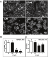
Effect of G-1 on the morphology, proliferation and viability of GPER-negative HEK-293 cells. A) HEK-293 cells were treated with or without increasing concentrations of G-1 for 16 h. Ctrl: normal HEK-293 cells (negative control); 0.5μM: 500nM of G-1 treated HEK-293 cells; 1μM: 1μM of G-1 treated HEK-293 cells; 2μM: 2μM of G-1 treated HEK-293 cells. Scale bar: 50μm. B) HEK-293 cells were incubated for 60 h with increasing concentrations of G-1 and cell numbers were determined. C) HEK-293 cells were incubated for 24 h with increasing concentrations of G-1 and cell viability (indicated by MTT metabolism) was determined. Bars represent means ± SEM, n=3. Bars with different letters are significantly (p<0.05) different from each other.
Figure 8
Figure 8
Effect of G-1 treatment on cell cycle progression in GPER negative HEK-293 cells. HEK-293 cells were incubated for 16 h with control media (Control) or different concentrations of G-1 (500nM, 1μM or 2μM). Cells were fixed and processed for flow cytometry. Results are from a representative experiment. Similar results were obtained in at least two additional experiments.
Figure 9
Figure 9
Effect of G-1 on the morphology and cell proliferation of MDA-MB 231 breast cancer cells. A) MDA-MB 231 cells were treated with G-1 for 16 h and the morphology of the MDA-MB 231 breast cancer cells was recorded. Ctrl: cultured normal MDA-MB 231 breast cancer cells as negative control; G-1: G-1 (2μM) treated MDA-MB 231 cells; G15: G15 (8 μM) treated MDA-MB 231 cells; G-1+G15: G15 (8μM) plus G-1 (2μM) treated MDA-MB 231 cells; Scale bar: 50μm. B) MDA-MB 231 breast cancer cells were treated with increasing concentrations of GPER antagonist G15 (0 – 8μM) for 4 h prior to treating with G-1 (2μM) for 36 h. Cell counts were determined and bars represent means ± SEM of at least 3 repeated experiments. Bars with different letters are significantly (p<0.05) different from each other.

References

    1. Heldring N, Pike A, Andersson S, Matthews J, Cheng G, Hartman J, Tujague M, Ström A, Treuter E, Warner M, Gustafsson JA. Estrogen receptors: how do they signal and what are their targets. Physiological Review. 2007;87:905–931. - PubMed
    1. Ho S-M. Estrogen, Progesterone and Epithelial Ovarian Cancer. Reproductive Biology and Endocrinology. 2003;1:73. - PMC - PubMed
    1. Thomas P, Pang Y, Filardo EJ, Dong J. Identity of an estrogen membrane receptor coupled to a G protein in human breast cancer cells. Endocrinology. 2005;146:624–632. - PubMed
    1. Filardo EJ, Quinn JA, Bland KI, Frackelton AR Jr. Estrogen-induced activation of Erk-1 and Erk-2 requires the G protein-coupled receptor homolog, GPR30, and occurs via trans-activation of the epidermal growth factor receptor through release of HB-EGF. Molecular Endocrinology. 2000;14:1649–1660. - PubMed
    1. Revankar CM, Cimino CF, Sklar LA, Arterburn JB, Prossnitz ER. A transmembrane intracellular estrogen receptor mediates rapid cell signaling. Science. 2005;307:1625–1630. - PubMed

LinkOut - more resources