Comparative Study
. 2012 Nov;132(5):3428-38.
doi: 10.1121/1.4754551.

Vibratory responses of synthetic, self-oscillating vocal fold models

Affiliations
Comparative Study

Vibratory responses of synthetic, self-oscillating vocal fold models

Preston R Murray et al. J Acoust Soc Am. 2012 Nov.

Abstract

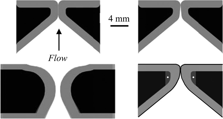
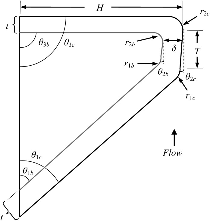
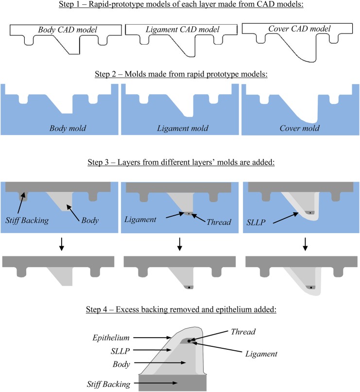
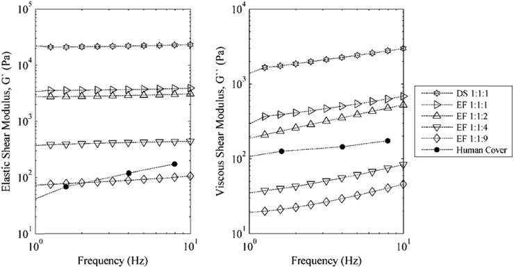
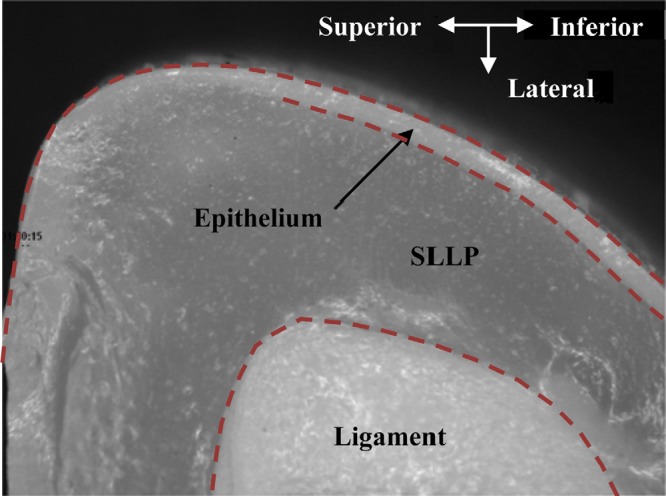
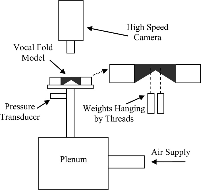
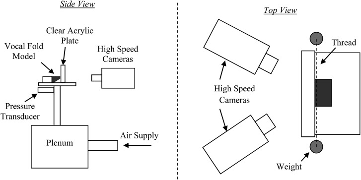
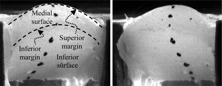
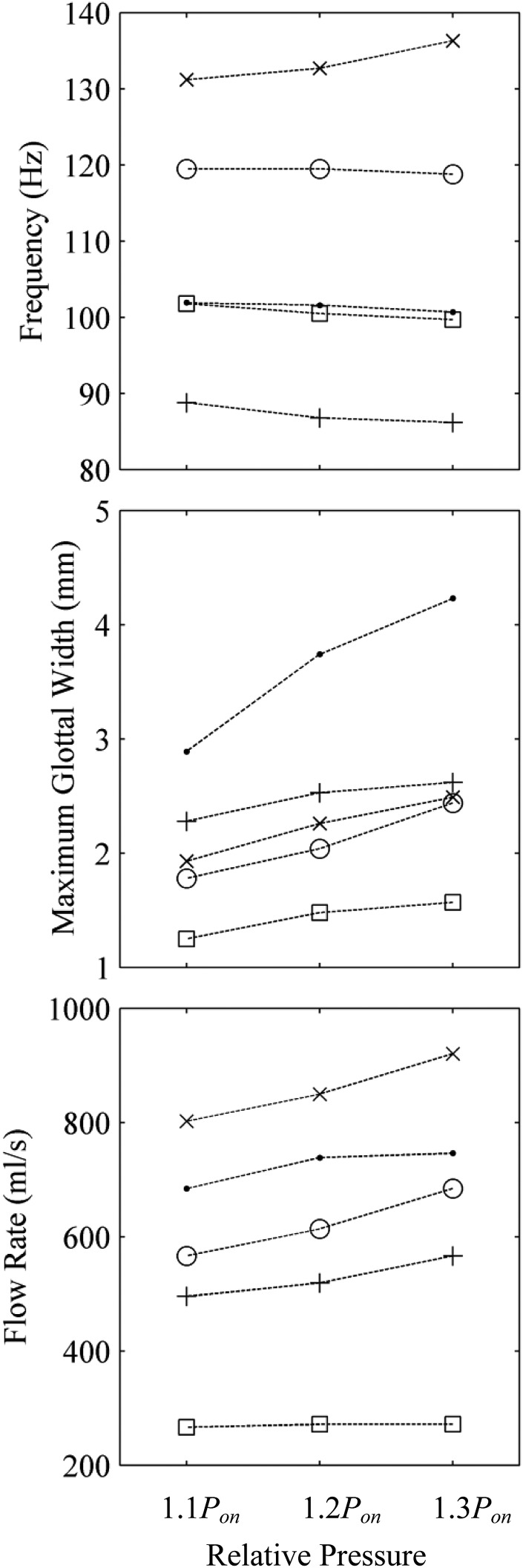
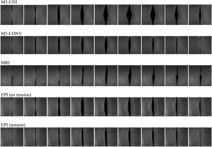
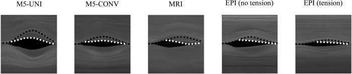
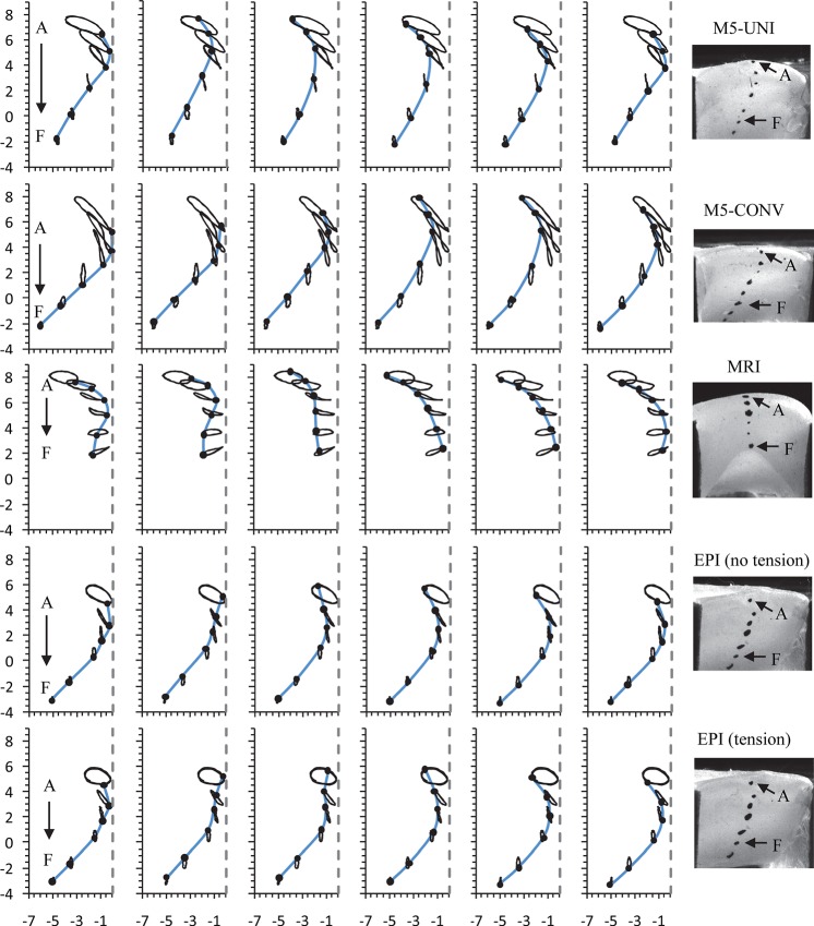
The flow-induced responses of four self-oscillating synthetic vocal fold models are compared. All models were life-sized and fabricated using flexible silicone compounds with material properties comparable to those of human vocal fold tissue. Three of the models had two layers of different stiffness to represent the body-cover grouping of vocal fold tissue. Two of the two-layer models were based on the "M5" geometry [Scherer et al., J. Acoust. Soc. Am. 109, 1616-1630 (2001)], while the third was based on magnetic resonance imaging data. The fourth model included several layers, including a thin epithelial layer, an exceedingly flexible superficial lamina propria layer, a ligament layer that included an anteriorly-posteriorly oriented fiber to restrict vertical motion, and a body layer. Measurements were performed with these models in full larynx and hemilarynx configurations. Data included onset pressure, vibration frequency, glottal flow rate, maximum glottal width, and medial surface motion, the latter two of which were acquired using high-speed imaging techniques. The fourth, multi-layer model exhibited onset pressure, frequency, and medial surface motion traits that are comparable to published human vocal fold data. Importantly, the model featured an alternating convergent-divergent glottal profile and mucosal wave-like motion, characteristics which are important markers of human vocal fold vibration.


Figures

Figure 1
Figure 1
Cross-sectional illustrations of four self-oscillating synthetic vocal fold models. Clockwise from the upper left-hand side: M5-Uniform (M5-UNI), M5-Convergent (M5-CONV), Epithelium (EPI), and Magnetic Resonance Imaging (MRI) models.
Figure 2
Figure 2
Geometric parameters for the M5-UNI, M5-CONV, and EPI models.
Figure 3
Figure 3
(Color online) EPI model fabrication process schematic, including cross sections of CAD models used for rapid prototyping, molds, and creation of the various layers. Shown at bottom is a finished model with all layers.
Figure 4
Figure 4
Elastic (G′) and viscous (G″) shear moduli for various ratios of silicone. Included are representative data of the human cover from Chan et al. (2007).
Figure 5
Figure 5
(Color online) Sample image of a coronal section of the EPI model showing the epithelium, cover, and ligament layers.
Figure 6
Figure 6
Full larynx experimental setup (not to scale).
Figure 7
Figure 7
Hemilarynx experimental setup (not to scale).
Figure 8
Figure 8
Stereo image pair of the M5-CONV model used for medial surface tracking. Direction of airflow is from the bottom to the top of the images.
Figure 9
Figure 9
(Top) Frequency, (middle) maximum glottal width, and (bottom) flow rate for the models at 110%, 120%, and 130% of their respective onset pressures. M5-UNI (•), M5-CONV (○), MRI (×), EPI without tension (+), and EPI with tension (□).
Figure 10
Figure 10
Superior view high-speed images of each model over one cycle. All images were obtained with the models vibrating at 120% of their respective onset pressures.
Figure 11
Figure 11
High-speed kymograms of one period for the four models. Estimated locations of the superior (black) and inferior (white) margins are shown by dotted lines.
Figure 12
Figure 12
(Color online) Medial surface motion at six phases of oscillation at 1.2Pon. Horizontal and vertical axes denote, respectively, medial-lateral and inferior–superior positions (mm). The vertical dashed line denotes the position of the clear acrylic plate. In each frame the thin solid lines are traces of the markers during oscillation, and the thick solid line is a spline interpolation between markers to approximate the position of the medial surface at the given phase. The left column corresponds to the phase near glottal opening.

References

    1. Abdel-Aziz, Y. I., and Karara, H. M. (1971). “ Direct linear transformation from comparator coordinates into object space coordinates in close-range photogrammetry,” in Proceedings of the Symposium on Close-Range Photogrammetry (American Society of Photogrammetry, Falls Church, VA), pp. 1–18.
    1. Baken, R. J., and Orlikoff, R. F. (2000). Clinical Measurement of Speech and Voice, 2nd ed. (Singular, San Diego: ), pp. 328, 358–363.
    1. Berry, D. A., Clark, M. J., Montequin, D. W., and Titze, I. R. (2001a). “ Characterization of the medial surface of the vocal folds,” Ann. Otol. Rhinol. Laryngol. 110(5 ), 470–477. - PubMed
    1. Berry, D. A., Montequin, D. W., and Tayama, N. (2001b). “ High-speed digital imaging of the medial surface of the vocal folds,” J. Acoust. Soc. Am. 110(5 ), 2539–2547.10.1121/1.1408947 - DOI - PubMed
    1. Berry, D. A., Zhang, Z., and Neubauer, J. (2006). “ Mechanisms of irregular vibration in a physical model of the vocal folds,” J. Acoust. Soc. Am. 120(3 ), EL36–EL42.10.1121/1.2234519 - DOI - PubMed

Publication types