

Tuning nanostructure dimensions with supramolecular twisting
- PMID: 23145959
- PMCID: PMC3586767
- DOI: 10.1021/jp3087978
Tuning nanostructure dimensions with supramolecular twisting
Abstract
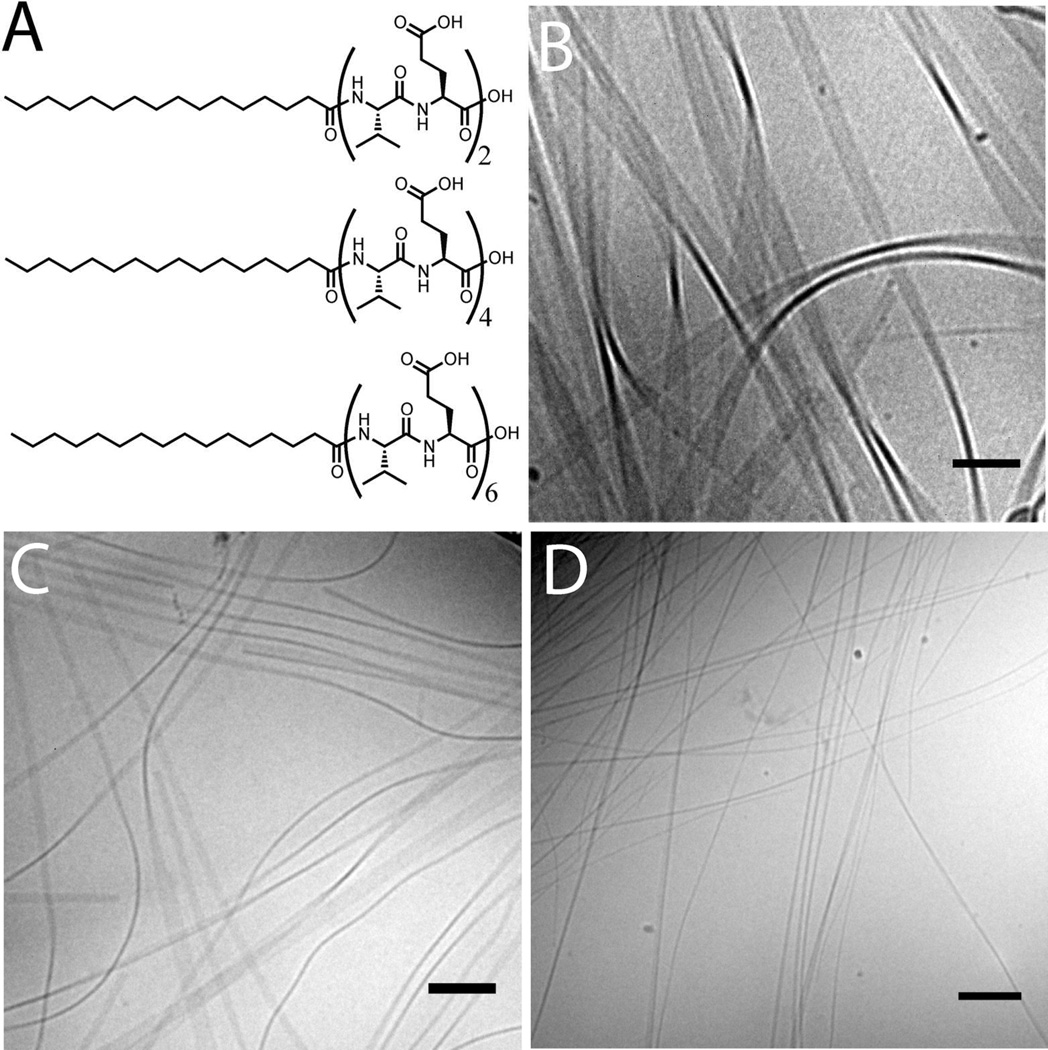
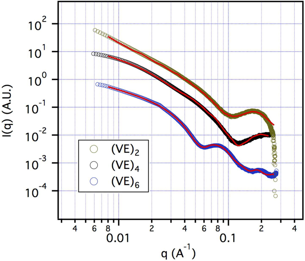
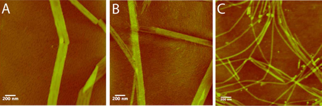
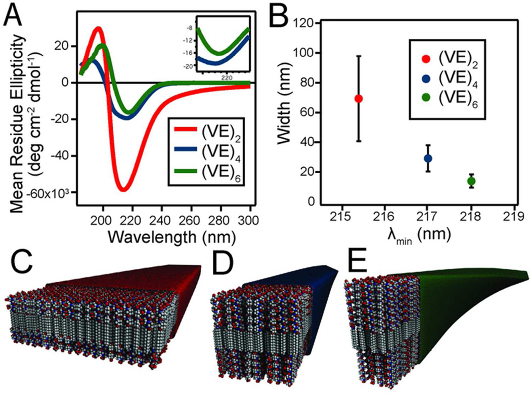
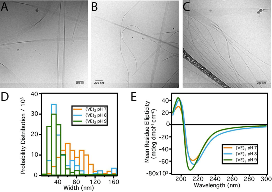
Peptide amphiphiles are molecules containing a peptide segment covalently bonded to a hydrophobic tail and are known to self-assemble in water into supramolecular nanostructures with shape diversity ranging from spheres to cylinders, twisted ribbons, belts, and tubes. Understanding the self-assembly mechanisms to control dimensions and shapes of the nanostructures remains a grand challenge. We report here on a systematic study of peptide amphiphiles containing valine-glutamic acid dimeric repeats known to promote self-assembly into belt-like flat assemblies. We find that the lateral growth of the assemblies can be controlled in the range of 100 nm down to 10 nm as the number of dimeric repeats is increased from two to six. Using circular dichroism, the degree of β-sheet twisting within the supramolecular assemblies was found to be directly proportional to the number of dimeric repeats in the PA molecule. Interestingly, as twisting increased, a threshold is reached where cylinders rather than flat assemblies become the dominant morphology. We also show that in the belt regime, the width of the nanostructures can be decreased by raising the pH to increase charge density and therefore electrostatic repulsion among glutamic acid residues. The control of size and shape of these nanostructures should affect their functions in biological signaling and drug delivery.
Figures
Similar articles
-
Supramolecular Interactions and Morphology of Self-Assembling Peptide Amphiphile Nanostructures.Nano Lett. 2021 Jul 28;21(14):6146-6155. doi: 10.1021/acs.nanolett.1c01737. Epub 2021 Jul 14. Nano Lett. 2021. PMID: 34259001
-
Supramolecular Assembly of Peptide Amphiphiles.Acc Chem Res. 2017 Oct 17;50(10):2440-2448. doi: 10.1021/acs.accounts.7b00297. Epub 2017 Sep 6. Acc Chem Res. 2017. PMID: 28876055 Free PMC article.
-
Molecular self-assembly into one-dimensional nanostructures.Acc Chem Res. 2008 Dec;41(12):1674-84. doi: 10.1021/ar8000926. Acc Chem Res. 2008. PMID: 18754628 Free PMC article.
-
A model for the controlled assembly of semiconductor peptides.Nanoscale. 2012 Nov 21;4(22):6940-7. doi: 10.1039/c2nr32140h. Nanoscale. 2012. PMID: 23034819 Review.
-
Methods to Characterize the Nanostructure and Molecular Organization of Amphiphilic Peptide Assemblies.Methods Mol Biol. 2018;1777:3-21. doi: 10.1007/978-1-4939-7811-3_1. Methods Mol Biol. 2018. PMID: 29744826 Review.
Cited by
-
Design of materials with supramolecular polymers.Prog Polym Sci. 2020 Dec;111:101310. doi: 10.1016/j.progpolymsci.2020.101310. Epub 2020 Oct 15. Prog Polym Sci. 2020. PMID: 33082608 Free PMC article. Review.
-
Design, Synthesis, and Nanostructure-Dependent Antibacterial Activity of Cationic Peptide Amphiphiles.ACS Appl Mater Interfaces. 2019 Jan 23;11(3):2790-2801. doi: 10.1021/acsami.8b17808. Epub 2019 Jan 10. ACS Appl Mater Interfaces. 2019. PMID: 30588791 Free PMC article.
-
Transition of Nano-Architectures Through Self-Assembly of Lipidated β3-Tripeptide Foldamers.Front Chem. 2020 Mar 31;8:217. doi: 10.3389/fchem.2020.00217. eCollection 2020. Front Chem. 2020. PMID: 32296680 Free PMC article.
-
Bioactive Supramolecular Polymers for Skin Regeneration Following Burn Injury.Biomacromolecules. 2025 Aug 11;26(8):5471-5482. doi: 10.1021/acs.biomac.5c01107. Epub 2025 Jul 16. Biomacromolecules. 2025. PMID: 40669455 Free PMC article.
-
Antiviral supramolecular polymeric hydrogels by self-assembly of tenofovir-bearing peptide amphiphiles.Biomater Sci. 2023 Jan 17;11(2):489-498. doi: 10.1039/d2bm01649d. Biomater Sci. 2023. PMID: 36449365 Free PMC article.
References
-
- De Greef TFA, Smulders MMJ, Wolffs M, Schenning APHJ, Sijbesma RP, Meijer EW. Chem. Rev. 2009;109:5687–5754. - PubMed
-
- Knowles TPJ, Buehler MJ. Nature Publishing Group. 2011:1–11.
Publication types
MeSH terms
Substances
Grants and funding
LinkOut - more resources
Full Text Sources
Other Literature Sources
