

Isolation of canine Anaplasma phagocytophilum strains from clinical blood samples using the Ixodes ricinus cell line IRE/CTVM20
- PMID: 23146170
- PMCID: PMC3757156
- DOI: 10.1016/j.vetmic.2012.10.021
Isolation of canine Anaplasma phagocytophilum strains from clinical blood samples using the Ixodes ricinus cell line IRE/CTVM20
Abstract
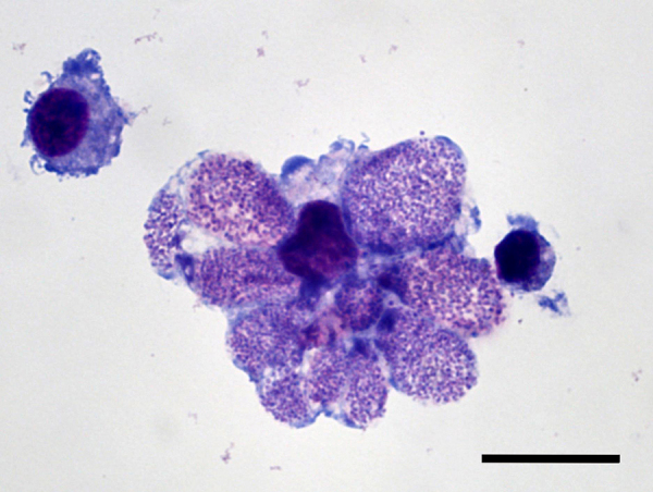
Anaplasma phagocytophilum is an intracellular tick-borne rickettsial pathogen, which causes granulocytic anaplasmosis in various species of livestock and companion animals and also in humans. Previously A. phagocytophilum has been isolated and propagated in cell lines derived from the tick Ixodes scapularis and in the human promyelocytic cell line HL60. In this study we used the Ixodes ricinus-derived cell line IRE/CTVM20 to isolate and propagate two new canine strains of A. phagocytophilum. Blood samples were collected by veterinarians from two dogs, one from Germany and the other from Austria. Suspicion of clinical canine granulocytic anaplasmosis was raised by the treating veterinarians and after confirmation of A. phagocytophilum infection by real-time PCR, buffy coat cells were isolated and co-cultivated with IRE/CTVM20 cells maintained at 28 °C in L15/L15B medium. In the tick cells, rickettsial inclusions were first recognised after 86 days of incubation. Electron microscopic examination of tick cells infected with one of the isolates revealed cytoplasmic vacuoles containing pleomorphic organisms with individual bacteria enveloped by a bilayer membrane. Sequencing of 16S rRNA genes confirmed the isolation of A. phagocytophilum and showed the highest identity to the A. phagocytophilum human HZ strain. The two A. phagocytophilum isolates were passaged several times in IRE/CTVM20 cells and transferred to the I. scapularis cell line ISE6. This confirms for the first time the successful establishment and continuous cultivation of this pathogen in I. ricinus cells as well as infectivity of these canine strains for I. scapularis cells.
Copyright © 2012. Published by Elsevier B.V.
Figures
Similar articles
-
Tissue-Specific Signatures in the Transcriptional Response to Anaplasma phagocytophilum Infection of Ixodes scapularis and Ixodes ricinus Tick Cell Lines.Front Cell Infect Microbiol. 2016 Feb 10;6:20. doi: 10.3389/fcimb.2016.00020. eCollection 2016. Front Cell Infect Microbiol. 2016. PMID: 26904518 Free PMC article.
-
Infection of Ixodes spp. tick cells with different Anaplasma phagocytophilum isolates induces the inhibition of apoptotic cell death.Ticks Tick Borne Dis. 2015 Sep;6(6):758-67. doi: 10.1016/j.ttbdis.2015.07.001. Epub 2015 Jul 6. Ticks Tick Borne Dis. 2015. PMID: 26183310
-
Anaplasma phagocytophilum in questing Ixodes ricinus ticks from Romania.Ticks Tick Borne Dis. 2015 Apr;6(3):408-13. doi: 10.1016/j.ttbdis.2015.03.010. Epub 2015 Mar 30. Ticks Tick Borne Dis. 2015. PMID: 25838178
-
[Equine granulocytic anaplasmosis (EGA): Case description and overview of the epidemiological situation with focus on Germany].Tierarztl Prax Ausg G Grosstiere Nutztiere. 2024 Dec;52(6):352-360. doi: 10.1055/a-2418-6540. Epub 2024 Dec 4. Tierarztl Prax Ausg G Grosstiere Nutztiere. 2024. PMID: 39631410 Review. German.
-
A review on the eco-epidemiology and clinical management of human granulocytic anaplasmosis and its agent in Europe.Parasit Vectors. 2019 Dec 21;12(1):599. doi: 10.1186/s13071-019-3852-6. Parasit Vectors. 2019. PMID: 31864403 Free PMC article. Review.
Cited by
-
Tick-borne pathogens induce differential expression of genes promoting cell survival and host resistance in Ixodes ricinus cells.Parasit Vectors. 2017 Feb 15;10(1):81. doi: 10.1186/s13071-017-2011-1. Parasit Vectors. 2017. PMID: 28202075 Free PMC article.
-
Tissue-Specific Signatures in the Transcriptional Response to Anaplasma phagocytophilum Infection of Ixodes scapularis and Ixodes ricinus Tick Cell Lines.Front Cell Infect Microbiol. 2016 Feb 10;6:20. doi: 10.3389/fcimb.2016.00020. eCollection 2016. Front Cell Infect Microbiol. 2016. PMID: 26904518 Free PMC article.
-
Uukuniemi Virus as a Tick-Borne Virus Model.J Virol. 2016 Jul 11;90(15):6784-98. doi: 10.1128/JVI.00095-16. Print 2016 Aug 1. J Virol. 2016. PMID: 27194760 Free PMC article.
-
Cultivation of the causative agent of human neoehrlichiosis from clinical isolates identifies vascular endothelium as a target of infection.Emerg Microbes Infect. 2019;8(1):413-425. doi: 10.1080/22221751.2019.1584017. Emerg Microbes Infect. 2019. PMID: 30898074 Free PMC article.
-
Use of Tick Cell Lines in Co-Infection Studies with a Preliminary Study of Co-Culture of Borrelia burgdorferi and Anaplasma phagocytophilum.Pathogens. 2025 Jan 15;14(1):78. doi: 10.3390/pathogens14010078. Pathogens. 2025. PMID: 39861039 Free PMC article.
References
-
- Bell-Sakyi L. Ehrlichia ruminantium grows in cell lines from four ixodid tick genera. J. Comp. Pathol. 2004;130:285–293. - PubMed
-
- Bell-Sakyi L., Zweygarth E., Blouin E.F., Gould E.A., Jongejan F. Tick cell lines: tools for tick and tick-borne disease research. Trends Parasitol. 2007;23:450–457. - PubMed
-
- Bjoersdorff A., Svendenius L., Owens J.H., Massung R.F. Feline granulocytic ehrlichiosis – a report of a new clinical entity and characterisation of the infectious agent. J. Small Anim. Pract. 1999;40:20–24. - PubMed
-
- Blouin E.F., Kocan K.M. Morphology and development of Anaplasma marginale (Rickettsiales: Anaplasmataceae) in cultured Ixodes scapularis (Acari: Ixodidae) cells. J. Med. Entomol. 1998;35:788–797. - PubMed
Publication types
MeSH terms
Substances
Grants and funding
LinkOut - more resources
Full Text Sources
Other Literature Sources
Molecular Biology Databases
Research Materials
