. 2013 Mar 1;434(1):78-83.
doi: 10.1016/j.ab.2012.11.004. Epub 2012 Nov 9.

A fluorogenic phospholipid for the detection of lysosomal phospholipase A2 activity

Affiliations

A fluorogenic phospholipid for the detection of lysosomal phospholipase A2 activity

Akira Abe et al. Anal Biochem. .

Abstract

Lysosomal phospholipase A2 (group XV PLA2, LPLA2) is a lysosomal enzyme linked to drug-induced phospholipidosis. We developed phospholipid "smart probes" based on the conversion of a quenched fluorogenic substrate to a fluorescent product. Due to the preference of LPLA2 for phosphatidylglycerol, three fluorogenic phosphatidylglycerols were synthesized. Two fluorogenic phosphatidylglycerols were conjugated with one FAM (fluorescein amidite) group and one DABCYL [4-(4-dimethylaminophenylazo)-benzoyl] group; the third substrate consisted of two FAM groups conjugated at the sn-1 and sn-2 positions. The sn-1 ester linkage was replaced with an amide linkage. 1-FAM-2-DABCYL-PG was degraded by recombinant LPLA2 and mouse serum but not by the serum obtained from LPLA2-deficient mice when 1,2-dioleoyl-PG/1-FAM-2-DABCYL-PG liposomes were used. The formation of 1-FAM-lyso-PG generated from 1-FAM-2-DABCYL-PG in the presence of LPLA2 was quantitatively determined by fluorescent measurements. The 1-FAM-2-DABCYL-PG incorporated into 1,2-dioleoyl-phosphatidylcholine/sulfatide liposomes was used to evaluate the effect of the cationic amphiphilic drugs amiodarone and fluoxetine on LPLA2 activity. The IC(50) values of amiodarone and fluoxetine estimated by fluorescent measurement were 10 and 19μM, respectively. These results indicate that 1-FAM-2-DABCYL-PG is a specific substrate for LPLA2 and a useful reagent for the detection of LPLA2 activity from multiple sources.


Figures

Fig. 1
Fig. 1
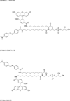
Fluorescent phosphatidylglycerol (PG) probe structures, including 1,2-bis-FAM-PG , 1-FAM-2-DABCYL-PG, and 1-DABCYL-2-FAM-PG.
Fig. 1
Fig. 1
Fluorescent phosphatidylglycerol (PG) probe structures, including 1,2-bis-FAM-PG , 1-FAM-2-DABCYL-PG, and 1-DABCYL-2-FAM-PG.
Fig. 2
Fig. 2
A. Degradation of fluorogenic phospholipids by recombinant mouse LPLA2. The reaction was carried out as described in the text and kept for 2.5, 5, 10 and 20 min at 37°C. B. Degradation of fluorogenic phospholipids by the serums obtained from wild type mouse and from LPLA2-deficient mouse. The reaction was kept for 90 min at 37°C in the presence or absence of 2% mouse serum. 1-FAM-2-DABCYL-PG, 1-DABCYL-2-FAM-PG and 1,2-bis-FAM-PG denote the respective liposomes. C. Degradation of 1-FAM-2-DABCYL-PG by the serums obtained from wild type mouse and from LPLA2-deficient mouse. The reaction was carried out as described under “Materials and Methods” using DOPG/1-FAM-2-DAB-PG (10:1, a molar ratio) liposomes and kept for 45 and 90 min at 37°C in the presence or absence of mouse serum.
Fig. 3
Fig. 3
Fluorescence measurement of LPLA2 activity using 1-FAM-2-DABCYL-PG. A. To obtain the standard curve of 1-FAM-lyso-PG (shown in panel A), known amounts of liposomes containing 1-FAM-2-DABCYL-PG were hydrolyzed in the presence of 0.5 N NaOH for 1 h at 37°C. After the reaction, the reaction mixture was neutralized with HCl. The reaction products such as 1-FAM-lyso-PG and other lipids were extracted as described in the materials and methods section. The fluorescence intensity of 1-FAM-lyso-PG recovered in the upper layer partitioned by adding chloroform, methanol and 0.9% NaCl was measured as described in the materials and methods section. B. The reaction by LPLA2 was carried out in the presence of liposomes (128 µM phospholipid) and 7.25 ng/ml of recombinant mouse LPLA2 in 500 µl of total volume as described in the materials and methods section and kept for 2, 4, 6, 8 and 10 min at 37°C after adding 5 µl of 0.9% NaCl or 725 ng/ml of recombinant mouse LPLA2. DOPG/1-FAM-2-DABCYL-PG (100:1, molar ratio, ■) liposomes and DOPC/sulfatide/1-FAM-2-DABCYL-PG (100:10:1, molar ratio, ●) liposomes were used. The fluorescent intensity of the reaction product recovered in the upper layer partitioned by adding chloroform, methanol and 0.9% NaCl was measured as described in the materials and methods section. The released reaction product, 1-FAM-lyso-PG, plotted in the figures were calculated from [(the fluorescent intensity in the presence of LPLA2)-(the fluorescent intensity in the absence of LPLA2)]/slope of Fig. 3A at each time point. In both panel A and panel B, error bars indicate S.D. (n = 4).
Fig. 4
Fig. 4
Quantitative measurement of LPLA2 activity using 1-FAM-2-DABCYL-PG. In panel A, the reaction mixture contained 49 mM sodium citrate (pH 4.5), 10 µg/ml BSA, liposomes (128 µM phospholipid) and 7.25 ng/ml of recombinant mouse LPLA2 in 500 µl of total volume. The liposomes consisted of DOPC/sulfatide/1-FAM-2-DABCYL-PG (100:10:1, molar ratio). The reaction was initiated by adding the LPLA2 and kept for 1, 2 and 4 min at 37°C. The released reaction product, 1-FAM-lyso-NAS, was determined as described in the legend of Fig. 3 and plotted against incubation time. In panel B, different concentrations of the LPLA2 were incubated with DOPC/sulfatide/1-FAM-2-DAB-PG liposomes for 2 min at 37°C in 500 µl of 49 mM Na-citrate (pH 4.5). The released reaction product, 1-FAM-lyso-PG, plotted in the figure was calculated as described in the legend of Fig. 3. In both panel A and panel B, error bars indicate S.D. (n = 4).
Fig. 5
Fig. 5
Inhibition study of LPLA2 activity by CAD using 1-FAM-2-DABCYL-PG. DOPC/sulfatide/NAS (100:10:30, molar ratio) and DOPC/sulfatide/1-FAM-2-DABCYL-PG (100:10:1, molar ratio) liposomes were pre-incubated with 0, 1, 3.33, 10, 33.3 and 100 µM amiodarone (●) and fluoxetine (■) for 5 min at 37°C. The reactions and measurements were carried out as described in the materials and methods section and the legend of Fig. 4. In panel A, the reaction was kept for 10 min in the presence of DOPC/sulfatide/NAS liposomes (127 µM phospholipid) and 14.5 ng/ml of recombinant mouse LPLA2 in 500 µl of total volume. The transacylase activity of LPLA2 was determined by 1-O-oleoyl-NAS produced from DOPC/sulfatide/NAS liposomes. The formation of 1-O-oleoyl-NAS in the absence of CADs was linearly proportional to the incubation time till 10 min (data not shown). In panel B, the reaction was kept for 4 min in the presence of DOPC/sulfatide/1-FAM-2-DABCYL-PG (128 µM phospholipid) and 7.25 ng/ml of recombinant mouse LPLA2 in 500 µl of total volume. LPLA2 activity was determined by the fluorescence measurement of the reaction product, 1-FAM-lyso-PG, released from DOPC/sulfatide/1-FAM-2-DABCYL-PG liposomes. In both panel A and panel B, error bars indicate S.D. (n = 4).

Similar articles

Cited by

References

    1. Shayman JA, Kelly R, Kollmeyer J, He Y, Abe A. Group XV phospholipase A, a lysosomal phospholipase A. Prog Lipid Res. 2011;50:1–13. - PMC - PubMed
    1. Abe A, Hiraoka M, Wild S, Wilcoxen SE, Paine R, 3rd, Shayman JA. Lysosomal phospholipase A2 is selectively expressed in alveolar macrophages. J Biol Chem. 2004;279:42605–42611. - PubMed
    1. Hiraoka M, Abe A, Lu Y, Yang K, Han X, Gross RW, Shayman JA. Lysosomal phospholipase A2 and phospholipidosis. Mol Cell Biol. 2006;26:6139–6148. - PMC - PubMed
    1. Abe A, Hiraoka M, Shayman JA. A role for lysosomal phospholipase A2 in drug induced phospholipidosis. Drug Metab Lett. 2007;1:49–53. - PubMed
    1. Shayman JA, Abe A. Drug induced phospholipidosis: An acquired lysosomal storage disorder. Biochimica et biophysica acta. 2012 - PMC - PubMed

Publication types

MeSH terms

LinkOut - more resources