Review
. 2012 Dec;13(12):1070-8.
doi: 10.1038/embor.2012.167. Epub 2012 Nov 13.

Lamins in development, tissue maintenance and stress

Affiliations
Review

Lamins in development, tissue maintenance and stress

Noam Zuela et al. EMBO Rep. 2012 Dec.

Abstract

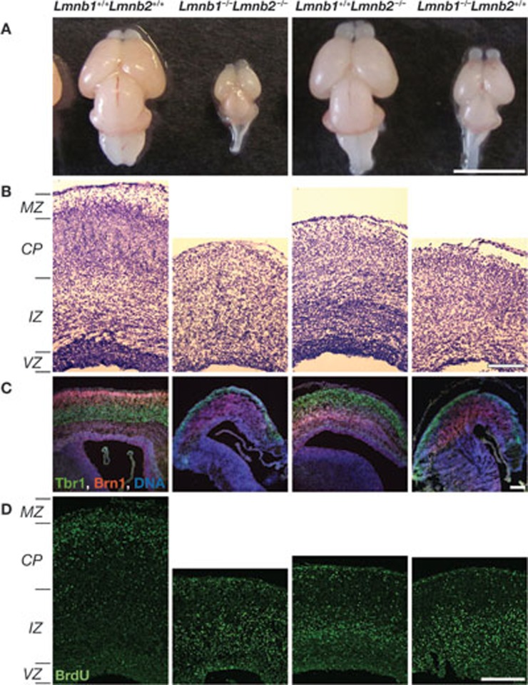
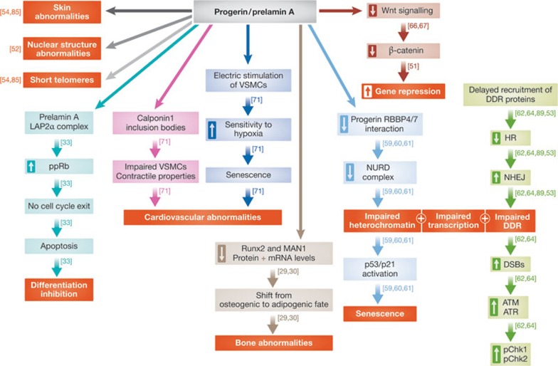
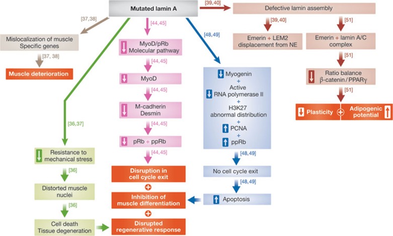
Lamins are nuclear intermediate filament proteins. They provide mechanical stability, organize chromatin and regulate transcription, replication, nuclear assembly and nuclear positioning. Recent studies provide new insights into the role of lamins in development, differentiation and tissue response to mechanical, reactive oxygen species and thermal stresses. These studies also propose the existence of separate filament networks for A- and B-type lamins and identify new roles for the different networks. Furthermore, they show changes in lamin composition in different cell types, propose explanations for the more than 14 distinct human diseases caused by lamin A and lamin C mutations and propose a role for lamin B1 in these diseases.


Conflict of interest statement

The authors declare that they have no conflict of interest.

Figures

Figure 1
Figure 1
B-type lamins are required for brain development. The genotype of the brain and the brain sections is indicated above the panels. (A) Mice mutant for lamin B have smaller brains; brains are from embryonic day (E)18.5 mouse embryos. (B) Staining of E18.5 neocortex coronal sections of mouse embryos for nucleic acids. Neurons from lamin B-null mice have layer organization and migration defects. (C) Layer-specific labelling of the neocortex with Tbr1 (green) for early-born deep layer (V,VI) and Brn1 (red) for late-born outer neuronal layers (II–IV). DNA is labelled in blue. (D) BrdU (green) labelling of mid-to-late-born neurons from E18.5 mice embryos. The data for this figure have been used with permission from figure 4 in [23]. BrdU, bromodeoxyuridine; Brn1, POU domain, class 3, transcription factor 3; CP, cortical plate; IZ, intermediate zone; MZ, marginal zone; Tbr1, T-box, brain, 1; VZ, ventricular zone.
Figure 2
Figure 2
A scheme depicting the different outcomes caused by the presence of progerin and prelamin in cells. The boxed hollow arrows show an increase and decrease in the mentioned phenotype. Each pathway is indicated by a different colour. The red boxes represent final outcomes. ATM, ataxia telangiectasia mutated; ATR, ataxia telangiectasia and Rad3-related; DDR, DNA damage response; DSB, double-strand break; HR, homologus recombination; LAP2α, lamina-associated polypeptide 2 isoform alpha; mRNA, microRNA; NHEJ, non-homologus end joining; NURD, nucleosome remodelling and histone deacetylation; pChk1/2, checkpoint kinase 1/2; ppRb, hyperphosphorylated retinoblastoma; RBBP4/7, retinoblastoma binding protein 4/7; Runx2, runt-related transcription factor 2; VSMC, vascular smooth muscle cell.
Figure 3
Figure 3
A scheme depicting the different outcomes caused by the presence of mutated lamin in cells. The boxed hollow arrows show an increase and decrease in the mentioned phenotype. Each pathway is indicated by a different colour. The red boxes represent final outcomes. LEM2, LAP2/emerin/MAN1 domain protein 2; MyoD, myogenic differentiation 1; NE, nuclear envelope; PCNA, proliferating cell nuclear antigen; PPARγ, peroxisome proliferator-activated receptor gamma; pRB, protein retinoblastoma 1; ppRb, hyperphosphorylated retinoblastoma protein.
Figure 4
Figure 4
Impact on nuclear shape by oxidative stress or by an ataxia telangiectasia cell mutation. Upper panel: wild-type fibroblast not treated (NT) or treated with H2O2 stained with lamin B1 antibodies. Lower panel: wild-type and ataxia telangiectasia primary cells stained with lamin B1 antibodies. Arrows indicate examples of alterations in nuclear morphology. The data have been taken with permission from figures 2 and 5 in [95].
None
Noam Zuela
None
Daniel Z Bar
None
Yosef Gruenbaum

Similar articles

Cited by

References

    1. Stuurman N, Heins S, Aebi U (1998) Nuclear lamins: their structure, assembly, and interactions. J Struct Biol 122: 42–66 - PubMed
    1. Aebi U, Cohn J, Buhle L, Gerace L (1986) The nuclear lamina is a meshwork of intermediate-type filaments. Nature 323: 560–564 - PubMed
    1. Goldberg MW, Huttenlauch I, Hutchison CJ, Stick R (2008) Filaments made from A- and B-type lamins differ in structure and organization. J Cell Sci 121: 215–225 - PubMed
    1. Heitlinger E, Peter M, Haner M, Lustig A, Aebi U, Nigg EA (1991) Expression of chicken lamin B2 in Escherichia coli: characterization of its structure, assembly, and molecular interactions. J Cell Biol 113: 485–495 - PMC - PubMed
    1. Ben-Harush K, Wiesel N, Frenkiel-Krispin D, Moeller D, Soreq E, Aebi U, Herrmann H, Gruenbaum Y, Medalia O (2009) The supramolecular organization of the C. elegans nuclear lamin filament. J Mol Biol 386: 1392–1402 - PubMed

Publication types