. 2012 Nov 21;4(161):161ra152.
doi: 10.1126/scitranslmed.3004190. Epub 2012 Nov 12.

Optogenetic and potassium channel gene therapy in a rodent model of focal neocortical epilepsy

Affiliations

Optogenetic and potassium channel gene therapy in a rodent model of focal neocortical epilepsy

Robert C Wykes et al. Sci Transl Med. .

Abstract

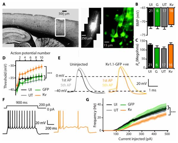
Neocortical epilepsy is frequently drug-resistant. Surgery to remove the epileptogenic zone is only feasible in a minority of cases, leaving many patients without an effective treatment. We report the potential efficacy of gene therapy in focal neocortical epilepsy using a rodent model in which epilepsy is induced by tetanus toxin injection in the motor cortex. By applying several complementary methods that use continuous wireless electroencephalographic monitoring to quantify epileptic activity, we observed increases in high frequency activity and in the occurrence of epileptiform events. Pyramidal neurons in the epileptic focus showed enhanced intrinsic excitability consistent with seizure generation. Optogenetic inhibition of a subset of principal neurons transduced with halorhodopsin targeted to the epileptic focus by lentiviral delivery was sufficient to attenuate electroencephalographic seizures. Local lentiviral overexpression of the potassium channel Kv1.1 reduced the intrinsic excitability of transduced pyramidal neurons. Coinjection of this Kv1.1 lentivirus with tetanus toxin fully prevented the occurrence of electroencephalographic seizures. Finally, administration of the Kv1.1 lentivirus to an established epileptic focus progressively suppressed epileptic activity over several weeks without detectable behavioral side effects. Thus, gene therapy in a rodent model can be used to suppress seizures acutely, prevent their occurrence after an epileptogenic stimulus, and successfully treat established focal epilepsy.


Figures

Fig. 1
Fig. 1
Tetanus toxin injection induces robust changes in EEG activity and neuronal excitability. (A) Representative control EEG (Ct, black) and EEG after tetanus toxin injection (TT, red), and EEG recorded during a secondary generalized seizure (below, red: i, onset; ii, evolution; iii, clonic phase; and iv, post-ictal period). (B) EEG power 6 to 7 days after tetanus toxin injection (Mann-Whitney: 70 to 120 Hz, P = 0.024; 120 to 160 Hz, P = 0.001; TT dose: mean, 14.2 ng; range, 10 to 35 ng; n = 58; Ct: n = 13). (C) Number of bursts in 1 hour in Ct and toxin-injected animals (Mann-Whitney P =0.01). (D) Coastline length (24-hour average; P =0.001). (E) High-frequency power (120 to 160 Hz) in Ct and after low and high toxin dose (low: 10 to 15 ng; high: 17.5 to 35 ng; Kruskal-Wallis P = 0.0001; Dunn’s multiple comparisons: Ct versus low, P <0.05; low versus high, P <0.001). (F) High-frequency power in Ct and in animals without (TT−) or with overt abnormalities (TT+; fig. S3) (Kruskal-Wallis P < 0.0001; Dunn’s multiple comparisons: Ct versus TT−, P < 0.01; TT− versus TT+, P < 0.01). (G) Automatically detected epileptiform events in a 12.5-ng 24-hour period (Mann-Whitney P < 0.0001). *P < 0.05; **P < 0.01; ***P < 0.001.
Fig. 2
Fig. 2
Injection of tetanus toxin increases excitability of adapting layer 5 pyramidal neurons. (A to C) Neurons in toxin-injected animals (TT) were assessed for (A) resting membrane potential (Vm; Mann-Whitney P = 0.01), (B) input resistance (RN; P = 0.02), and (C) current threshold (Ithresh; P = 0.04). (D) Representative traces from Ct and TT neurons evoked by −200- and +50-pA current injections. TT neurons often showed rebound action potentials (7 of 16 versus 0 of 21 in Ct neurons, Fisher’s exact test P = 0.0011). When the hyperpolarizing current was adjusted to reach the same voltage as in Ct neurons, 4 of 16 TT neurons showed rebound action potentials (P < 0.05). (E) Sample traces showing response to 0.3-ms depolarizing current injection sufficient to evoke an action potential. This elicited two or more action potentials in 5 of 14 TT neurons and 0 of 19 Ct neurons (P = 0.0084). (F and G) After depolarization parameters in neurons firing only once, showing longer time to peak (P = 0.04) (F) and decay time constant (P = 0.04) (G) in tetanus toxin-treated animals. *P < 0.05; **P < 0.01.
Fig. 3
Fig. 3
Acute optogenetic attenuation of epileptic activity. (A) Schematic of the implanted headstage for simultaneous EEG recording and optical stimulation. (B) Immunohistochemical staining of cells in the focus showing GFP-containing transduced neurons (left) and CaMKIIa staining of the same neurons (middle). Right, merged images. (C) Representative EEG traces before, during, and after 561-nm laser illumination. (D) Mean high-frequency (HF) power in animals injected with tetanus toxin (TT) and NpHR lentivirus (left, n = 6), showing a significant decrease upon laser stimulation (n = 6; Wilcoxon matched pairs signed-rank test, P = 0.03; gray, individual experiments; purple, mean ± SEM). Baseline high-frequency power was lower in animals injected with NpHR lentivirus alone (middle; n = 5; unpaired two-tailed t test, P < 0.001) and unaffected by laser illumination (yellow, mean ± SEM). Laser illumination had no effect on high-frequency power in control animals that were injected with TT together with either GFP expressing control virus or fluorescent beads (right; n = 8; red, mean ± SEM). n.s., not significant. (E) Mean coastline length, plotted as in (D), was decreased by illumination in animals injected with TT and NpHR (Wilcoxon matched pairs signed-rank test, P = 0.03) but not in control animals injected with TT and GFP. (F) Automatically detected epileptiform events in the same animals, plotted as in (D), were decreased upon laser illumination (paired t test after log transform, P = 0.048). Event counts in animals injected with TT together with GFP or beads were unaffected by laser illumination (before, 50 ± 25; during, 47 ± 30; after, 51 ± 40). *P < 0.05.
Fig. 4
Fig. 4
In vivo injection of Kv1.1 lentivirus attenuates excitability of adapting layer 5 pyramidal neurons. (A) GFP expression in a restricted area of motor cortex after injection of Kv1.1-GFP lentivirus. (B and C) Subthreshold properties in neurons from uninjected animals (UI) and animals injected with GFP only lentivirus (G), and in GFP-negative untransduced (UT) and neighboring GFP-positive (Kv) neurons from Kv1.1-GFP–injected animals [resting membrane potential (RMP) (B) and resting input resistance (RN) (C)]. (D to G) Neuronal excitability in neurons overexpressing Kv1.1. (D) Action potential threshold in a train of spikes (P < 0.0005 for an effect of group, linear mixed model analysis). (E) First and fifth action potentials showing depolarized threshold but no difference in accommodation (spike amplitude, rise time, decay, and width at half-maximum). (F) Representative traces from neighboring untransduced (black) and Kv1.1-overexpressing (orange) neurons in response to +200-pA current injection. (G) Frequency-current relationship showing a significant decrease in firing rate in neurons overexpressing Kv1.1 (P < 0.0001 for difference between groups, log-inear mixed model). All recordings at 36 ± 1°C, 7 to 20 days after virus injections. ***P < 0.001.
Fig. 5
Fig. 5
Kv1.1 lentivirus injection with tetanus toxin prevents focal neocortical epileptogenesis. (A) Effect of coinjection of Kv1.1-GFP lentivirus with tetanus toxin (TT+Kv) on high-frequency power (EEG analyzed over 24 hours, 7 days after injection; Kruskal-Wallis P < 0.0001, SEM concealed by symbols). (B) Effect of coinjection of Kv1.1 and tetanus toxin on EEG coastline (P < 0.001). (C) Sample coastline analysis from 800 s of EEG recorded from three representative animals 7 days after injection (each point represents the coastline length of a 2-s segment of EEG); arrows indicate expanded EEG traces (top): Ct (black), control; TT (red), TT injected animal; TT+Kv (blue), TT + Kv1.1–injected animal. (D) Effect of Kv1.1 and tetanus toxin coinjection on epileptiform events measured by automated event classification. Four categories of events are shown with representative library templates (traces, see also fig. S4; HF short, HF long, HF low, and HF spike defined in fig. S4). Animals that received Kv1.1 had fewer epileptiform events than those that received tetanus toxin only (all events aggregated, log-linear mixed model P < 0.001). Kv1.1 injection reduced epileptiform events to rates indistinguishable from Ct animals that did not receive tetanus toxin (P = 0.511). Data are means ± SEM. Gray area indicates quarantine period after virus injection. *P < 0.05; **P < 0.01; ***P < 0.001.
Fig. 6
Fig. 6
Kv1.1 lentivirus treats focal neocortical epilepsy. (A) EEG coastline in tetanus toxin–treated animals relative to the value at day 7, just before delayed injection (arrow) of Kv1.1-GFP lentivirus (blue) or GFP-only lentivirus (red). Gray bar indicates quarantine period (linear mixed model, P = 0.004). Below: Numbers of animals at different time points (another control group without tetanus toxin but injected with GFP lentivirus exhibited no change in coastline length). (B) Sample EEG traces from two animals at days 7 and 35 (red, GFP; blue, Kv1.1-GFP). (C) Effect of Kv1.1 injection after established epileptogenesis on high-frequency power (unpaired t test for change in power versus GFP, P < 0.05). (D) Effect of Kv1.1 injection after established epileptogenesis on the number of epileptiform events (log-linear mixed model for treatment with Kv1.1 versus GFP, P < 0.0001). Red, tetanus toxin followed by GFP; blue, tetanus toxin followed by Kv1.1; black, GFP only. *P < 0.05; **P < 0.01; ***P < 0.001.

Comment in

Similar articles

Cited by

References

    1. Ngugi AK, Bottomley C, Kleinschmidt I, Sander JW, Newton CR. Estimation of the burden of active and life-time epilepsy: A meta-analytic approach. Epilepsia. 2010;51:883–890. - PMC - PubMed
    1. Kwan P, Schachter SC, Brodie MJ. Drug-resistant epilepsy. N. Engl. J. Med. 2011;365:919–926. - PubMed
    1. Annegers JF, Hauser WA, Elveback LR. Remission of seizures and relapse in patients with epilepsy. Epilepsia. 1979;20:729–737. - PubMed
    1. Jasper’s Basic Mechanisms of the Epilepsies—NCBI Bookshelf. http://www.ncbi.nlm.nih.gov/books/NBK50785/
    1. Rosenow F, Lüders H. Presurgical evaluation of epilepsy. Brain. 2001;124:1683–1700. - PubMed

Publication types

MeSH terms