. 2013 Jan;81(1):381-9.
doi: 10.1128/IAI.00950-12. Epub 2012 Nov 12.

Alveolar epithelial cells are critical in protection of the respiratory tract by secretion of factors able to modulate the activity of pulmonary macrophages and directly control bacterial growth

Affiliations

Alveolar epithelial cells are critical in protection of the respiratory tract by secretion of factors able to modulate the activity of pulmonary macrophages and directly control bacterial growth

Olga D Chuquimia et al. Infect Immun. 2013 Jan.

Abstract

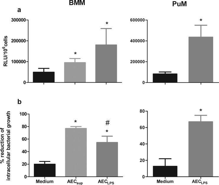
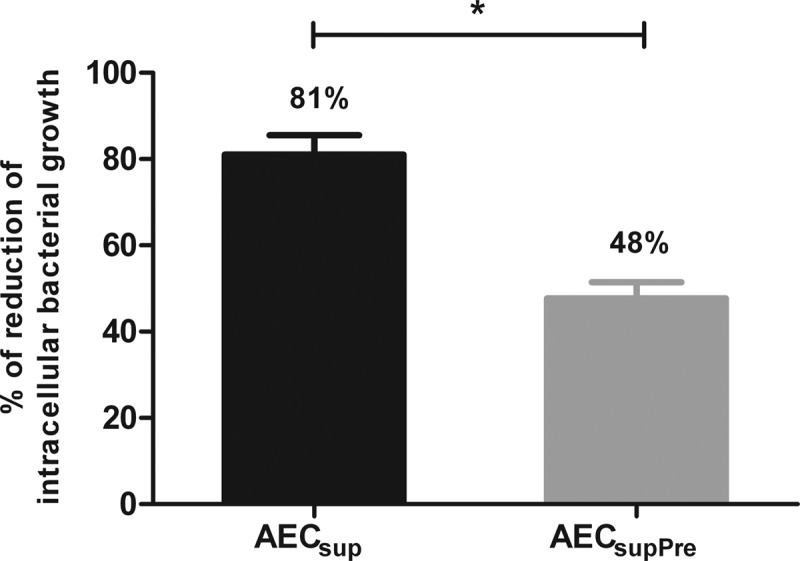
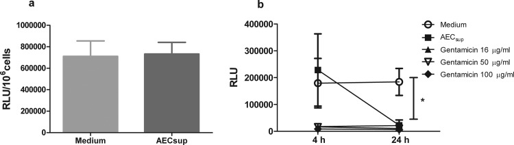
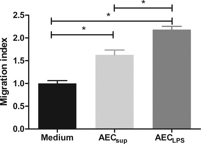
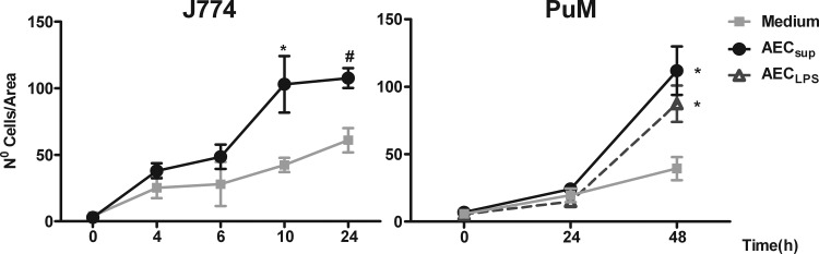
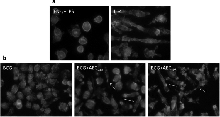
The respiratory epithelium is a physical and functional barrier actively involved in the clearance of environmental agents. The alveolar compartment is lined with membranous pneumocytes, known as type I alveolar epithelial cells (AEC I), and granular pneumocytes, type II alveolar epithelial cells (AEC II). AEC II are responsible for epithelial reparation upon injury and ion transport and are very active immunologically, contributing to lung defense by secreting antimicrobial factors. AEC II also secrete a broad variety of factors, such as cytokines and chemokines, involved in activation and differentiation of immune cells and are able to present antigen to specific T cells. Another cell type important in lung defense is the pulmonary macrophage (PuM). Considering the architecture of the alveoli, a good communication between the external and the internal compartments is crucial to mount effective responses. Our hypothesis is that being in the interface, AEC may play an important role in transmitting signals from the external to the internal compartment and in modulating the activity of PuM. For this, we collected supernatants from AEC unstimulated or stimulated in vitro with lipopolysaccharide (LPS). These AEC-conditioned media were used in various setups to test for the effects on a number of macrophage functions: (i) migration, (ii) phagocytosis and intracellular control of bacterial growth, and (iii) phenotypic changes and morphology. Finally, we tested the direct effect of AEC-conditioned media on bacterial growth. We found that AEC-secreted factors had a dual effect, on one hand controlling bacterial growth and on the other hand increasing macrophage activity.


Figures

Fig 1
Fig 1
Phagocytosis and intracellular bacterial growth control by BMM and PuM upon treatment with AEC-derived media. BMM and PuM from TLR4−/− mice were pretreated with either AECsup or AECLPS for 20 to 24 h or left untreated. Cells were infected with GFP-BCG. (a) Phagocytosis. Four hours after infection, phagocytosis was evaluated by determining RLU. Data are shown as RLU/106 cells. (b) Intracellular bacterial growth control. After infection, cells were treated with either AECsup or AECLPS for 48 h or left untreated. Bacterial growth was evaluated by determining RLU. Data are shown as percent reduction of phagocytosed bacteria evaluated as RLU. Values are means ± SD from 3 independent experiments. The differences between groups of BMM were analyzed using a one-way ANOVA followed by Bonferroni's multiple comparison test. *, significantly different from medium control; #, significantly different from AECsup; P < 0.05. The differences between groups of PuM were analyzed using an unpaired Student t test. *, significant differences, P < 0.05.
Fig 2
Fig 2
Continuous presence of AEC-derived media is required for optimal control of intracellular bacterial growth. PuM from WT mice were either continuously treated or pretreated only with AECsup and washed before infection (AECsupPre). The bacterial growth was measured by determining RLU. Data are shown as percent reduction of phagocytosed bacteria evaluated as RLU. Values are means ± SD from 3 independent experiments. The differences between groups were analyzed using an unpaired Student t test. *, significant differences, P < 0.05.
Fig 3
Fig 3
Opsonization and direct bacterial killing upon treatment with AEC-derived media. (a) Opsonization. GFP-BCG was pretreated with AECsup for 30 min. BMM were infected with either pretreated or untreated GFP-BCG for 4 h. Phagocytosis was evaluated by determining RLU. (b) Direct killing. GFP-BCG was treated with AECsup or gentamicin for 4 and 24 h or kept in medium. Then, bacteria were centrifuged and cultured in medium without antibiotics for 72 h. Bacterial growth was measured by determining RLU. Values are means ± SD from 3 independent experiments. The differences between the groups and time points were analyzed using a two-way ANOVA followed by Bonferroni's multiple comparison test. *, significant differences, P < 0.001.
Fig 4
Fig 4
Total lung cell migration toward media from unstimulated AEC (AECsup) and LPS-stimulated AEC (AECLPS). Total lung cells from TLR4−/− mice were placed in a Transwell insert in control medium or AEC-derived media in the lower chamber. After 2 h of incubation, cells were removed from the lower chamber and relative cell numbers were determined using a flow cytometer. Values are means ± SD, n = 3. The differences between the groups were analyzed using a one-way ANOVA followed by Bonferroni's multiple comparison test. *, significant differences, P < 0.05. The data are representative of 4 independent experiments.
Fig 5
Fig 5
Wound-healing assay performed on J774 cells and PuM from TLR4−/− mice. J774 cells were grown to confluence on tissue culture plates, the monolayers were scratched to create a wound, and the cells were cultured for the indicated time in media from unstimulated (AECsup) or LPS-stimulated AEC (AECLPS). After being cultured, the cells were stained with DAPI and photographed. The number of cells in the scratch wound was evaluated in three different fields within a defined area. Values are means ± SEM, n = 4 experiments. Differences between mean values were calculated using an unpaired Student t test for J774 or one-way ANOVA for PuM, followed by Bonferroni's multiple comparison test. *, significantly different from medium, P < 0.05.
Fig 6
Fig 6
Cytoskeleton staining of M1- or M2-differentiated BMM (a) or BCG-infected PuM (b). BMM were differentiated by adding IFN-γ and LPS (M1) or IL-4 (M2) for 48 h. PuM were infected with BCG for 4 h. After thorough washing and treatment with gentamicin for 30 min, cells were cultured further for 48 h in RPMI or media from untreated AEC (AECsup) or LPS-stimulated AEC (AECLPS). After being cultured, cells were stained with FITC-phalloidin and analyzed by microscopy.

References

    1. Bals R. 2000. Epithelial antimicrobial peptides in host defense against infection. Respir. Res. 1:141–150 - PMC - PubMed
    1. Ryu JH, Kim CH, Yoon JH. 2010. Innate immune responses of the airway epithelium. Mol. Cells 30:173–183 - PubMed
    1. Williams MC. 2003. Alveolar type I cells: molecular phenotype and development. Annu. Rev. Physiol. 65:669–695 - PubMed
    1. Fereol S, Fodil R, Pelle G, Louis B, Isabey D. 2008. Cell mechanics of alveolar epithelial cells (AECs) and macrophages (AMs). Respir. Physiol. Neurobiol. 163:3–16 - PubMed
    1. Castranova V, Rabovsky J, Tucker JH, Miles PR. 1988. The alveolar type II epithelial cell: a multifunctional pneumocyte. Toxicol. Appl. Pharmacol. 93:472–483 - PubMed

Publication types

MeSH terms

Substances