Review
. 2013 Jan 23;425(2):199-213.
doi: 10.1016/j.jmb.2012.11.001. Epub 2012 Nov 9.

Design principles of a universal protein degradation machine

Affiliations
Review

Design principles of a universal protein degradation machine

Mary E Matyskiela et al. J Mol Biol. .

Abstract

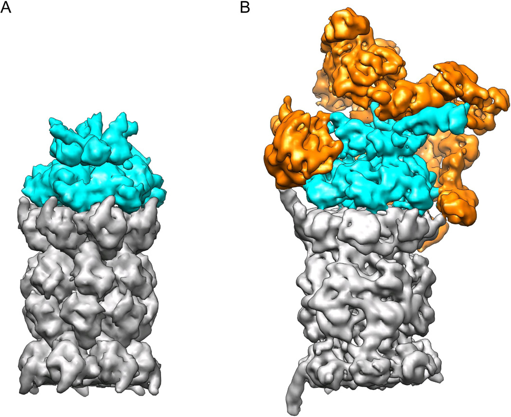
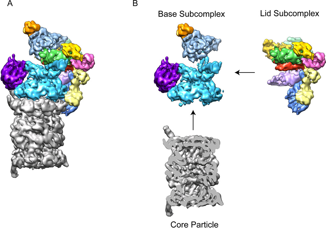
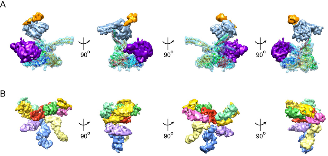
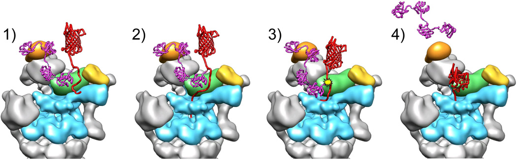
The 26S proteasome is a 2.5-MDa, 32-subunit ATP-dependent protease that is responsible for the degradation of ubiquitinated protein targets in all eukaryotic cells. This proteolytic machine consists of a barrel-shaped peptidase capped by a large regulatory particle, which contains a heterohexameric AAA+ unfoldase as well as several structural modules of previously unknown function. Recent electron microscopy (EM) studies have allowed major breakthroughs in understanding the architecture of the regulatory particle, revealing that the additional modules provide a structural framework to position critical, ubiquitin-interacting subunits and thus allow the 26S proteasome to function as a universal degradation machine for a wide variety of protein substrates. The EM studies have also uncovered surprising asymmetries in the spatial arrangement of proteasome subunits, yet the functional significance of these architectural features remains unclear. This review will summarize the recent findings on 26S proteasome structure and discuss the mechanistic implications for substrate binding, deubiquitination, unfolding, and degradation.


Figures

Figure 1
Figure 1. The 26S proteasome is a heteromeric and asymmetric ATP-dependent protease with higher complexity than its prokaryotic ancestors
A) Model of the homo-hexameric archaeal unfoldase PAN (cyan) bound to the core particle (grey). The separate crystal structures of the N-ring (3H43), the ATPases (3H4M), and core particle (3H4P) were docked together and rendered with a surface resolution of 4 Å to match more closely the EM structure of the 26S proteasome. B) Cryo-EM reconstruction of the 26S proteasome at subnanometer resolution . The core particle (grey) and AAA+ unfoldase subunits (cyan) resemble the archaeal PAN-20S protease in size and shape, but bear distinct asymmetries due to their heteromeric ring architectures. The additional structural modules that do not share homology with other compartmental peptidases and have likely been added to accommodate ubiquitin signaling are shown in orange. They include the lid sub-complex, the torroidal subunits Rpn1 and Rpn2, and the ubiquitin receptors.
Figure 2
Figure 2. The 26S proteasome can be separated into three stably associated subcomplexes
A) Cryo-EM reconstruction of the eukaryotic proteasome holoenzyme with the core particle in grey and the regulatory particle multicolored . B) Individual sub-complexes of the 26S proteasome. The regulatory particle can be separated into two stable sub-complexes, the lid and the base. The base consists of the AAA+ ATPase subunits Rpt1-6, (cyan), two large torroidal subunits, Rpn1 (purple) and Rpn2 (light blue), and the ubiquitin receptor Rpn13 (orange). The lid contains six PCI-domain subunits (multicolored) along with the essential DUB Rpn11 (green) and the ubiquitin receptor Rpn10 (yellow). A central cross-section of the core particle (grey) is shown to allow visualization of the barrel-shaped central cavity.
Figure 3
Figure 3. Subunit architecture of the lid and base sub-complexes
A) Multiple views of the base sub-complex. A molecular model based on the PAN crystal structure has been docked into the EM density for Rpt1-6 to highlight each subunit of the heterohexamer , . The N-terminal segment of Rpt1 (dark blue) forms a minimal coiled coil with Rpt2 (light blue) that provides a docking site for Rpn1 (purple) at the periphery of the unfoldase. Rpt 6 (red) forms a long coiled coil with Rpt3 (green) that positions Rpn2 (light blue) above the AAA+ ring. Rpn2 in turn holds Rpn13 (orange) high above the pore of the unfoldase. The coiled coil formed by the N-terminal segments of Rpt4 (yellow) and Rpt5 (orange) does not appear to interact with any other proteasome subunits, but may stabilize the UIM of Rpn10. B) Multiple views of the lid subcomplex in its holoenzyme-bound conformation. The first view (far left) shows the base-facing, inner surface of the lid. The PCI-domain subunits in this orientation, from left to right, are Rpn12 (light green), Rpn3 (yellow), Rpn7 (purple), Rpn6 (blue), Rpn5 (light yellow) and Rpn9 (pink). The central green density was initially identified as Rpn11, but may be an MPN domain dimer of Rpn11 and Rpn8. In turn, the central density depicted in red, initially proposed to be Rpn8, may be a helix bundle formed by the C-termini of all lid subunits . The ubiquitin receptor Rpn10 (yellow, right) is positioned high above the unfoldase pore through interactions with Rpn11 and Rpn9 .
Figure 4
Figure 4. Mechanistic model for substrate degradation by the 26S proteasome
1) A chain of four or more ubiquitins (purple) binds to an ubiquitin receptor (orange) on the proteasome, tethering the substrate (red). 2) Structures in the subjacent pore of the AAA+ unfoldase (cyan) engage the flexible initiation site of the substrate. 3) Translocation commences and helps to position the isopeptide bond of the ubiquitin-modified lysine in the active site of Rpn11 (green) for deubiquitination. 4) The substrate is unfolded and translocated through the AAA+ ring and into the core particle.

References

    1. Sauer RT, Baker TA. AAA+ proteases: ATP-fueled machines of protein destruction. Annu Rev Biochem. 80:587–612. - PubMed
    1. Sauer RT, et al. Sculpting the proteome with AAA(+) proteases and disassembly machines. Cell. 2004;119:9–18. - PMC - PubMed
    1. Martin A, Baker TA, Sauer RT. Pore loops of the AAA+ ClpX machine grip substrates to drive translocation and unfolding. Nat Struct Mol Biol. 2008;15:1147–1151. - PMC - PubMed
    1. Maillard RA, et al. ClpX(P) generates mechanical force to unfold and translocate its protein substrates. Cell. 2011;145:459–469. - PMC - PubMed
    1. Aubin-Tam ME, Olivares AO, Sauer RT, Baker TA, Lang MJ. Single-molecule protein unfolding and translocation by an ATP-fueled proteolytic machine. Cell. 2011;145:257–267. - PMC - PubMed

Publication types

LinkOut - more resources