. 2012 Dec;71(12):1100-12.
doi: 10.1097/NEN.0b013e31827733c8.

Gene dosage-dependent association of DYRK1A with the cytoskeleton in the brain and lymphocytes of down syndrome patients

Affiliations

Gene dosage-dependent association of DYRK1A with the cytoskeleton in the brain and lymphocytes of down syndrome patients

Karol Dowjat et al. J Neuropathol Exp Neurol. 2012 Dec.

Abstract

The triplication of the DYRK1A gene encoding proline-directed serine/threonine kinase and located in the critical region of Down syndrome (DS) has been implicated in cognitive deficits and intellectual disability of individuals with DS. We investigated the effect of abnormal levels of this kinase on the cytoskeleton in brain and peripheral tissues of DS subjects. In DS tissues, the predictable approximately equal to 1.5-fold enhancement of the levels of DYRK1A protein was demonstrated. An association of DYRK1A with all 3 major cytoskeleton networks was identified using immunoprecipitation. We concentrated on the actin cytoskeleton because its association with DYRK1A was the most affected by the enzyme levels. As measured by coimmunoprecipitation in DS tissues, but not in fragile X lymphocytes, actin association with DYRK1A was reduced. This reduced association was dependent on the state of phosphorylation of cytoskeletal proteins and was present only in cells overproducing DYRK1A kinase; therefore, the effect was attributable to the DYRK1A gene dosage. Alterations of DYRK1A-actin assemblies were detected in newborn and infant groups, thereby linking DYRK1A overexpression with abnormal brain development of DS children. The identification of the actin cytoskeleton as one of cellular targets of DYRK1A action provides new insights into a gene dosage-sensitive mechanism by which DYRK1A could contribute to the pathogenesis of DS. In addition, the presence of this DS-specific cytoskeleton anomaly in lymphocytes attests to the systemic nature of some features of DS. To our knowledge, this is the first study conducted in human tissue that shows DYRK1A association with the cytoskeleton.


Figures

Figure 1
Figure 1
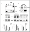
Association of DYRK1A with the cytoskeleton as revealed by immunoprecipitation and immunostaining of brain tissue and cultured mouse neuroblastoma neuro-2a (N2a) cells. Left panels: Frozen frontal cortex tissue of a control subject was homogenized in RIPA buffer and subjected to immunoprecipitation (IP) with anti-DYRK1A (H143) antibody. Immunoprecipitates were analyzed by Western blotting (WB) with antibodies against the indicated cytoskeletal proteins. All major structural cytoskeletal proteins were detected in the DYRK1A IP. Right panels: High magnification image of fetal (20-week gestation) Down syndrome (DS) white matter immunostained with anti-DYRK1A antibody (7F3). Merge images are of double immunofluorescent labeling of N2a cells with DYRK1A (H143) and either NF-H (N52), α-tubulin (B-5-1-2) or β-actin (AC-15) antibodies.
Figure 2
Figure 2
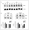
Quantification of DYRK1A and β-actin in immunoprecipitates of pooled frontal cortex tissue of control and Down syndrome (DS) subjects. (A) Specificity of DYRK1A and actin co-immunoprecipitation (IP). NIH3T3 cell lysate was used for IP with uncoated (Beads), anti-glutathione S-transferase (GST) antibody –coated, and anti-DYRK1A R420 antibody (DYRK1A) – coated Dynabeads. The levels of DYRK1A and β-actin in crude lysate and precipitates were detected by immunoblotting. (B) Analysis of DYRK1A and β-actin co-IP. Pools were made by mixing 0.1 g of tissue powdered in liquid N2 (n = 6 for control and n = 12 for DS) of each brain. Parallel samples of brain lysates were immunoprecipitated with the rabbit polyclonal anti-DYRK1A antibody directed against the C-terminus (H143) of DYRK1A polypeptide and monoclonal β-actin antibody. IP and Western blot (WB) analysis of lysates and immunoprecipitates were conducted as described in Materials and Methods. 20 μg of each lysate and 10 μl of IP sample were taken for analysis. Blots were probed with 1:5000 dilutions of DYRK1A 8D9 and β-actin monoclonal antibodies. In the DS sample, IP with actin antibody was also done with lysate denatured by boiling. Representative Western blots and the results of quantification are shown. The results are expressed as percent of values recorded for the control pool. Each bar represents the mean value of 4 IP experiments conducted on 2 independently prepared pools. Error bars indicate SEM. CTR: control lysate; DS: Down syndrome lysate; DS 100°: boiled DS lysate. *Denotes the differences with statistical significance at the level of p < 0.05. (C) Patterns of DYRK1A-immunoreactive bands that co-IP with actin. DYRK1A and β–actin was independently IP with each respective antibody from control lysate and then compared by Western blotting. Running time for SDS-PAGE was extended to permit the separation of multiple DYRK1A bands approximately 90 kDa.
Figure 3
Figure 3
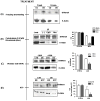
Immunoblotting profiles of DYRK1A immunoprecipitates in developing brains of control and Down syndrome (DS) subjects. Pools of frontal cortex tissue of newborns (n = 4 for control and n = 3 for DS), infants (n = 9 for control and n = 8 for DS), and adults (n = 7 for control and n = 14 for DS) were subjected to immunoprecipitation with R420 rabbit anti-DYRK1A antibody, and analyzed by immunoblotting with DYRK1A (8D9) and β-actin antibodies. Others details are as described in the legend to Figure 2. (A, B) Representative immunoblots of crude lysates (A) and immunoprecipitates (B) are shown. Results of densitometric measurements are expressed as a percent of immunoreactivity recorded for controls in each group. Each bar represents mean value ± SEM of 4 experiments conducted on 2 independently prepared pools. The differences between control and DS for all age groups were significant at *p < 0.05.
Figure 4
Figure 4
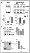
Quantification of DYRK1A and β-actin in immunoprecipitates of pooled lymphoblastoid cell lines (LCLs) of healthy (CTR), Down syndrome (DS) and fragile X (FraX) subjects. (A) Levels of DYRK1A and β–actin in crude lysates. RIPA lysates were prepared from LCLs established from 3 control and 4 DS donors Samples of each lysate were immunoblotted with DYRK1A 8D9 and s- actin antibodies. (B) Immunoblot quantification of DYRK1A and β-actin in DYRK1A IP (R420). Preparation of pooled control (5 cases), DS (6 cases) and FraX (2 cases) LCLs, DYRK1A IP, and analysis of immunoprecipitates were as described in Materials and Methods. Samples were immunoblotted with DYRK1A 8D9 and β-actin antibodies. Plots of densitometric measurements are expressed as a percent of immunoreactivity recorded for the control pool. Each bar represents mean value ± SEM of 4 experiments conducted on 2 independently prepared pools. *Denotes the differences with statistical significance at the level of p < 0.05
Figure 5
Figure 5
Conditions affecting DYRK1A and β–actin co-immunoprecipitation (IP) in control and Down syndrome (DS) lymphoblastoid cell lines (LCLs). (A) Effect of freezing. DYRK1A IP was performed with lysates prepared from freshly harvested cells of control AG09387 and DS405 and cells after freezing for 2 hours at -20°C. DYRK1A and β–actin complexes in IP were measured by immunoblotting. (B) Effect of cytochalasin D (CytD) or nocodazole (Noc) treatment on DYRK1A and β–actin co-IP. Equal aliquots of control (AG09387) cells lysate were incubated at 30°C with CytD or Noc before IP with DYRK1A antibody followed by immunoblot analysis for DYRK1A and β-actin. Load. crude lysate,(–) IP from lysate without treatment, CytD: IP from lysate treated with CytD, and Noc: IP from lysate treated with Noc. The results were quantified and shown as a bar graph. (C) Effect of phosphatase inhibitor okadaic acid (OKA). Lysates of control (GM07045) and DS (DS206) LCLs were prepared with or without 200 nM OKA; and then IP with anti-DYRK1A (R420) antibody. The representative immunoblot is shown together with the plot of densitometric measurements where each bar represents values for DS expressed as percent of corresponding untreated (- OKA) or OKA-treated (+ OKA) control. (D) Effect of incubating with ATP. DYRK1A IP was performed with lysates of control and DS LCLs as described. DYRK1A-actin complexes bound to Dynabeads were subsequently incubated for 30 min at 30°C with or without ATP in kinase buffer. After extensive washing, DYRK1A and β–actin retained on Dynabeads were then analyzed by immunoblotting. KB: incubated only with kinase buffer, ATP: incubated with ATP in kinase buffer.
Figure 6
Figure 6
Effect of exogenous DYRK1A on the formation and stability of DYRK1A-actin complexes in NIH 3T3 fibroblasts. (A) Effect of phosphatase inhibitors (PPIs) in DYRK1A over-expressing cells. NIH 3T3 fibroblasts were transfected with the wild-type (WT) DYRK1A or the cloning vector (VEC) for 48 hours before harvesting. Cells were frozen at -20°C for 2 hours, lysed in RIPA with (+) or without (-) PPIs, and processed for IP. Aliquots of cell lysates containing 400 μg of total protein were taken for IP with R420 DYRK1A antibody and immunoprecipitates were probed with DYRK1A (8D9), s-tubulin, and β-actin antibodies. As a control, crude lysates without IP (lysates) were also probed with DYRK1A (8D9) and β-actin antibodies. The intensity of immunoreactive bands was quantified and reported as independent values in arbitrary units. Representative Western blots and plots of densitometric quantification of DYRK1A and actin are shown. Each bar represents mean value ± SEM of 3 independent experiments. *Denotes the differences with statistical significance at the level of p < 0.05. (B) Effect of over-expression of kinase-deficient mutant. Cells were transfected with the cloning vector (VEC), wild-type (WT) or mutant (K188R) DYRK1A and processed with PPIs for IP and subsequent Western blotting as in (A). Quantification and analysis of the intensity of immunoreactive bands were also performed as described above. (C) Effect of exogenous recombinant DYRK1A on Dynabeads immobilized DYRK1A-actin complexes. Complexes bound to DYRK1A antibody-coated Dynabeads were prepared from untransfected NIH 3T3 lysate as described earlier. Beads were then incubated in kinase buffer for 30 minutes at 30°C, with or without ATP or recombinant truncated wild-type DYRK1A. The coated beads were then extensively washed with PBST and DYRK1A and actin that remained on the beads were quantified by immunoblotting. KB: incubated with kinase buffer only, ATP: incubated with ATP, and WT: incubated with ATP and DYRK1A.

Similar articles

Cited by

References

    1. Tejedor F, Zhu XR, Kaltenbach E, et al. minibrain: a new protein kinase family involved in postembryonic neurogenesis in Drosophila. Neuron. 1995;14:287–301. - PubMed
    1. Himpel S, Tegge W, Frank R, et al. Specificity determinants of substrate recognition by the protein kinase DYRK1A. J Biol Chem. 2000;275:2431–8. - PubMed
    1. Guimerá J, Casas C, Pucharcòs C, et al. A human homologue of Drosophila minibrain (MNB) is expressed in the neuronal regions affected in Down syndrome and maps to the critical region. Hum Mol Genet. 1996;5:1305–10. - PubMed
    1. Rahmani Z, Blouin J-L, Creau-Goldberg N, et al. Critical role of the D21S55 region on chromosome 21 in the pathogenesis of Down syndrome. Proc Nat Acad Sci U S A. 1989;86:5958–62. - PMC - PubMed
    1. Belichenko NP, Belichenko PV, Kleschevnikov AM, et al. The "Down syndrome critical region" is sufficient in the mouse model to confer behavioral, neurophysiological, and synaptic phenotypes characteristic of Down syndrome. J Neurosci. 2009;29:5938–48. - PMC - PubMed

Publication types

MeSH terms

Substances