. 2013 Feb;131(2):351-9.
doi: 10.1093/toxsci/kfs316. Epub 2012 Nov 11.

Metabolically competent human skin models: activation and genotoxicity of benzo[a]pyrene

Affiliations

Metabolically competent human skin models: activation and genotoxicity of benzo[a]pyrene

Joep Brinkmann et al. Toxicol Sci. 2013 Feb.

Abstract

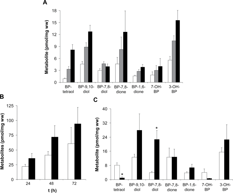
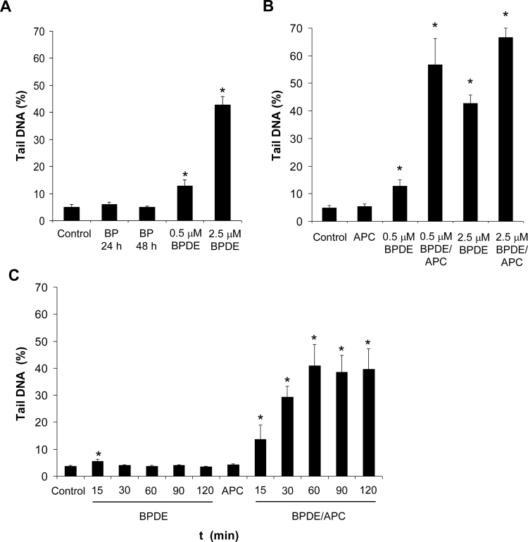
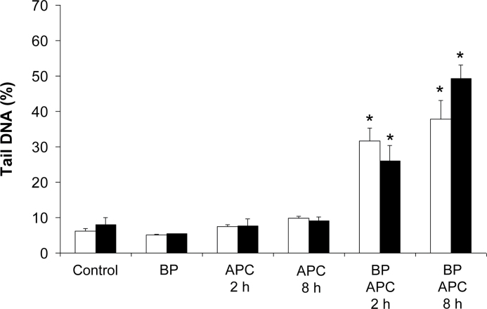
The polycyclic aromatic hydrocarbon (PAH) benzo[a]pyrene (BP) is metabolized into a complex pattern of BP derivatives, among which the ultimate carcinogen (+)-anti-BP-7,8-diol-9,10-epoxide (BPDE) is formed to certain extents. Skin is frequently in contact with PAHs and data on the metabolic capacity of skin tissue toward these compounds are inconclusive. We compared BP metabolism in excised human skin, commercially available in vitro 3D skin models and primary 2D skin cell cultures, and analyzed the metabolically catalyzed occurrence of seven different BP follow-up products by means of liquid chromatography coupled to tandem mass spectrometry (LC-MS/MS). All models investigated were competent to metabolize BP, and the metabolic profiles generated by ex vivo human skin and skin models were remarkably similar. Furthermore, the genotoxicity of BP and its derivatives was monitored in these models via comet assays. In a full-thickness skin, equivalent BP-mediated genotoxic stress was generated via keratinocytes. Cultured primary keratinocytes revealed a level of genotoxicity comparable with that of direct exposure to 50-100 nM of BPDE. Our data demonstrate that the metabolic capacity of human skin ex vivo, as well as organotypic human 3D skin models toward BP, is sufficient to cause significant genotoxic stress and thus cutaneous bioactivation may potentially contribute to mutations that ultimately lead to skin cancer.


Figures

FIG. 1.
FIG. 1.
Metabolism of BP in cultured skin cells: differences between NHEK and primary fibroblasts. (A) Time-dependent generation of BP metabolites in cultured NHEK. NHEK were exposed to 3.5µM BP, levels of generated BP metabolites were determined after incubation for 24h (□), 48h (■), and 72h (■) and normalized for cell wet weights (ww). (B) Quantitative comparison of BP metabolism in NHEK (□) and primary fibroblasts (■). Cells were incubated with 3.5µM BP and the sum of seven analyzed metabolites was used to quantify overall BP metabolism. (C) Qualitative comparison of BP metabolism in NHEK (□) and primary fibroblasts (■) after 72h of incubation with 3.5µM BP. Data are means ± SEM (n = 5; *p < 0.05 vs. NHEK).
FIG. 2.
FIG. 2.
Metabolism of BP in in vitro 3D skin models and human skin ex vivo. (A) Comparison of metabolite profiles generated by EpiDerm skin models (□), EpiDermFT skin models (■), and human skin ex vivo (■). Skin models and dermatomized human skin (~500 µm) received a topical dose of 50 nmol/cm2 BP dissolved in acetone. Metabolites were determined 48h thereafter and detected metabolite levels were normalized for model area. (B) Quantitative comparison of BP metabolism in NHEK, EpiDerm models, and human skin. This graph compares the sum of the seven analyzed metabolites expressed as pmol/10cm2 for NHEK and pmol/cm2 for EpiDerm models and skin. Data are means ± SEM (n = 3 for skin and skin models, n = 5 for NHEK).
FIG. 3.
FIG. 3.
Inhibition of DNA polymerases by APC significantly increases the sensitivity for detecting BPDE-induced genotoxicity in NHEK cultures. (A) BP-induced DNA damage is not readily detectable in NHEK. Confluent NHEK cultures were incubated with 3.5µM BP for 24 and 48h. Alternatively, BPDE was added to cell cultures at concentrations of 0.5 and 2.5µM and allowed to react for 2h. DNA damage was analyzed by a standard alkaline comet assay and quantified based on the % of DNA detected in the comet tails. (B) APC significantly decreases the detection limit for BPDE-induced genotoxicity. Confluent NHEK cultures were incubated with 500nM or 2.5µM BPDE for 2h in the presence (BPDE/APC) or absence (BPDE) of APC (5 µg/ml). (C) Confluent NHEK cultures were incubated with 100nM BPDE for 15, 30, 60, 90, and 120min in the presence (BPDE/APC) or absence (BPDE) of APC (5 µg/ml). Data are means ± SEM (n = 3; *p < 0.05 vs. control).
FIG. 4.
FIG. 4.
BP-exposed NHEK cultures generate DNA damage, whereas dermal fibroblasts do not. Confluent NHEK (A) and confluent dermal fibroblast cultures (B) were exposed to 3.5µM BP for 24 and 48h. APC (5 µg/ml) was added for the last 4h of the incubations. BPDE was used to quantify DNA damage generated by BP and allowed to react with the cells in culture for 2h in the presence of APC. Data are means ± SEM (n = 3; *p < 0.05 vs. control).
FIG. 5.
FIG. 5.
Both keratinocytes and fibroblasts of the EpiDermFT reconstructed 3D skin model generate DNA damage upon BP exposure. EpiDermFT models received a topical dose of 50 nmol/cm2 BP and were subsequently incubated for 48h. APC (5 µg/ml) was added to culture media during the last 2 or 8h of the incubation periods. The epidermal and dermal layers of the models were mechanically separated and cells were isolated by trypsin digestion. DNA damage was analyzed in EpiDermFT-derived keratinocytes (epidermal layer, ■) and fibroblasts (dermal layer, ■). Data are means ± SEM (n = 3; *p < 0.05 vs. APC exposure only).

References

    1. Boffetta P., Jourenkova N., Gustavsson P. (1997). Cancer risk from occupational and environmental exposure to polycyclic aromatic hydrocarbons. Cancer Causes Control 8, 444–472 - PubMed
    1. Bosetti C., Boffetta P., La Vecchia C. (2007). Occupational exposures to polycyclic aromatic hydrocarbons, and respiratory and urinary tract cancers: A quantitative review to 2005. Ann. Oncol. 18, 431–446 - PubMed
    1. Cavalieri E. L., Rogan E. G. (1992). The approach to understanding aromatic hydrocarbon carcinogenesis. The central role of radical cations in metabolic activation. Pharmacol. Ther. 55, 183–199 - PubMed
    1. Cavalieri E. L., Rogan E. G. (1995). Central role of radical cations in metabolic activation of polycyclic aromatic hydrocarbons. Xenobiotica 25, 677–688 - PubMed
    1. Chakravarti D., Venugopal D., Mailander P. C., Meza J. L., Higginbotham S., Cavalieri E. L., Rogan E. G. (2008). The role of polycyclic aromatic hydrocarbon-DNA adducts in inducing mutations in mouse skin. Mutat. Res. 649, 161–178 - PMC - PubMed

Publication types