. 2013 May;37(5):1100-8.
doi: 10.1002/jmri.23906. Epub 2012 Nov 12.

In vivo validation of 4D flow MRI for assessing the hemodynamics of portal hypertension

Affiliations

In vivo validation of 4D flow MRI for assessing the hemodynamics of portal hypertension

Alejandro Roldán-Alzate et al. J Magn Reson Imaging. 2013 May.

Abstract

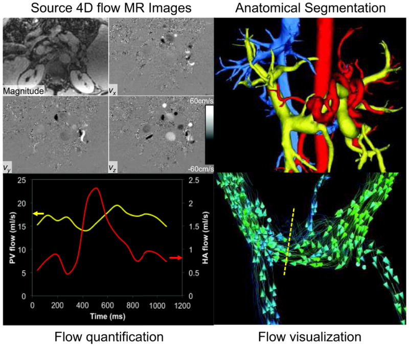
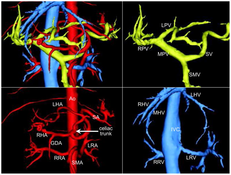
Purpose: To implement and validate in vivo radial 4D flow MRI for quantification of blood flow in the hepatic arterial, portal venous, and splanchnic vasculature of healthy volunteers and patients with portal hypertension.

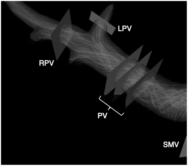
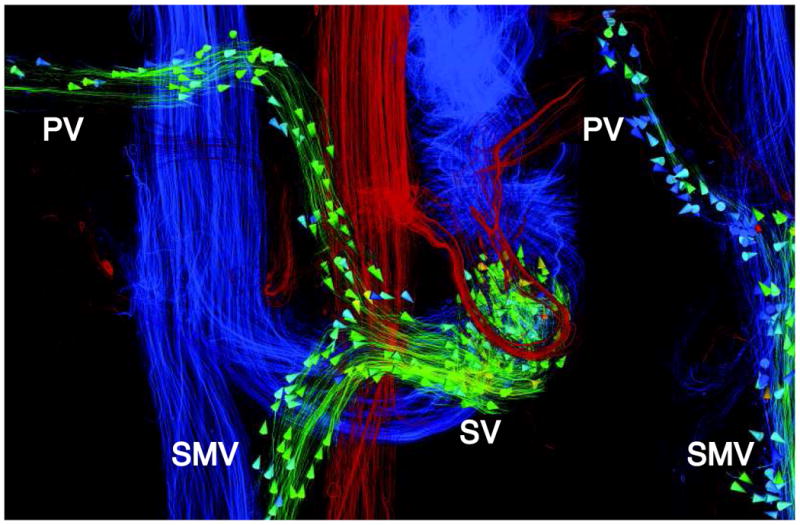
Materials and methods: Seventeen patients with portal hypertension and seven subjects with no liver disease were included in this Health Insurance Portability and Accountability Act (HIPAA)-compliant and Institutional Review Board (IRB)-approved study. Exams were conducted at 3T using a 32-channel body coil with large volumetric coverage and 1.4 mm isotropic true spatial resolution. Using postprocessing software, cut-planes orthogonal to vessels were used to quantify flow (L/min) in the hepatic and splanchnic vasculature.

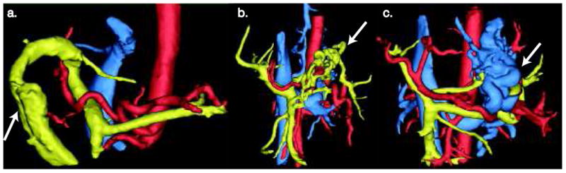
Results: Flow quantification was successful in all cases. Portal vein and supraceliac aorta flow demonstrated high variability among patients. Measurements were validated indirectly using internal consistency at three different locations within the portal vein (error = 4.2 ± 3.9%) and conservation of mass at the portal confluence (error = 5.9 ± 2.5%) and portal bifurcation (error = 5.8 ± 3.1%).

Conclusion: This work demonstrates the feasibility of radial 4D flow MRI to quantify flow in the hepatic and splanchnic vasculature. Flow results agreed well with data reported in the literature, and conservation of mass provided indirect validation of flow quantification. Flow in patients with portal hypertensions demonstrated high variability, with patterns and magnitude consistent with the hyperdynamic state that commonly occurs in portal hypertension.


Figures

Figure 1
Figure 1
Visualization of abdominal hemodynamics using 4D flow MRI. Workflow starts with source magnitude and velocity images (upper left, axial plane), which are combined into an anatomical PC angiogram (PCA) using complex difference processing. The PCA is segmented into color-coded vascular territories (Upper right), followed by streamline or particle trace visualization (bottom right). Note the cut-lanes perpendicular to the direction of the flow in the vessels of interest. Flow in the vessel of interest, in this case blood flow in the hepatic artery, is measured from flow waveforms using a Matlab-based tool (bottom left).
Figure 2
Figure 2
Conservation of mass at the portal confluence and within the main portal vein for indirect validation of flow measurement accuracy. Flow in the portal vein (QPV) should be approximately equal to the flow in the splenic (QSV) and superior mesenteric vein (QSMV) added.
Figure 3
Figure 3
The hepatic vasculature is complex and variable. Segmented 4D flow MR images provide 3D visualization of vascular anatomy. Note the replaced LHA, a normal variant, and visualization of the RRA and LRA. (MPV=main portal vein, RPV=rt. PV, LPV=lt. PV, SV=splenic vein, SMV = superior mesenteric vein, Ao=aorta, RRA=rt. renal artery, LRA=lt. RA, RHA=rt. hepatic artery, LHA=lt. hepatic artery, SA=splenic artery, SMA=superior mesenteric artery, IVC= inferior vena cava, RRV=rt. renal vein, LRV=lt. renal vein, RHV=rt. hepatic vein. MHV=middle HV, LHV=lt. HV).
Figure 4
Figure 4
Anatomical variations of the abdominal vasculature in patients with portal hypertension. a) Enlarged parahumbilical vein draining the portal vein (white arrow) b) Complex anatomy of the splenic vein (white arrow). c) Spleno-renal shunt (white arrow). Arterial circulation (Red), portal venous circulation (Yellow) and systemic venous circulation (Blue).
Figure 5
Figure 5
Summary of the flow results in the abdominal aorta (QAo) portal vein (QPV), hepatic artery (QHA). High variability of blood flow in the PV in patients with portal hypertension correlate well with the variability in blood flow in the abdominal aorta. Thus, variability in portal venous and hepatic arterial flow may be explained by differences in cardiac output due to variable presentation of a hyperdynamic state in patients with portal hypertension.
Figure 6
Figure 6
Portal vein and hepatic artery flow normalized to abdominal aortic flow (QPV/QAo) and QPV/QAo). Individual contributions to total liver inflow (QPV/ (QPV + QHA) and QHA/ (QPV + QHA)) show good agreement with data reported in the literature. Healthy controls are represented by triangles and circles represent patients with portal hypertension. (*) PH patient with reversed (hepato-fugal) QPV (HA fraction and PV fraction were not included for this patient since they are meaningless parameters in the setting of hepatofugal flow). (**) PH patient with markedely reduced QPV and reversed QSV.
Figure 7
Figure 7
Physiological variation in blood flow through the portal vein due to the increase resitance in two patients with portal hypertension. a) Reversed (hepato-fugal) flow is seen in the portal and splenic veins (** in Fig.6). Conservation of mass analysis showed good agreement (4.57%) between QPV and QSMV + QSV. b) Reversed QSV with reduced QPV and normal QSMV (* in Fig.6). Reversed flow can also be seen in the coronary vein in this patient.
Figure 8
Figure 8
Conservation mass validation. a. Portal confluence flow (QPV ∼ QSMV + QSV). b. Portal bifurcation (QPV ∼ QRPV + QLPV). Dashed line represents unity.

Similar articles

Cited by

References

    1. Centers for Desease Control and Prevention. 2009
    1. Bhathal PS, Grossman HJ. Reduction of the increased portal vascular resistance of the isolated perfused cirrhotic rat liver by vasodilators. J Hepatol. 1985;1(4):325–337. - PubMed
    1. Zardi EM, Dobrina A, Uwechie V, et al. Postmeal portal flow variations in HCV-related chronic hepatitis and liver cirrhosis with and without hyperdynamic syndrome. In Vivo. 2008;22(4):509–512. - PubMed
    1. Moller S, Hobolth L, Winkler C, Bendtsen F, Christensen E. Determinants of the hyperdynamic circulation and central hypovolaemia in cirrhosis. Gut. 2011;60(9):1254–1259. - PubMed
    1. Burkart DJ, Johnson CD, Ehman RL, Weaver AL, Ilstrup DM. Evaluation of portal venous hypertension with cine phase-contrast MR flow measurements: high association of hyperdynamic portal flow with variceal hemorrhage. Radiology. 1993;188(3):643–648. - PubMed

Publication types

MeSH terms