. 2013 Jan;19(1):36-50.
doi: 10.1261/rna.035469.112. Epub 2012 Nov 12.

The association of a La module with the PABP-interacting motif PAM2 is a recurrent evolutionary process that led to the neofunctionalization of La-related proteins

Affiliations

The association of a La module with the PABP-interacting motif PAM2 is a recurrent evolutionary process that led to the neofunctionalization of La-related proteins

Rémy Merret et al. RNA. 2013 Jan.

Abstract

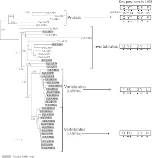
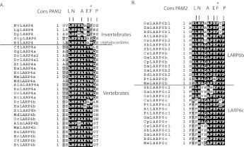
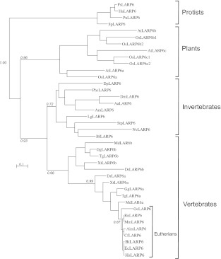
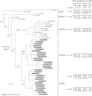
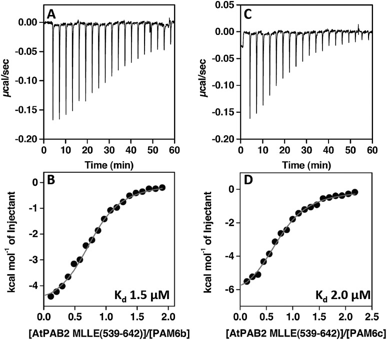
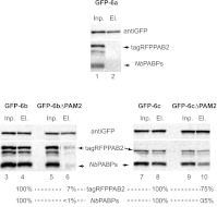
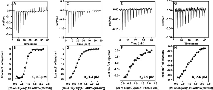
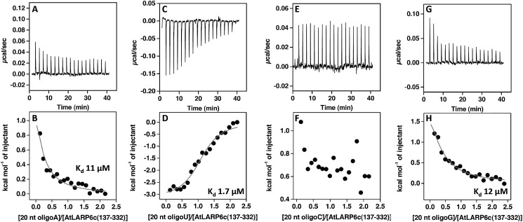
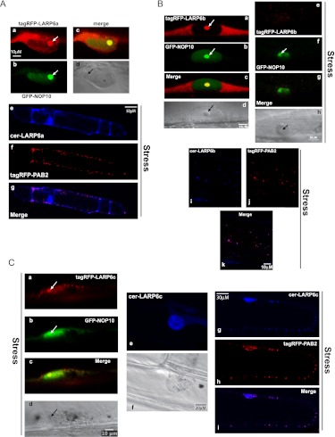
La-related proteins (LARPs) are largely uncharacterized factors, well conserved throughout evolution. Recent reports on the function of human LARP4 and LARP6 suggest that these proteins fulfill key functions in mRNA metabolism and/or translation. We report here a detailed evolutionary history of the LARP4 and 6 families in eukaryotes. Genes coding for LARP4 and 6 were duplicated in the common ancestor of the vertebrate lineage, but one LARP6 gene was subsequently lost in the common ancestor of the eutherian lineage. The LARP6 gene was also independently duplicated several times in the vascular plant lineage. We observed that vertebrate LARP4 and plant LARP6 duplication events were correlated with the acquisition of a PABP-interacting motif 2 (PAM2) and with a significant reorganization of their RNA-binding modules. Using isothermal titration calorimetry (ITC) and immunoprecipitation methods, we show that the two plant PAM2-containing LARP6s (LARP6b and c) can, indeed, interact with the major plant poly(A)-binding protein (PAB2), while the third plant LARP6 (LARP6a) is unable to do so. We also analyzed the RNA-binding properties and the subcellular localizations of the two types of plant LARP6 proteins and found that they display nonredundant characteristics. As a whole, our results support a model in which the acquisition by LARP4 and LARP6 of a PAM2 allowed their targeting to mRNA 3' UTRs and led to their neofunctionalization.


Figures

FIGURE 1.
FIGURE 1.
Detailed phylogenetic analysis of the LARP4 family. The phylogenetic tree was obtained using La module (LAM-RRM) sequences of 43 LARP4 proteins from protist, invertebrate, and vertebrate species (see Supplemental Table S1 for protein sequences). Selected informative statistical supports (approximate likelihood-ratio test [aLRT] data) are indicated. (Gray) Proteins presenting a putative PAM2 (see Fig. 2A). Key positions of the LAM, known to form the RNA-binding pocket in the context of the genuine La protein (Bayfield et al. 2010), are indicated on the right (dots indicate conserved amino acids, underlined amino acids are for possible conservative changes, and gray amino acids with an asterisk are for possible nonconservative changes). The numbering refers to the position of the amino acids in the human genuine LAM. Species codes are the following: (Ps) Phytophtora species; (Es) Ectocarpus siliculosus; (Ng) Naegleria gruberi; (Pp) Polysphondylium pallidum; (Dd) dictyostelium discoideum; (Ci) Ciona intestinalis; (Nv) Nematostella vectensis; (Hm) Hydra magnipapillata; (Ce) Caenorhabditis elegans; (Ta) Trichoplax adhaerens; (Dp) Daphnia pullex; (Dm) Drosophila melanogaster; (Aa) Aedes aegypti; (Am) Apis mellifera; (Phc) Pediculus humanus corporis; (Bf) Branchiostoma floridae; (Stp) Strongylocentrotus purpuratus; (Lg) Lottia gigantae; (Dr) Danio rerio; (Gg) Gallus gallus; (Xt) Xenopus tropicalis; (Md) Monodelphis domestica; (Aim) Ailuropoda melanoleuca; (Cf) Canis familiaris; (Mm) Mus musculus; (Rn) Rattus norvegicus; (Hs) Homo sapiens; (Oc) Oryctolagus cuniculus; (Bt) Bos taurus; (Ec) Equus caballus.
FIGURE 2.
FIGURE 2.
Alignment of the putative PAM2 present in LARP4 (A) and LARP6 (B) proteins. The most conserved positions of the eukaryote PAM2 are indicated above each alignment. (*) The position of an invariant phenylalanine proposed to be essential for binding to the PABP MLLE motif (Kozlov et al. 2010). A dark line separates PAM2 from invertebrate and vertebrate LARP4. The conserved phenylalanine was replaced by a tryptophan starting from cephalocordates, thus before the gene duplication events that happened early in the vertebrate lineage. For LARP6, PAM2s are only found for vascular plant proteins that belong to LARP6b or LARP6c subgroups.
FIGURE 3.
FIGURE 3.
Detailed phylogenetic analysis of the LARP6 family. The phylogenetic tree was obtained using La module (LAM-RRM) sequences of 39 LARP6 proteins form protist, plant, invertebrate, and vertebrate species (see Supplemental Table S1 for protein sequences). Selected informative statistical supports (approximate likelihood-ratio test [aLRT] data) are indicated. Species codes are as in Figure 1 with the following additions: (Ha) Hyaloperonospora arabidopsidis; (Pu) Pythium ultimum; (Sp) Saprolegnia parasitica; (At) Arabidopsis thaliana; (Os) Oryza sativa.
FIGURE 4.
FIGURE 4.
Phylogenetic analysis of the LARP6 family in the green lineage. The phylogenetic tree was obtained using La module (LAM-RRM) sequences of 46 LARP6 proteins from green algae, mosses, and vascular plant species (see Supplemental Table S1 for protein sequences). Selected informative statistical supports (approximate likelihood-ratio test [aLRT] data) are indicated. (Gray) Proteins presenting a putative PAM2 (see Fig. 2B). Key positions of the LAM, known to form the RNA-binding pocket in the context of the genuine La protein (Bayfield et al. 2010), are indicated on the right (dots indicate conserved amino acids, underlined amino acids are for possible conservative changes, and gray amino acids with an asterisk are for possible nonconservative changes). The numbering refers to the position of the amino acids in the genuine human LAM. Species codes are (Ot) Ostreococcus tauri; (Mi) Micromonas; (Vc) Volvox carteri; (Cr) Chlamydomonas reinhardii; (Php) Physcomitrella patens; (Sm) Selaginella moellendorfii; (At) Arabidopsis thaliana; (Cp) Carica papaya; (Gm) Glycine max; (Pt) Populus trichocarpa; (Me) Manihot esculenta; (Cs) Cucumis sativis; (Bd) Brachypodium distachyon; (Os) Oryza sativa; (Sb) Sorghum bicolor; (Zm) Zea mays.
FIGURE 5.
FIGURE 5.
Calorimetric analysis of the interaction between the AtPAB2 MLLE domain (539–642) and the peptides PAM6b and PAM6c. Raw titration data showing the thermal effect of injecting an AtPAB2 MLLE solution into a calorimetric cell containing peptide PAM6b (A) or peptide PAM6c (C). The normalized heat for the titrations, shown in B and D, respectively, was obtained by integrating the raw data and subtracting the heat of the protein dilution. The gray lines in B and D represent the best fit derived by a nonlinear least-squares procedure based on an independent binding sites model. The dissociation constant Kd is indicated for each interaction, and the thermodynamic parameters are shown in Supplemental Table S2.
FIGURE 6.
FIGURE 6.
Plant PAM2-containing AtLARP6b and AtLARP6c can form a complex with AtPAB2. GFP-tagged versions of AtLARP6a, 6b, 6c, 6bΔPAM2, or 6cΔPAM2 were, respectively, coexpressed in N. benthamiana leaves with AtPAB2 fused to the tagRFP label (a fluorescent tag from E. quadricolor that is not recognized by anti-GFP antibodies) (Merzlyak et al. 2007). Immunoprecipitations were performed using the anti-GFP antibodies. Western blot analyses using either anti-GFP or anti-PAB2 antibodies on the input (Inp; 3.5% of the total input) or eluate (El; 50% of the eluate) fractions are shown. The positions of the GFP fusions (GFP), tagRFPPAB2, and N. benthamiana PABP (NbPABPs) are indicated. The intensities of the GFP, tagRFPPAB2, and NbPABPs signals in eluate fractions of the 6b, 6bΔPAM2 and 6c, 6cΔPAM2 have been quantified and compared. Each eluate signal (GFP, tagRFPPAB2, NbPABPs) has been quantified and divided by its corresponding value in the input. Then the tagRFP and NbPABP obtained values have been divided by the GFP signal from the same eluate. This gives normalization to the pull-down efficiency. We arbitrarily set the eluate values obtained for the pull-down experiments with the full-length proteins to 100% and give the values of the remaining signals in the eluates fractions of the ΔPAM2 pull-downs.
FIGURE 7.
FIGURE 7.
RNA-binding properties of AtLARP6a. ITC experiments were conducted with four RNA targets: 20-nt oligo(A) (A,B), 20-nt oligo(U) (C,D), 20-nt oligo(C) (E,F), and 20-nt oligo(G) (G,H). Integration of the raw titration data (A,C,E,G) generated sigmoid-shaped curves (B,D,F,H, respectively) centered on a 1:1 molar ratio that are interpolated by an equivalent and independent binding model (gray lines represent the best fit). The dissociation constant Kd is indicated for each RNA target, and the thermodynamic parameters are shown in Supplemental Table S2. All the RNA sequences tested bind to AtLARP6a, but 20-nt oligo(A) interacts approximately fivefold stronger than 20-nt oligo(U) and ninefold stronger than 20-nt oligo(C) and 20-nt oligo(G).
FIGURE 8.
FIGURE 8.
RNA-binding properties of AtLARP6c. ITC experiments were performed with four RNA targets: 20-nt oligo(A) (A,B), 20-nt oligo(U) (C,D), 20-nt oligo(C) (E,F), and 20-nt oligo(G) (G,H). Integration of the raw titration data (A,C,G) generated sigmoid-shaped curves (B,D,H, respectively) centered on a 1:1 molar ratio that are interpolated by an equivalent and independent binding model (gray lines represent the best fit). Nonetheless, the fit is poorer for 20-nt oligo(A) and 20-nt oligo(G) (B,H, respectively) because of the weaker interaction. No interaction could be detected by ITC with 20-nt oligo(C) (F). The dissociation constant Kd is indicated, and the thermodynamic parameters are shown in Supplemental Table S2. AtLARP6c shows a clear preference for 20-nt oligo(U), binding to 20-nt oligo(A), and 20-nt oligo(G) with a sevenfold lower affinity.
FIGURE 9.
FIGURE 9.
Subcellular localization of plant LARP6. TagRFP-labeled AtLARP6a (A), 6b (B), or 6c (C) proteins were transiently expressed in onion epidermal cells, and their subcellular localization was analyzed by confocal microscopy. tagRFP-tagged versions of the AtLARP6a, 6b, or 6c were, respectively, coexpressed with GFP-NOP10 and Cerulean (cer)–tagged versions of AtLARP6a, 6b, or 6c were, respectively, coexpressed with a tagRFP-labeled AtPAB2 protein in onion epidermal cells, and the subcellular localization of each fusion protein was analyzed and compared in stress situations (hypoxia). Arrows point to the nucleoli.

Similar articles

Cited by

References

    1. Anderson P, Kedersha N 2009. RNA granules: Post-transcriptional and epigenetic modulators of gene expression. Nat Rev Mol Cell Biol 10: 430–436 - PubMed
    1. Anderson JT, Paddy MR, Swanson MS 1993. PUB1 is a major nuclear and cytoplasmic polyadenylated RNA-binding protein in Saccharomyces cerevisiae. Mol Cell Biol 13: 6102–6113 - PMC - PubMed
    1. Anisimova M, Gascuel O 2006. Approximate likelihood-ratio test for branches: A fast, accurate, and powerful alternative. Syst Biol 55: 539–552 - PubMed
    1. Bayfield MA, Maraia RJ 2009. Precursor-product discrimination by La protein during tRNA metabolism. Nat Struct Mol Biol 16: 430–437 - PMC - PubMed
    1. Bayfield MA, Yang R, Maraia RJ 2010. Conserved and divergent features of the structure and function of La and La-related proteins (LARPs). Biochim Biophys Acta 1799: 365–378 - PMC - PubMed

Publication types

MeSH terms