. 2012 Dec 28;287(53):44425-34.
doi: 10.1074/jbc.M112.415935. Epub 2012 Nov 12.

An ultra-specific avian antibody to phosphorylated tau protein reveals a unique mechanism for phosphoepitope recognition

Affiliations

An ultra-specific avian antibody to phosphorylated tau protein reveals a unique mechanism for phosphoepitope recognition

Heather H Shih et al. J Biol Chem. .

Abstract

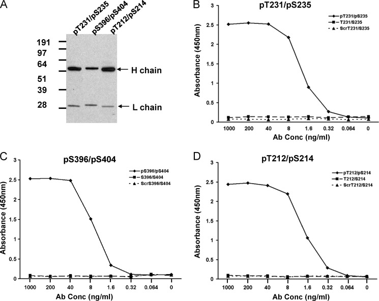
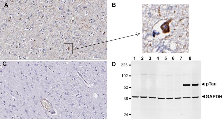
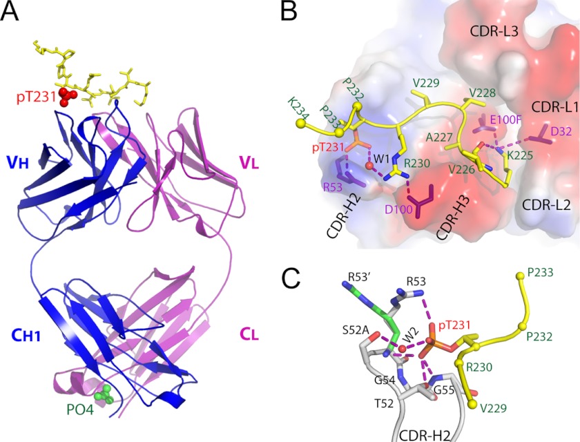
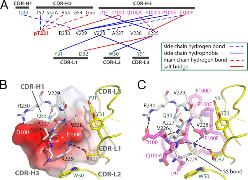
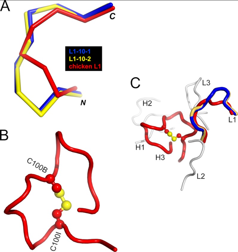
Highly specific antibodies to phosphoepitopes are valuable tools to study phosphorylation in disease states, but their discovery is largely empirical, and the molecular mechanisms mediating phosphospecific binding are poorly understood. Here, we report the generation and characterization of extremely specific recombinant chicken antibodies to three phosphoepitopes on the Alzheimer disease-associated protein tau. Each antibody shows full specificity for a single phosphopeptide. The chimeric IgG pT231/pS235_1 exhibits a K(D) of 0.35 nm in 1:1 binding to its cognate phosphopeptide. This IgG is murine ortholog-cross-reactive, specifically recognizing the pathological form of tau in brain samples from Alzheimer patients and a mouse model of tauopathy. To better understand the underlying binding mechanisms allowing such remarkable specificity, we determined the structure of pT231/pS235_1 Fab in complex with its cognate phosphopeptide at 1.9 Å resolution. The Fab fragment exhibits novel complementarity determining region (CDR) structures with a "bowl-like" conformation in CDR-H2 that tightly and specifically interacts with the phospho-Thr-231 phosphate group, as well as a long, disulfide-constrained CDR-H3 that mediates peptide recognition. This binding mechanism differs distinctly from either peptide- or hapten-specific antibodies described to date. Surface plasmon resonance analyses showed that pT231/pS235_1 binds a truly compound epitope, as neither phosphorylated Ser-235 nor free peptide shows any measurable binding affinity.


Figures

FIGURE 1.
FIGURE 1.
Expression and binding activity of chimeric IgG molecules converted from scFv. A, Western blot analysis of chimeric IgG molecules converted from scFv clones pT231/pS235_1, pS396/pS404_1, and pT212/pS214_1, respectively. The heavy and light chains for each IgG are indicated by arrows. B–D, ELISA analysis of each purified IgG molecule for binding to the phosphopeptide, non-phosphopeptide, and scrambled (Scr) peptide.
FIGURE 2.
FIGURE 2.
Binding of pathological phospho-tau by antibody pT231/pS235_1 in human AD and Tg4510 transgenic mouse brains. A–C, immunohistochemical staining of human AD brain (A and B) and healthy brain (C). B, magnification of the boxed area in A. D, Western blot analysis of Tg4510 transgenic and wild-type brain lysates with pT231/pS235_1 IgG (plus control anti-GAPDH antibody). Each lane represents a different animal, and the lysate samples are 3-month-old wild-type mice (lanes 1 and 2), 3-month-old transgenic mice (lanes 3 and 4), 6-month-old wild-type mice (lanes 5 and 6), and 6-month-old transgenic mice (lanes 7 and 8).
FIGURE 3.
FIGURE 3.
Structure of anti-phospho-tau Fab (pT231/pS235_1) in complex with phosphoepitope pT231/pS235. A, schematic view showing the complex. The Fab heavy chain (VH + CH1) and light chain (VL + CL) are shown in blue and purple, respectively. The phosphoepitope is shown as yellow sticks on top with phosphorylated Thr-231 (pT231) highlighted as red spheres. The solvent PO43− ion is labeled and shown as green spheres. B, transparent electrostatic surface view of the CDRs showing the strong electrostatic interactions between CDR residues and the phosphoepitope. Positively charged areas are shown in blue, and negatively charged areas are shown in red. The backbone of the bound phosphoepitope is shown as a schematic in yellow; the side chains are shown as a stick model in atomic colors (carbon, yellow; nitrogen, blue; oxygen, red; and phosphorus, orange). The side chains of Pro-232, Pro-233, and Lys-234 are omitted for clarity, with Cα shown as spheres. The residues within CDRs that form strong electrostatic interactions with the phosphoepitope are shown as purple sticks. A water molecule is labeled W1. Hydrogen bonds are shown as dashed purple lines. C, interaction details between CDR-H2 and pThr-231. Two conformations of the H/Arg-53 side chain are shown as R53 and R53′, respectively. Hydrogen bonds are shown as dashed purple lines. A water molecule is labeled W2.
FIGURE 4.
FIGURE 4.
Fab-phosphoepitope interaction details. A, schematic representation of all contacts between pT231/pS235_1 Fab and the phosphoepitope pT231/pS235 in the Fab-phosphoepitope co-crystal structure. B and C, interaction details between 225KVAVVR230 and the Fab fragment, with the same orientation in both panels. B, CDR-H3 is shown as an electrostatic surface model, with positively charged areas in blue and negatively charged areas in red. C, CDR-H3 is shown as a schematic in magenta and as sticks in atomic colors (carbon, magenta; nitrogen, blue; and oxygen, red). The CDRs from the light chain are shown as a schematic in yellow, with side chains shown as sticks (carbon, yellow; and oxygen, red). Peptide 225KVAVVR230 is shown as gray sticks (carbon, gray; nitrogen, blue; and oxygen, red). Gln-33 from CDR-H1 is shown as green sticks (carbon, green). Hydrogen bonds are shown as dashed black lines. The disulfide bond within CDR-H3 is shown and labeled SS bond.
FIGURE 5.
FIGURE 5.
Chicken-specific conformations of CDR-L1 and CDR-H3 from anti-phospho-tau Fab (pT231/pS235_1). A, ribbon view showing the superposition of chicken CDR-L1 (red) with canonical mammalian CDR-L1–10-1 (blue; Protein Data Bank code 1YQV) and CDR-L1–10-2 (yellow; code 1AY1), respectively. CDR-L1–10-1 and CDR-L1–10-2 represent two clusters of mammalian CDR-L1 conformations and show the best structural similarity to chicken CDR-L1. B, schematic view showing the conformation of chicken CDR-H3 stabilized by the intramolecular disulfide bond shown as a ball and stick model in atomic colors (carbon, red; and sulfur, yellow). Two cysteines are labeled C100B and C100I, respectively. C, schematic view showing chicken CDRs with the same coloring scheme as in A and B. CDR-L2, CDR-L3, CDR-H1, and CDR-H2 are shown in gray.

References

    1. Manning G., Whyte D. B., Martinez R., Hunter T., Sudarsanam S. (2002) The protein kinase complement of the human genome. Science 298, 1912–1934 - PubMed
    1. Nairn A. C., Detre J. A., Casnellie J. E., Greengard P. (1982) Serum antibodies that distinguish between the phospho- and dephospho-forms of a phosphoprotein. Nature 299, 734–736 - PubMed
    1. Ross A. H., Baltimore D., Eisen H. N. (1981) Phosphotyrosine-containing proteins isolated by affinity chromatography with antibodies to a synthetic hapten. Nature 294, 654–656 - PubMed
    1. Grundke-Iqbal I., Iqbal K., Quinlan M., Tung Y. C., Zaidi M. S., Wisniewski H. M. (1986) Microtubule-associated protein tau. A component of Alzheimer paired helical filaments. J. Biol. Chem. 261, 6084–6089 - PubMed
    1. Grundke-Iqbal I., Iqbal K., Tung Y. C., Quinlan M., Wisniewski H. M., Binder L. I. (1986) Abnormal phosphorylation of the microtubule-associated protein tau (tau) in Alzheimer cytoskeletal pathology. Proc. Natl. Acad. Sci. U.S.A. 83, 4913–4917 - PMC - PubMed

Publication types

MeSH terms

Associated data