. 2013 Jan 11;288(2):1365-73.
doi: 10.1074/jbc.M112.434142. Epub 2012 Nov 12.

High mobility group Box 1 inhibits human pulmonary artery endothelial cell migration via a Toll-like receptor 4- and interferon response factor 3-dependent mechanism(s)

Affiliations

High mobility group Box 1 inhibits human pulmonary artery endothelial cell migration via a Toll-like receptor 4- and interferon response factor 3-dependent mechanism(s)

Eileen M Bauer et al. J Biol Chem. .

Abstract

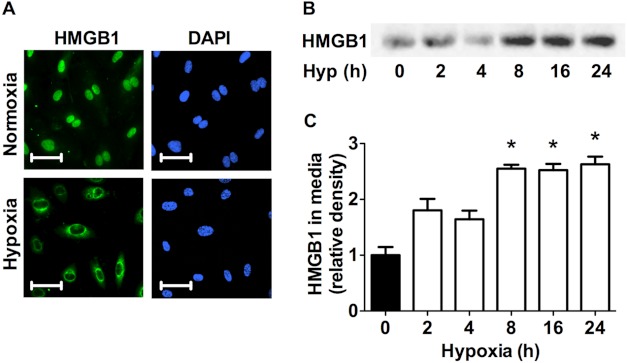
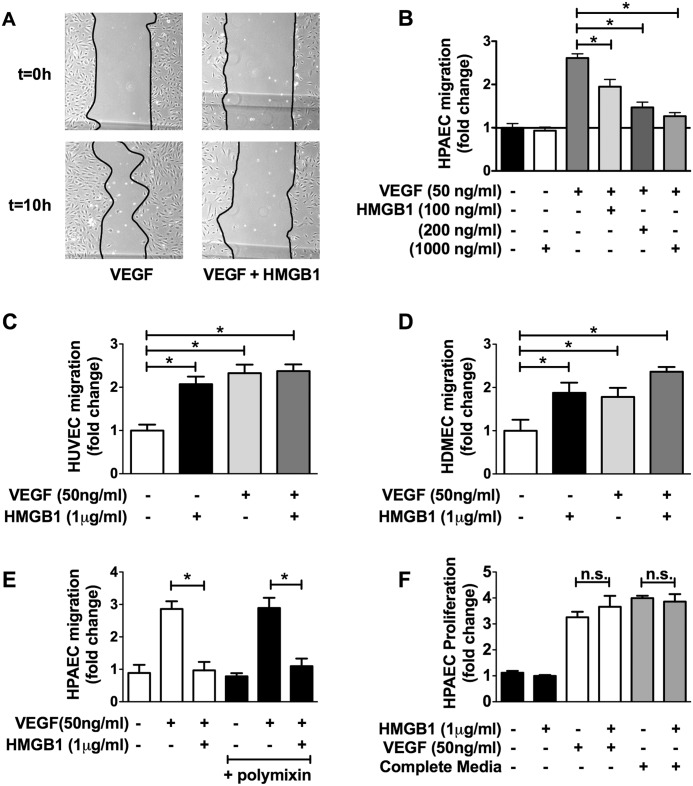
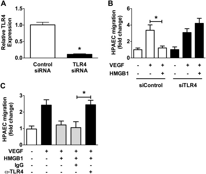
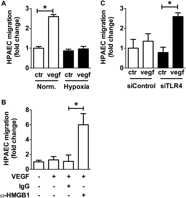
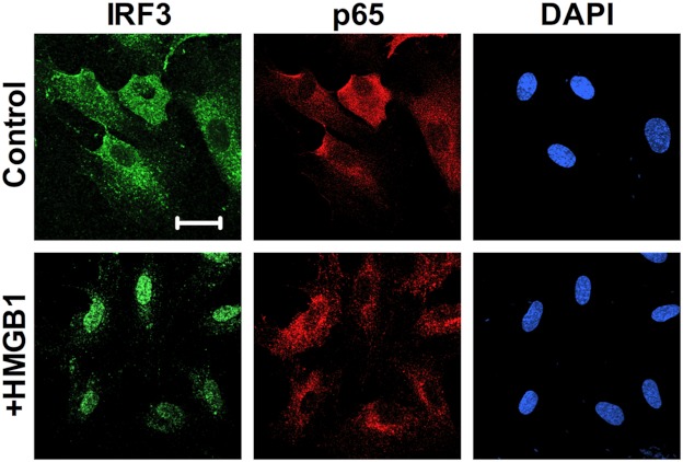
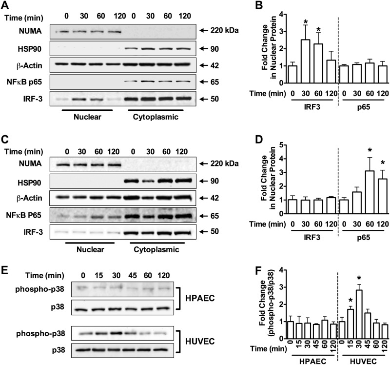
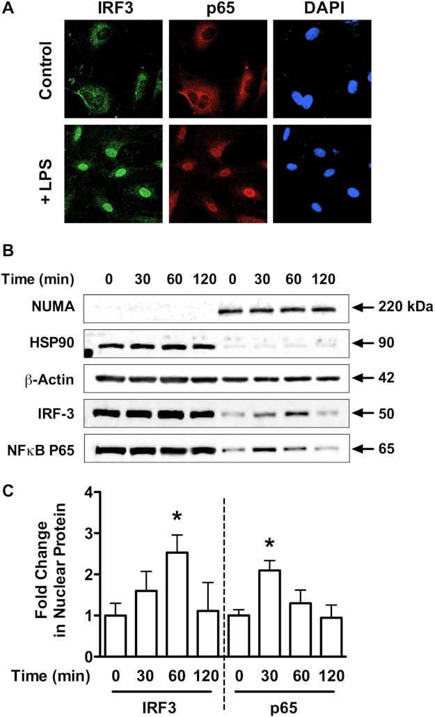
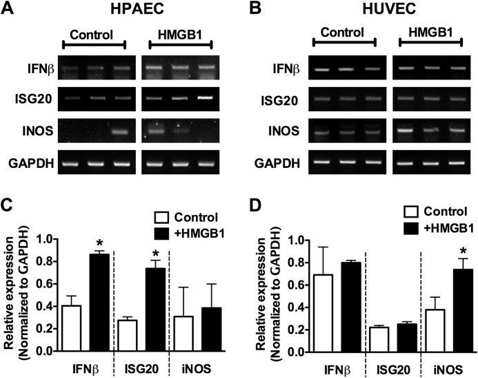
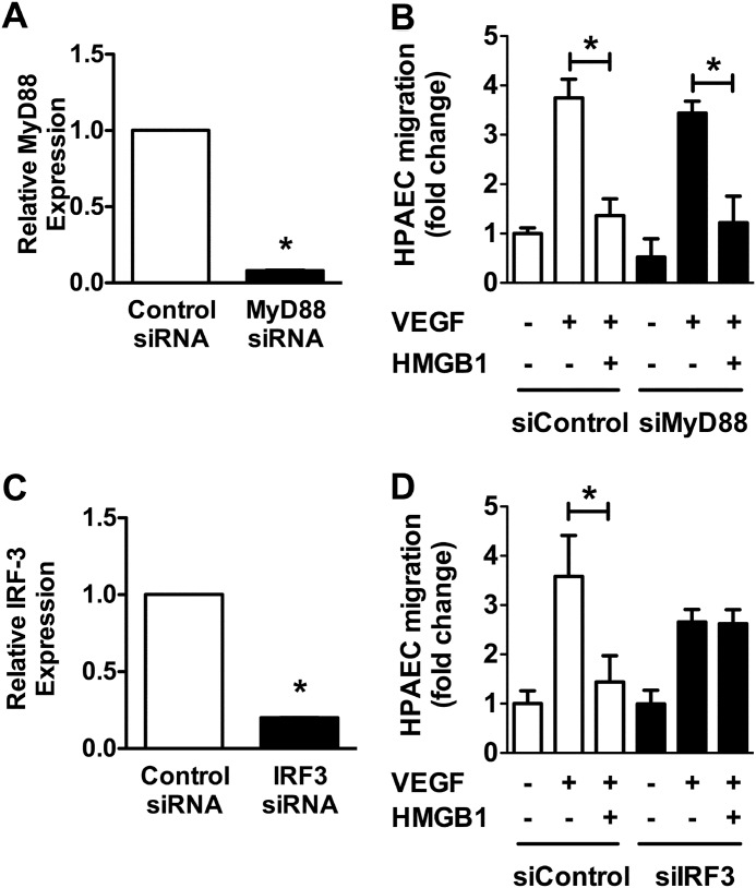
In pulmonary hypertension the loss of precapillary arterioles results from vascular injury causing endothelial dysfunction. Endothelial cell migration and proliferation are critical for vascular regeneration. This study focused on the effect of high mobility group box 1 protein (HMGB1) on these critical processes. HMGB1 had no effect on human pulmonary artery endothelial cell (HPAEC) proliferation. In contrast, treatment of HPAECs with HMGB1 dose-dependently inhibited VEGF-stimulated HPAEC migration. The effect of HMGB1 on HPAEC migration was TLR4-dependent because it was reversed by TLR4 siRNA or TLR4-neutralizing antibody. Exposure of HPAECs to hypoxia caused translocation and release of HMGB1 and inhibition of HPAEC migration. The effect of hypoxia on HPAEC migration was mediated by HMGB1 because HMGB1-neutralizing antibody but not control IgG restored HPAEC migration. Likewise, TLR4 siRNA but not control siRNA reversed the inhibitory effect of hypoxia in HPAECs. The canonical TLR4 signaling pathway requires the adaptor protein MyD88 and leads to downstream NFκB activation. Interestingly, HMGB1 failed to stimulate NFκB translocation to the nucleus, but instead activated an alternative pathway characterized by activation of interferon response factor 3 (IRF3). This was in contrast to human umbilical vein endothelial cells in which HMGB1 stimulated nuclear translocation of NFκB but not IRF3. IRF3 siRNA, but not MyD88 siRNA, reversed the inhibitory effect of HMGB1 on HPAEC migration. These data demonstrate that HMGB1 inhibits HPAEC migration, a critical process for vascular regeneration, via TLR4- and IRF3-dependent mechanisms.


Figures

FIGURE 1.
FIGURE 1.
Hypoxia induces HMGB1 release in HPAECs. A, HPAECs exposed to 8 h of hypoxia (1%) were assessed for HMGB1 release by immunofluorescent staining (HPAECs, green; DAPI, blue; scale bars, 25 μm). Photomicrographs are representative of three independent experiments. B and C, Western blot analysis (B) and quantitative densitometry (C) of HMGB1 accumulation in the cell media of HPAECs exposed to hypoxia for the indicated times are shown. Blots are representative of four independent experiments. *, p < 0.05; error bars, S.E.
FIGURE 2.
FIGURE 2.
HMGB1 inhibits VEGF-induced HPAEC migration. A, representative images of cell migration assay of HPAECs treated for 10 h with VEGF (50 ng/ml) or VEGF + HMGB1 (1 μg/ml). B, serum-starved HPAECs stimulated with VEGF (50 ng/ml) in the absence/presence of HMGB1 (100 ng/ml–1 μg/ml) and assessed for cellular migration using the monolayer wound assay. C and D, effect of HMGB1 (1 μg/ml) on VEGF (50 ng/ml)-stimulated HUVEC migration (C) and HDMEC migration (D). E, effect of polymyxin on HMGB1-inhibited HPAEC migration (1000 ng/ml). F, effect of HMGB1 on HPAEC proliferation using [3H]thymidine incorporation. Data represent the mean ± S.E. (error bars) of three independent experiments. Analysis of variance; *, p < 0.05.
FIGURE 3.
FIGURE 3.
TLR4 mediates the effects of HMGB1 of HPAEC migration. A, quantitative PCR for TLR4 in control and TLR4 siRNA-treated HPAECs. B, HPAECs were transfected with TLR4 siRNA (25 nm) or control siRNA (25 nm) and VEGF-induced cell migration assessed in the presence/absence of HMGB1 (1 μg/ml). (C) HPAECs were treated with TLR4 blocking antibody (20 μg/ml) or control IgG (20 μg/ml) and VEGF-induced cell migration assessed in the presence/absence of HMGB1 (1 μg/ml). Data represent the mean ± S.E. (error bars) of three independent experiments. Analysis of variance; *, p < 0.05.
FIGURE 4.
FIGURE 4.
Hypoxia-induced HMGB1 release leads to inhibition of HPAEC migration via TLR4. VEGF-stimulated cell migration was assessed in HPAECs exposed to (A) hypoxia (1% O2), (B) αHMGB1-neutralizing antibody (10 μg/ml) or IgG control antibody, (C) hypoxia when transfected with siRNA against TLR4 (25 nm) or control siRNA (25 nm). Data represent the mean ± S.E. (error bars) of three to four independent experiments. Analysis of variance; *, p < 0.05.
FIGURE 5.
FIGURE 5.
HMGB1 stimulates nuclear translocation of IRF3 in HPAECs. A, HPAECs exposed for 2 h to HMGB1 (1 μg/ml) were assessed for IRF3 nuclear translocation by immunofluorescent staining against IRF3 (green; nuclear stain DAPI, blue; scale bar, 50 μm). Images are representative of three independent experiments.
FIGURE 6.
FIGURE 6.
Differential signaling in HPAECs versus HUVECs. Western blot analysis for NUMA (150 kDa, nuclear marker), HSP90 (90 kDa, cytoplasmic marker), β-actin (42 kDa), NFκB-p65 (65 kDa), and IRF3 (45 kDa) in cytoplasmic and nuclear fractions from HPAECs (A) and HUVECs (C) treated with HMGB1 (1 μg/ml) for the indicated times. Blots are representative of three independent experiments and are quantified in B and D, respectively. E, Western blot analysis for phospho-p38 MAPK and total p38 MAPK in HPAECs and HUVECs for the indicated times. Blots are representative of three independent experiments and are quantified in F. Graphs represent the mean ± S.E. (error bars) of three independent experiments. Analysis of variance; *, p < 0.05 versus unstimulated control.
FIGURE 7.
FIGURE 7.
LPS stimulates MyD88-dependent and -independent TLR4 signaling in HPAECs. A, HPAECs exposed for 1 h to LPS (1 μg/ml) were assessed for IRF3 nuclear translocation by immunofluorescent staining against IRF3 (green; nuclear stain DAPI, blue; scale bar, 25 μm). Images are representative of three independent experiments. B, Western blot analysis is shown of NUMA (150 kDa, nuclear marker), HSP90 (90 kDa, cytoplasmic marker), β-actin (42 kDa), NFκB-p65 (65 kDa), and IRF3 (50 kDa) in cytoplasmic and nuclear fractions from HPAECs treated with LPS (1 μg/ml) for the indicated times. Blots are representative of three independent experiments and are quantified in C. Data represent the mean ± S.E. (error bars) of three independent experiments. Analysis of variance; *, p < 0.05 versus unstimulated control.
FIGURE 8.
FIGURE 8.
Differential gene induction by HMGB1 in HPAECs versus HUVECs. A and B, HPAECs (A) and HUVECs (B) were stimulated with HMGB1 (1 μg/ml) for 4 h and then assessed for the induction of ISG20, IFNβ, or iNOS mRNA by RT-PCR. C and D, the relative expression of each gene is quantified in the histograms and normalized to GAPDH mRNA expression. Data represent the mean ± S.E. (error bars) of three independent experiments. Analysis of variance; *, p < 0.05 versus unstimulated control.
FIGURE 9.
FIGURE 9.
Inhibition of HPAEC migration by HMGB1 is IRF3-dependent. A, relative MyD88 expression assessed by quantitative RT-PCR after treating HPAECs with control or MyD88siRNA. B, VEGF-stimulated cell migration assessed in HPAECs treated with siRNA against MyD88 (25 nm) or with control siRNA (25 nm) in the presence/absence of HMGB1 (1 μg/ml). C, relative IRF-3 expression using quantitative PCR after treating HPAECs with scramble or IRF-3 siRNA. D, VEGF-stimulated cell migration assessed in HPAECs treated with siRNA against IRF3 (25 nm) or with control siRNA (25 nm) in the presence/absence of HMGB1 (1 μg/ml). In all panels, data represent the mean ± S.E. (error bars) of three independent experiments. Analysis of variance; *, p < 0.05.

References

    1. Evankovich J., Cho S. W., Zhang R., Cardinal J., Dhupar R., Zhang L., Klune J. R., Zlotnicki J., Billiar T., Tsung A. (2010) High mobility group box 1 release from hepatocytes during ischemia and reperfusion injury is mediated by decreased histone deacetylase activity. J. Biol. Chem. 285, 39888–39897 - PMC - PubMed
    1. Park J. S., Svetkauskaite D., He Q., Kim J. Y., Strassheim D., Ishizaka A., Abraham E. (2004) Involvement of Toll-like receptors 2 and 4 in cellular activation by high mobility group box 1 protein. J. Biol. Chem. 279, 7370–7377 - PubMed
    1. Park J. S., Gamboni-Robertson F., He Q., Svetkauskaite D., Kim J. Y., Strassheim D., Sohn J. W., Yamada S., Maruyama I., Banerjee A., Ishizaka A., Abraham E. (2006) High mobility group box 1 protein interacts with multiple Toll-like receptors. Am. J. Physiol. Cell Physiol. 290, C917–924 - PubMed
    1. Huang W., Tang Y., Li L. (2010) HMGB1, a potent proinflammatory cytokine in sepsis. Cytokine 51, 119–126 - PubMed
    1. Ohmori H., Luo Y., Kuniyasu H. (2011) Non-histone nuclear factor HMGB1 as a therapeutic target in colorectal cancer. Expert Opin. Ther. Targets 15, 183–193 - PubMed

Publication types

MeSH terms