

Pancreatitis in cats
- PMID: 23148855
- PMCID: PMC7105028
- DOI: 10.1053/j.tcam.2012.09.001
Pancreatitis in cats
Abstract
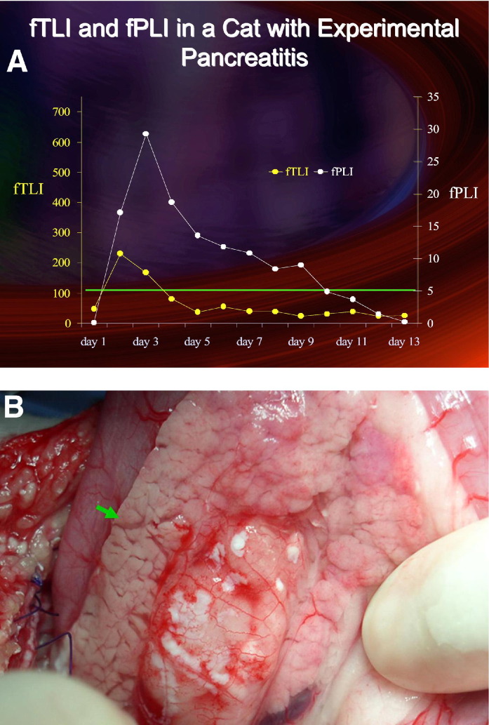
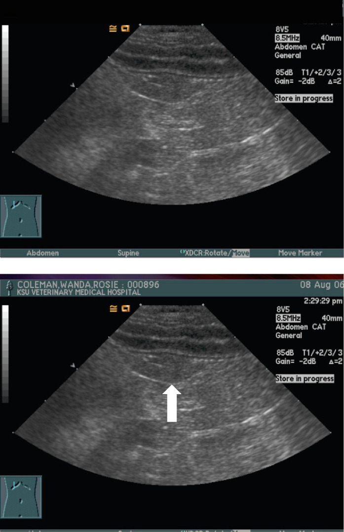
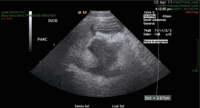
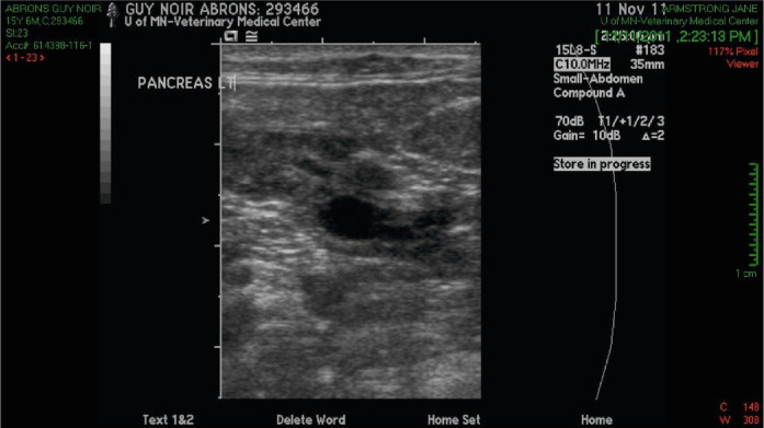
Pancreatitis was considered a rare disease in the cat until a couple of decades ago when several retrospective studies of severe acute pancreatitis were published. It was apparent that few of the diagnostic tests of value in the dog were helpful in cats. With increasing clinical suspicion, availability of abdominal ultrasonography, and introduction of pancreas-specific blood tests of increasing utility, it is now accepted that acute pancreatitis is probably almost as common in cats as it is in dogs, although the etiology(s) remain more obscure. Pancreatitis in cats often co-exists with inflammatory bowel disease, less commonly with cholangitis, and sometimes with both. Additionally, pancreatitis may trigger hepatic lipidosis, while other diseases, such as diabetes mellitus, may be complicated by pancreatitis. Therapy is similar to that used in dogs, with added emphasis on early nutritional support to prevent hepatic lipidosis. Less is known about chronic pancreatitis than the acute form, but chronic pancreatitis is more common in cats than it is in dogs and may respond positively to treatment with corticosteroids.
Copyright © 2012. Published by Elsevier Inc.
Figures
Similar articles
-
Pancreatitis in cats: is it acute, is it chronic, is it significant?J Feline Med Surg. 2014 May;16(5):395-406. doi: 10.1177/1098612X14523186. J Feline Med Surg. 2014. PMID: 24794036 Free PMC article. Review.
-
Review of feline pancreatitis part two: clinical signs, diagnosis and treatment.J Feline Med Surg. 2001 Sep;3(3):125-32. doi: 10.1053/jfms.2001.0130. J Feline Med Surg. 2001. PMID: 11876629 Free PMC article. Review.
-
Feline comorbidities: What do we really know about feline triaditis?J Feline Med Surg. 2020 Nov;22(11):1047-1067. doi: 10.1177/1098612X20965831. J Feline Med Surg. 2020. PMID: 33100169 Free PMC article. Review.
-
Acute pancreatitis in cats with hepatic lipidosis.J Vet Intern Med. 1993 Jul-Aug;7(4):205-9. doi: 10.1111/j.1939-1676.1993.tb01008.x. J Vet Intern Med. 1993. PMID: 8246208
-
Pancreatitis and diabetes in cats.Vet Clin North Am Small Anim Pract. 2013 Mar;43(2):303-17. doi: 10.1016/j.cvsm.2012.12.001. Vet Clin North Am Small Anim Pract. 2013. PMID: 23522174 Review.
Cited by
-
Longitudinal evaluation of serum pancreatic enzymes and ultrasonographic findings in diabetic cats without clinically relevant pancreatitis at diagnosis.J Vet Intern Med. 2015 Mar-Apr;29(2):589-96. doi: 10.1111/jvim.12565. J Vet Intern Med. 2015. PMID: 25818213 Free PMC article.
-
A retrospective study of 157 hospitalized cats with pancreatitis in a tertiary care center: Clinical, imaging and laboratory findings, potential prognostic markers and outcome.J Vet Intern Med. 2018 Nov;32(6):1874-1885. doi: 10.1111/jvim.15317. Epub 2018 Oct 13. J Vet Intern Med. 2018. PMID: 30315665 Free PMC article.
-
Feline abdominal ultrasonography: What's normal? What's abnormal? The pancreas.J Feline Med Surg. 2020 Mar;22(3):241-259. doi: 10.1177/1098612X20903599. J Feline Med Surg. 2020. PMID: 32093577 Free PMC article. Review.
-
Pancreatitis in cats: is it acute, is it chronic, is it significant?J Feline Med Surg. 2014 May;16(5):395-406. doi: 10.1177/1098612X14523186. J Feline Med Surg. 2014. PMID: 24794036 Free PMC article. Review.
-
Evaluation of Clinicopathological Data, the Specific Feline Pancreatic Lipase Assay, and Abdominal Ultrasound as Severity Determinants in Cats with Pancreatitis.Vet Sci. 2023 Mar 10;10(3):209. doi: 10.3390/vetsci10030209. Vet Sci. 2023. PMID: 36977248 Free PMC article.
References
-
- Mansfield C. Acute pancreatitis in dogs: advances in understanding, diagnostics, and treatment. Top Companion Anim Med. 2012;27:123–132. - PubMed
-
- Watson P. Chronic Pancreatitis in Dogs. Top Companion Anim Med. 2012;27:133–139. - PubMed
-
- Steiner J.M., Williams D.A. Feline exocrine pancreatic disorders. Vet Clin North Am Small Anim Pract. 1999;29:551–575. - PubMed
Publication types
MeSH terms
LinkOut - more resources
Full Text Sources
Medical
Miscellaneous
