

Three-dimensional mapping of hippocampal and amygdalar structure in euthymic adults with bipolar disorder not treated with lithium
- PMID: 23149020
- PMCID: PMC3594485
- DOI: 10.1016/j.pscychresns.2012.08.002
Three-dimensional mapping of hippocampal and amygdalar structure in euthymic adults with bipolar disorder not treated with lithium
Abstract
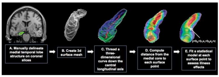
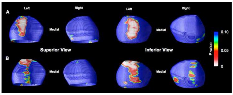
Structural neuroimaging studies of the amygdala and hippocampus in bipolar disorder have been largely inconsistent. This may be due in part to differences in the proportion of subjects taking lithium or experiencing an acute mood state, as both factors have recently been shown to influence gray matter structure. To avoid these problems, we evaluated euthymic subjects not currently taking lithium. Thirty-two subjects with bipolar type I disorder and 32 healthy subjects were scanned using magnetic resonance imaging. Subcortical regions were manually traced, and converted to three-dimensional meshes to evaluate the main effect of bipolar illness on radial distance. Statistical analyses found no evidence for a main effect of bipolar illness in either region, although exploratory analyses found a significant age by diagnosis interaction in the right amygdala, as well as positive associations between radial distance of the left amygdala and both prior hospitalizations for mania and current medication status. These findings suggest that, when not treated with lithium or in an acute mood state, patients with bipolar disorder exhibit no structural abnormalities of the amygdala or hippocampus. Future studies, nevertheless, that further elucidate the impact of age, course of illness, and medication on amygdala structure in bipolar disorder are warranted.
Published by Elsevier Ireland Ltd.
Figures

Similar articles
-
Hippocampal and amygdala volumes in an older bipolar disorder sample.Int Psychogeriatr. 2013 Jan;25(1):54-60. doi: 10.1017/S1041610212001469. Epub 2012 Aug 29. Int Psychogeriatr. 2013. PMID: 22929183
-
Increased hippocampal, thalamus and amygdala volume in long-term lithium-treated bipolar I disorder patients compared with unmedicated patients and healthy subjects.Bipolar Disord. 2017 Feb;19(1):41-49. doi: 10.1111/bdi.12467. Epub 2017 Feb 27. Bipolar Disord. 2017. PMID: 28239952
-
Lithium treatment and hippocampal subfields and amygdala volumes in bipolar disorder.Bipolar Disord. 2015 Aug;17(5):496-506. doi: 10.1111/bdi.12295. Epub 2015 Mar 24. Bipolar Disord. 2015. PMID: 25809287
-
The Amygdala in Schizophrenia and Bipolar Disorder: A Synthesis of Structural MRI, Diffusion Tensor Imaging, and Resting-State Functional Connectivity Findings.Harv Rev Psychiatry. 2019 May/Jun;27(3):150-164. doi: 10.1097/HRP.0000000000000207. Harv Rev Psychiatry. 2019. PMID: 31082993 Review.
-
Limbic changes identified by imaging in bipolar patients.Curr Psychiatry Rep. 2008 Dec;10(6):505-9. doi: 10.1007/s11920-008-0080-8. Curr Psychiatry Rep. 2008. PMID: 18980734 Review.
Cited by
-
Brain Structural Effects of Psychopharmacological Treatment in Bipolar Disorder.Curr Neuropharmacol. 2015;13(4):445-57. doi: 10.2174/1570159x13666150403231654. Curr Neuropharmacol. 2015. PMID: 26412064 Free PMC article. Review.
-
NCAN Cross-Disorder Risk Variant Is Associated With Limbic Gray Matter Deficits in Healthy Subjects and Major Depression.Neuropsychopharmacology. 2015 Oct;40(11):2510-6. doi: 10.1038/npp.2015.86. Epub 2015 Mar 24. Neuropsychopharmacology. 2015. PMID: 25801500 Free PMC article.
-
Effects of ANK3 variation on gray and white matter in bipolar disorder.Mol Psychiatry. 2017 Sep;22(9):1345-1351. doi: 10.1038/mp.2016.76. Epub 2016 May 31. Mol Psychiatry. 2017. PMID: 27240527 Free PMC article.
-
Effects of lithium on cortical thickness and hippocampal subfield volumes in psychotic bipolar disorder.J Psychiatr Res. 2015 Feb;61:180-7. doi: 10.1016/j.jpsychires.2014.12.008. Epub 2014 Dec 23. J Psychiatr Res. 2015. PMID: 25563516 Free PMC article.
-
Sex differences in amygdala shape: Insights from Turner syndrome.Hum Brain Mapp. 2016 Apr;37(4):1593-601. doi: 10.1002/hbm.23122. Epub 2016 Jan 28. Hum Brain Mapp. 2016. PMID: 26819071 Free PMC article.
References
-
- Allen JS, Bruss J, Brown CK, Damasio H. Normal neuroanatomical variation due to age: the major lobes and a parcellation of the temporal region. Neurobiology of Aging. 2005;26:1245–1260. - PubMed
-
- Altshuler LL, Bartzokis G, Grieder T, Curran J, Jimenez T, Leight K, Wilkins J, Gerner R, Mintz J. An MRI study of temporal lobe structures in men with bipolar disorder or schizophrenia. Biological Psychiatry. 2000;48:147–162. - PubMed
-
- Altshuler LL, Bartzokis G, Grieder T, Curran J, Mintz J. Amygdala enlargement in bipolar disorder and hippocampal reduction in schizophrenia: an MRI study demonstrating neuroanatomic specificity. Archives of General Psychiatry. 1998;55:663–664. - PubMed
-
- Altshuler LL, Bookheimer SY, Proenza MA, Townsend J, Sabb F, Firestine A, Bartzokis G, Mintz J, Mazziotta J, Cohen MS. Increased amygdala activation during mania: a functional magnetic resonance imaging study. American Journal of Psychiatry. 2005;162:1211–1213. - PubMed
-
- Bartzokis G, Mintz J, Marx P, Osborn D, Gutkind D, Chiang F, Phelan CK, Marder SR. Reliability of in vivo volume measures of hippocampus and other brain structures using MRI. Magnetic Resonance Imaging. 1993;11:993–1006. - PubMed
Publication types
MeSH terms
Grants and funding
- R21 EB001561/EB/NIBIB NIH HHS/United States
- RR00865/RR/NCRR NIH HHS/United States
- F31 MH078556/MH/NIMH NIH HHS/United States
- MH01848/MH/NIMH NIH HHS/United States
- MH078556/MH/NIMH NIH HHS/United States
- R21 RR019771/RR/NCRR NIH HHS/United States
- EB01651/EB/NIBIB NIH HHS/United States
- MH075944/MH/NIMH NIH HHS/United States
- P41 RR013642/RR/NCRR NIH HHS/United States
- R21 MH075944/MH/NIMH NIH HHS/United States
- K24 MH001848/MH/NIMH NIH HHS/United States
- RR13642/RR/NCRR NIH HHS/United States
- AG016570/AG/NIA NIH HHS/United States
- C06 RR012169/RR/NCRR NIH HHS/United States
- M01 RR000865/RR/NCRR NIH HHS/United States
- RR019771/RR/NCRR NIH HHS/United States
- P50 AG016570/AG/NIA NIH HHS/United States
LinkOut - more resources
Full Text Sources
Other Literature Sources
Medical
