. 2013 Jan 15;373(2):322-37.
doi: 10.1016/j.ydbio.2012.11.003. Epub 2012 Nov 10.

Neuroblast lineage-specific origin of the neurons of the Drosophila larval olfactory system

Affiliations

Neuroblast lineage-specific origin of the neurons of the Drosophila larval olfactory system

Abhijit Das et al. Dev Biol. .

Abstract

The complete neuronal repertoire of the central brain of Drosophila originates from only approximately 100 pairs of neural stem cells, or neuroblasts. Each neuroblast produces a highly stereotyped lineage of neurons which innervate specific compartments of the brain. Neuroblasts undergo two rounds of mitotic activity: embryonic divisions produce lineages of primary neurons that build the larval nervous system; after a brief quiescence, the neuroblasts go through a second round of divisions in larval stage to produce secondary neurons which are integrated into the adult nervous system. Here we investigate the lineages that are associated with the larval antennal lobe, one of the most widely studied neuronal systems in fly. We find that the same five neuroblasts responsible for the adult antennal lobe also produce the antennal lobe of the larval brain. However, there are notable differences in the composition of larval (primary) lineages and their adult (secondary) counterparts. Significantly, in the adult, two lineages (lNB/BAlc and adNB/BAmv3) produce uniglomerular projection neurons connecting the antennal lobe with the mushroom body and lateral horn; another lineage, vNB/BAla1, generates multiglomerular neurons reaching the lateral horn directly. lNB/BAlc, as well as a fourth lineage, vlNB/BAla2, generate a diversity of local interneurons. We describe a fifth, previously unknown lineage, BAlp4, which connects the posterior part of the antennal lobe and the neighboring tritocerebrum (gustatory center) with a higher brain center located adjacent to the mushroom body. In the larva, only one of these lineages, adNB/BAmv3, generates all uniglomerular projection neurons. Also as in the adult, lNB/BAlc and vlNB/BAla2 produce local interneurons which, in terms of diversity in architecture and transmitter expression, resemble their adult counterparts. In addition, lineages lNB/BAlc and vNB/BAla1, as well as the newly described BAlp4, form numerous types of projection neurons which along the same major axon pathways (antennal tracts) used by the antennal projection neurons, but which form connections that include regions outside the "classical" olfactory circuit triad antennal lobe-mushroom body-lateral horn. Our work will benefit functional studies of the larval olfactory circuit, and shed light on the relationship between larval and adult neurons.


Figures

Figure 1
Figure 1. Lineage specific labeling of adult olfactory interneurons by GAL4 lines
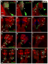
Panels A–D show z-projection of frontal confocal sections showing cell bodies and proximal (antennal lobe) branches of labeled neurons; A′-D′ depict posterior levels, showing distal (posterior) branches of labeled neurons. Diagrams A″-E″ show schematic representation of lineages and their projections. (A and A′) GAL4-GH146 labels 3 projection neuron clusters around the adult antennal lobe: antero-dorsal projection neurons belonging to the adNB/BAmv3 lineage, lateral projection neurons belonging to the lNB/BAlc lineage, and ventral projection neurons corresponding to the vNB/BAla1 lineage. They send out axonal tracts to the higher olfactory centers, the calyx of the mushroom body (MB) and the lateral horn of the protocerebrum through two antenno-cerebral tracts (ACTs): lNB/BAlc neurons send their axonal projections through the inner antenno-cerebral tract (iACT) and vNB/BAla1 neurons through the medial antenno-cerebral tract (mACT). (B and B′) Acj6-GAL4 labels two sets of projection neurons: adNB/BAmv3 neurons, and lateral atypical projection neurons of the lNB/BAlc lineage (B) sending axonal projections through the iACT and oACT (outer antenno-cerebral tract white arrow in B′) respectively. (C and C′) GAL4-MZ699 labels the ventral projection neurons of the vNB/BAla1 lineage, sending axons to the lateral horn through the mACT. (D and D′) GAL4-OK371 labels local interneurons belonging to the ventrolateral local interneuron (vlLN/BAla2) lineage; several of these local interneurons send axons to the contralateral antennal lobe through the antennal lobe commissure (white arrow in D′). Inset in D shows MARCM clone in BAla2 visualized by OK371. Neuron on contralateral side (asterisk) is not part of the clone. (E and E′) GAL4-VGN9281 labels several projection neurons of the BAlp4 lineage, located ventro-lateral to the antennal lobe. They send their dendrites mostly to the ventro-medial glomeruli of the antennal lobe and also to the SOG (white arrow in E). The axon tract of these projection neurons follow the iACT (white arrow in E′) at least initially, before terminating in the protocerebral neuropil. Note: Cell bodies of neurons belonging to specific lineages are circled by lines rendered in colors representing distinct lineages: BAmv3: cyan, BAlc: magenta, BAla1: blue, BAla2: red and BAlp4: yellow. Scale bar represents 10 μm.
Figure 2
Figure 2. Antennal lobe-associated lineages in the late larval brain
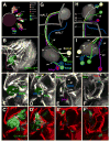
(A) Schematic diagram showing lineage-specific expression of the 8 GAL4 lines used in this study. Dark magenta colored circle (at the middle) represents the larval antennal lobe neuropil and cell body clusters (smaller circles around the lobe) denote primary projection neurons and local interneurons belonging to different lineages. All GAL4 lines are listed on the right hand side with specific color codes to indicate the lineages where they are expressed. Some GAL4 lines are restricted to very few lineages, for example OK371 is seen to express only in the BAla2 lineage, GH146 is restricted to BAmv3 and BAlc, LN2 expression is seen in BAlc and BAla2 lineages; whereas some lines show much broader expression, for example, 0.3Cha line is expressed in primary local interneurons and projection neurons belonging to all 5 lineages. (BF) Characteristic axonal projection of the five antennal lobe-associated lineages. B: Overview. Z-projection of anterior part of confocal stack of a third instar larval brain hemisphere, expressing membrane tagged GFP in antennal lobe projection neurons driven by GAL4-GH146 (green) in the background of anti-neurotactin immunolabeling to show the developing secondary neurons and their axonal tracts. Midline (ml) to the left. Rectangular frame indicates the area of the brain, centered on the antennal lobe, which is shown in panels C–F. These panels show enlargements of z-projections of the same hemisphere at different antero-posterior levels. The levels are indicated by hatched lines in the digital model presented in panel H. Outline of antennal lobe is shaded (green). The five antennal lobe-associated lineages are indicated in colors that are used throughout the following panels of this figure. C: anterior surface of antennal lobe, BAmv3 tract enters the antennal lobe at its anterior-dorsal surface (ad); BAlc tract enters laterally (al); BAla1 and BAla2 run close to each other and enter ventro-laterally (av). D, E: Level 5–10 micron into the antennal lobe. The tracts of BAmv3 and BAlc have approached each other and turn dorsally to form the anterior root of the antennal tract (ar in E). The tracts of BAla1 and BAla2 also turn upward (D); BAla2 (local interneurons) does not leave the antennal lobe, whereas BAla1 forms the posterior root of the ACT (pr in E). The fifth lineage, BAlp4, enters the antennal lobe near its posterior-ventral surface (pv in E). It projects straight dorsally and joins BAla1 in the posterior root of the ACT. F: level shortly posterior to the antennal lobe. Tracts of BAmv3, BAlc and BAlp4 have come close and form the inner ACT (iACT); axons of BAla1 veer off laterally and form the mACT. (GI) Digital 3-dimentional models of the five antennal lobe-associated lineages and projection pattern of their SATs with respect to the antennal lobe (gray). Round spheres indicate location of cell body clusters of the lineages; pipes show projection of axon tracts of the corresponding lineages. The same color code is used as in panels C–F. Aside from antennal lobe, the lobes, peduncle, and calyx of the mushroom body are shown for reference. G: anterior view; medial to the left (ml: midline), dorsal up; H: dorsal view; medial to the left, anterior up; I: lateral view; anterior to the left, dorsal up. Hatched lines in H indicate the anterior-posterior levels represented by photographs in panels C–F.
Figure 3
Figure 3. Development of the fifth antennal lobe-associated lineage (BAlp4) through larval and pupal stages
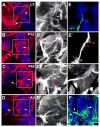
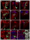
(A–D) Identification of the BAlp4 lineage and its axon tracts through development (L3, 18 hrs APF, 48 hrs APF, and adult brain). Anti-Neurotactin (Anti-Nrt, BP106) is used to label all secondary lineages and their SATs in the larval brain (red in A–D and gray in A′-D′) and neuropils are labeled by anti-DN-Cadherin (blue). The boxed area in A–D shows the antennal lobe region of the brain in each panel, shown at higher magnification in A′-D′. The BAlp4 lineage is identifiable as a cell cluster located ventral and lateral of the antennal lobe, sending its axon tract straight dorsally along the posterior surface of the antennal lobe. This tract can be followed throughout development into the adult. The BAlp4 tract forms a characteristic ventrally directed branch (red arrowhead) towards the SOG, visibly most clearly at pupal stages (C′, C″). (E and E′) Neuroblast clone of the BAlp4 lineage in L3 showing cell body location and axon tract. Note tuft of filopodia (red arrowhead) which foreshadow the ventral branch depicted in C-C″. (F) The GAL4-VGN9281 driver line labels a subset of neurons of the BAlp4 lineage (yellow arrowheads) in the adult brain, sending primary neurites (white arrowheads) into the ventro-medial glomeruli of the antennal lobe (red arrowhead). Ventral branch (red arrow) exits the antennal lobe towards the SOG.
Figure 4
Figure 4. Spatial relationship between primary and secondary neurons of the same lineage
(A and B) Neuroblast clones of the BAmv3 lineage induced at early (A) and late (B) embryo and recovered in L3. In A, both primary projection neurons (yellow arrowhead) and secondary projection neurons (blue arrowhead) are present within the clone (induced at early embryo); whereas in B, the neuroblast clone contains only secondary projection neurons. The larval antennal lobe is innervated only by the primaries (A) and not by the secondaries. The primary axon tract (PAT), containing axons of differentiated primary BAmv3 neurons, projects via the iACT and forms branches in the calyx (CX) and lateral horn (LH) neuropils (outlined by yellow dotted lines in A, A′); the secondary axon tract (SAT) follows the same route through the iACT, but does not form terminal arbors in these neuropils (yellow dotted line in B). (C and D) Single cell clones of primary and secondary projection neurons in 3rd instar larval brain. The primary projection neuron sends terminal neurite branches to a single antennal lobe glomerulus (red arrow) and the axon terminates in the MB calyx and lateral horn neuropils (C, C′). The single secondary projection neuron has no branches in the antennal lobe; instead a few immature branches are observed in the prospective adult antennal lobe region (demarcated by cyan circular line, dorsal to the larval antennal lobe demarcated by yellow dotted line, in D, D′). No terminal branches of the SAT reach within the calyx or lateral horn (yellow dotted lines in D, D′). (E) BAmv3 neuroblast clone induced in early embryo, labeled by GAL4-GH146, showing close association between primary and secondary projection neuron cell bodies and also between PAT and SAT (yellow arrowhead) running parallel to each other. Only the primary neurons form mature axon terminals in the neuropils of the MB calyx and the lateral horn (yellow dotted lines). (F–H) BAmv3 projection neurons labeled by GAL4-GH146 (green) in the background of neurotactin immunolabeling (red) at late larval stage. GAL4-GH146 expresses strongly in primary neurons (yellow arrowhead) and less strongly in secondary neurons (cyan arrowhead). By contrast, only secondary neurons are Neurotactin (Nrt) immunoreactive. The PAT and SAT run parallel to each other (H-H″) en route to the higher center neuropils; the SAT is Nrt+ve (cyan arrowheads) and the PAT is Nrt-ve (yellow arrowheads). Abbreviations: AL= antennal lobe; CX=calyx; LH= lateral horn; PAT=primary axon tract; SAT=secondary axon tract. Scale bar represents 10 μm.
Figure 5
Figure 5. BAmv3 produces projection neurons of the larval antennal lobe
(A–C) Larval projection neurons of the BAmv3 lineage labeled by GAL4-GH146. The neuropile is labeled by anti-Brp (red). A, B: First instar larva; z-projection of anterior brain hemisphere, including antennal lobe (AL in A), and of posterior hemisphere with calyx (CX) and lateral horn (LH). Medial to the left, dorsal up. C: anterior part of third instar larval brain. Note the increase in cell number in BAmv3 lineage in third instar larval stage due to addition of secondary projection neurons which form a dense cluster of small cell bodies (arrowhead in C). (D–F) Third instar larva, z-projection of one brain hemisphere, medial to the left, dorsal up. Three types of single cell MARCM clones of the BAmv3 projection neurons: uniglomerular (D), biglomerular (E), and large-field or poly-glomerular projection neuron (F) (blue arrows in the antennal lobe). All single cell clones send their axon through the iACT (yellow arrow) and only one or two glomeruli in the MB calyx are innervated (cyan arrows in calyx) by a single projection neuron irrespective of their innervation pattern in the antennal lobe glomeruli. Abbreviations: AL= antennal lobe; CX= calyx of mushroom body; CPLd= centro-posterior-lateral compartment, dorsal domain Scale bar represents 10 μm (for all panels).
Figure 6
Figure 6. Primary neurons of the BAla1 lineage
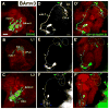
All panels show z-projections of third instar larval brain hemispheres; medial to the left, dorsal up. Neuropile is labeled by anti-Brp (red). (A–A′) GAL4–MZ699 labeled single cell clone of a projection neuron innervating 2–3 glomeruli of the posterior antennal lobe; the axonal projection ascends through the mACT (white arrow) to terminate in the CPLd (larval forerunner of the lateral horn; yellow dotted line) at its boundary with the MB calyx. (B–D) Same clone as in A; B shows z-projection of anterior part of brain hemisphere, including cell body and proximal arbors in antennal lobe; C contains a segment of the axon crossing over the peduncle (p); D shows terminal arborization in the form of two glomeruli located at the boundary between CX and CPLd. (E–E′) A single projection neuron labeled by GAL4-LN2 line sends oligo-glomerular projections to the larval antennal lobe (yellow dotted line), as well as to the SOG (yellow arrow). The axon tract initially follows the iACT, but then deviates from this tract to form terminal branches (yellow arrowhead) around the proximal peduncle and the dorsal lobe of the MB (dl, blue dotted line). (F–G) Same clone as in E; F shows z-projection of anterior brain hemisphere with cell body and projections to antennal lobe and SOG; G shows terminal axonal boutons in the neuropile surrounding the peduncle and dorsal lobe (yellow arrowhead). (H–K) Period-GAL4 labels the extra-antennal lobe BAla1 projection neurons. H: First instar larval brain, lateral view, anterior to the left, dorsal up. I–K: first instar larval brain; medial to the left, dorsal up. Anterior level (I), central level (J), posterior level (K). Labeled BAla1 neurons do not form branches in the antennal lobe (AL); axons follow the mACT and from branches in the BPM compartment posterior to the antennal lobe and in the vicinity of the peduncle (yellow arrowheads in H and K). Abbreviations: AL= antennal lobe; SOG= suboesophageal ganglion; CX= calyx of mushroom body; dl= dorsal lobe of mushroom body; ml= medial lobe of mushroom body; p= peduncle of mushroom body; CPL= centro-posterior-lateral compartment ; CPLd= centro-posterior-lateral compartment, dorsal domain; BPM= baso-posterior medial compartment; LAL= lateral accessory lobe. Scale bars represent 10 μm (for all panels).
Figure 7
Figure 7. BAla2 lineage produces primary local interneurons of diverse architectures
All panels show z-projections of third instar larval brain hemispheres; medial to the left, dorsal up. Neuropile is labeled by anti-Brp (red). The antennal lobe is outlined by yellow dotted line; SOG by blue dotted line. Small white arrowheads point at entry point of BAla2 neuronal fibers into antennal lobe. (A–C) local interneurons labeled by GAL4-OK371. Neuron with multiglomerular projections restricted mainly to the central glomeruli (A); multiglomerular projections restricted to peripheral glomeruli (B); oligoglomerular local interneurons with projections to a few glomeruli located centrally in the antennal lobe (C). In the first two cases, neurites extend to the SOG as well (white arrows). (D) local interneuron labeled by 0.3Cha-GAL4, with arbors thinly branching throughout the antennal lobe, as well as the SOG (white arrow). Antennal lobe neurites are denser in the medial side of the lobe (magenta arrowhead). (E) GAL4-LN2 labeled single multiglomerular local interneuron with arbors occupying the ventro-lateral side of the larval antennal lobe (cyan arrowhead) which also sends some projections to the SOG (white arrow). (F) Another type of single local interneuron clone labeled by GAL4-LN2 with oligo-glomerular antennal lobe projections mostly restricted to a few ventro-medial glomeruli (blue arrowhead) and prominent branches to the SOG (white arrow). Abbreviations: AL= antennal lobe; LAL= lateral accessory lobe; SOG= suboesophageal ganglion. Scale bar represents 10 μm (for all panels).
Figure 8
Figure 8. Diverse primary interneuron types formed by the BAlc lineage
All panels show z-projections of third instar larval brain hemispheres; medial to the left, dorsal up. Neuropile is labeled by anti-Brp (red). The antennal lobe is outlined by yellow dotted line; SOG by blue dotted line. White arrowheads mark entry points of BAlc fibres. Panels of upper row (A–C) show three different types of local interneurons belonging to the BAlc lineage. (A) A pan-antennal lobe local interneuron labeled by GAL4-LN2, with all neurites restricted to the antennal lobe. (B) Another pan-antennal lobe local interneuron labeled by the same driver with few projections to the SOG (white arrow). (C) An oligo-glomerular local interneuron labeled by 0.3Cha-GAL4 with strong projections to the SOG (white arrow). Panels of lower three rows (D–F, G–I, J–L) show three types of atypical projection neurons. Left panel of each row represents z-projection of entire brain hemisphere; panels in center show partial z-projection of anterior brain, including antennal lobe and SOG; panels at the right represent posterior brain. (D–F) Atypical projection neuron labeled by Acj6-GAL4 sending a small number of branches to the larval antennal lobe; significant amount of neurites are detected in the ipsilateral and contralateral SOG (white arrows). The long axon projects via oACT to the CPLd (forerunner of lateral horn). (G–I) Another type of projection neuron has sparse branches in the posterior antennal lobe; it projects its axon along the iACT, and forms terminal branches medial and dorsal of the peduncle in a domain called the central posterior intermediate compartment (CPI; forerunner of inferior protocerebrum of adult brain). (J–L) A third type of BAlc projection neuron with bilateral antennal lobe innervation. It innervates the ipsilateral antennal lobe with sparse neurites, the axon tract ascends through the iACT to form widespread branches in the higher centers, crosses the midline to send out branches to the contralateral higher centers, and finally descends through the iACT towards the contralateral antennal lobe, where it sends out pan-glomerular branches, some even terminating in the SOG neuropil. This neuron corresponds to the contralaterally projecting serotonin-immunoreactive deutocerebral (CSD) interneuron. Abbreviations: AL= antennal lobe; BPL= baso-posterior lateral compartment; BPM= baso-posterior medial compartment; CPI= centro-posterior intermediate compartment; CPLd= centro-posterior-lateral compartment, dorsal domain; CX= calyx of mushroom body; dl= dorsal lobe of mushroom body; LAL= lateral accessory lobe; oACT= outer antenno-cerebral tract; iACT= inner antenno-cerebral tract; ml= medial lobe of mushroom body; p= peduncle of mushroom body; SOG= suboesophageal ganglion. Scale bars represent 10 μm, correspond to specific rows.
Figure 9
Figure 9. Projection neurons of the BAlp4 lineage
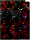
All panels except J–O show z-projections of third instar larval brain hemispheres; medial to the left, dorsal up. J–O represent adult brain hemisphere. Neuropile is labeled by anti-Brp (red). The antennal lobe (AL) is outlined by yellow dotted line; SOG by cyan dotted line. All four rows of panels (A–C, D–F, G–I, J–L) are organized such that left panel of each row represents z-projection of entire brain hemisphere; panels in center show partial z-projection of anterior brain, including antennal lobe and SOG; panels at the right represent posterior brain. (A–C) A full BAlp4 NB clone induced at early embryonic stage and recovered at the late third instar larval stage showing cell bodies (both primaries and secondaries) located ventro-lateral of the antennal lobe, sparse proximal projections in the antennal lobe, and profuse branching in the SOG; axons project through the iACT to the CPI and CPL compartments of the protocerebrum. (D–F) A single BAlp4 projection neuron clone labeled by 0.3Cha-GAL4 line sending neurites into larval antennal lobe and SOG; the axon termini are largely observed in the CPI compartment. (G–I) Two primary projection neurons of BAlp4 lineage are labeled by GAL4-VGN9281 line. Antennal lobe and SOG received very sparse neurite projections whereas the axons follow the iACT to project to CPI and CPL compartments. (J–O) Adult brain showing BAlp4 lineage labeled partly by GAL4-VGN9281. The cell bodies are located latero-ventral to the antennal lobe, sending oligo-glomerular dendritic innervation to the posterior antennal lobe and to the SOG; axons terminate in the inferior protocerebrum of the brain, terminal varicosities are shown in M–O. Abbreviations: AL= antennal lobe; SOG= suboesophageal ganglion; CX= calyx of mushroom body; dl= dorsal lobe of mushroom body; ml= medial lobe of mushroom body; p= peduncle of mushroom body; CPI= central posterior intermediate compartment; CPL= centro-posterior-lateral compartment ; CPLd= centro-posterior-lateral compartment, dorsal domain; BPL= baso-posterior lateral compartment; LAL= lateral accessory lobe. Scale bars = 10 μm, correspond to specific rows.
Figure 10
Figure 10. A schematic depicting comparison of anatomical features of the primary and secondary interneurons born from each of the five olfactory interneuron lineages
(A) BAmv3 lineage produces olfactory projection neurons, primaries and secondaries are nearly identical. They are mostly uniglomerular in nature (with minor variations in both cases); axons follow the iACT to reach the MB calyx and lateral horn of the protocerebrum. (B) The BAlc interneurons in both larval and adult stages are similar. (i) Local interneurons (orange) of the larva often invade the neighbouring SOG neuropil, but in the adult they are restricted to the antennal lobe. (ii) Projection neurons (light green) are uniglomerular in the adult, axons follow the iACT to reach the MB calyx and lateral horn; in larva they are oligo-glomerular and follow the iACT where they terminate in the CPI compartment surrounding the peduncle of the MB. (iii) Atypical projection neurons (dark green) of both the adult and larval brain follow the oACT to the reach the CPLd/LH and CPI/IP compartments. (C) Larval and adult BAla1 neurons show considerable differences. In the adult, they are uniglomerular and multiglomerular projection neurons that project through the mACT. In the larval brain the primaries are oligoglomerular or multiglomerular. Their axons project through either the mACT or iACT. Only a fraction of larval BAla1 primaries connect AL and CPLd/LH; most neurons have atypical connections with the SOG, CPI/IP, and BPM/VMC compartments. (D) Both primaries and secondaries of the BAla2 lineage are either oligoglomerular or multiglomerular local interneurons, though the primaries often connect with the larval SOG and secondaries often cross-over to connect with the contra-lateral adult antennal lobe. (E) BAlp4 neurons are projection neurons in both larval and adult brains and have comparable features. They send oligoglomerular or sparse branches to the antennal lobe as well as prominent branches to the SOG; axons follow the iACT and form terminal connections in the protocerebrum.

Similar articles

Cited by

References

    1. Axel R. The molecular logic of smell. Sci Am. 1995;273:154–159. - PubMed
    1. Brierley DJ, Blanc E, Reddy OV, Vijayraghavan K, Williams DW. Dendritic targeting in the leg neuropil of Drosophila: the role of midline signalling molecules in generating a myotopic map. PLoS Biol. 2009;7:e1000199. - PMC - PubMed
    1. Brochtrup A, Hummel T. Olfactory map formation in the Drosophila brain: genetic specificity and neuronal variability. Curr Opin Neurobiol 2010 - PubMed
    1. Cardona A, Saalfeld S, Arganda I, Pereanu W, Schindelin J, Hartenstein V. Identifying neuronal lineages of Drosophila by sequence analysis of axon tracts. J Neurosci. 2010;30:7538–7553. - PMC - PubMed
    1. Carlsson MA, Diesner M, Schachtner J, Nassel DR. Multiple neuropeptides in the Drosophila antennal lobe suggest complex modulatory circuits. J Comp Neurol. 2010;518:3359–3380. - PubMed

Publication types

LinkOut - more resources