Clinical Trial
. 2013 Mar;33(3):365-71.
doi: 10.1038/jcbfm.2012.171. Epub 2012 Nov 14.

Assessment of leptomeningeal collaterals using dynamic CT angiography in patients with acute ischemic stroke

Affiliations
Clinical Trial

Assessment of leptomeningeal collaterals using dynamic CT angiography in patients with acute ischemic stroke

Bijoy K Menon et al. J Cereb Blood Flow Metab. 2013 Mar.

Abstract

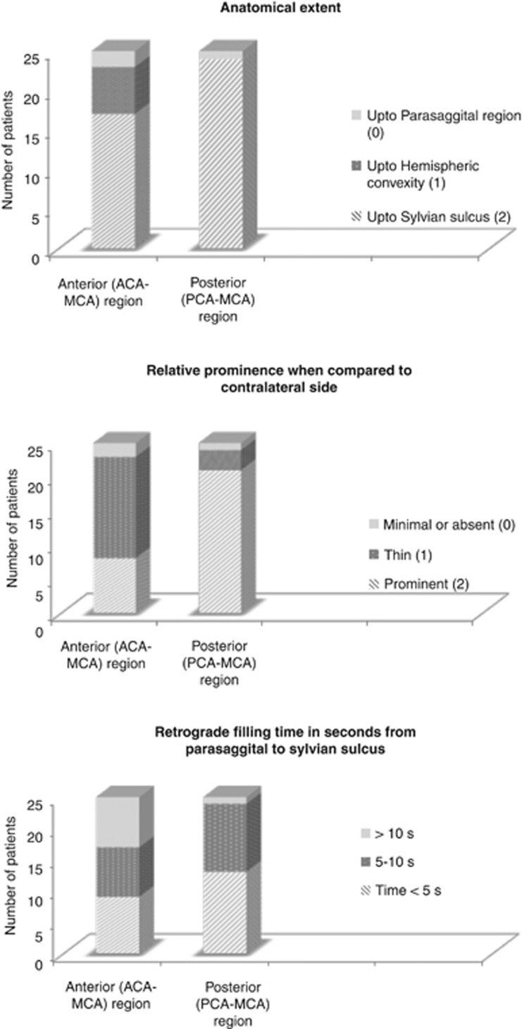
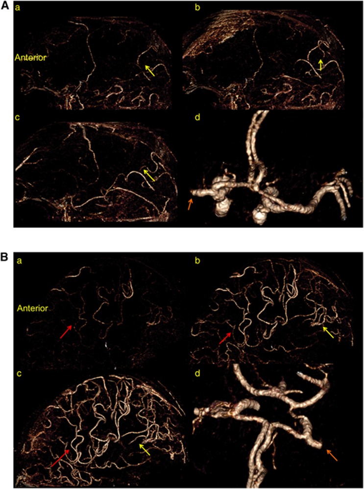
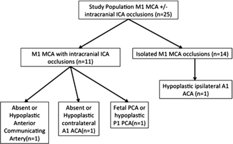
Whole-brain dynamic time-resolved computed tomography angiography (CTA) is a technique developed on the new 320-detector row CT scanner capable of generating time-resolved cerebral angiograms from skull base to vertex. Unlike a conventional cerebral angiogram, this technique visualizes pial arterial filling in all vascular territories, thereby providing additional hemodynamic information. Ours was a retrospective study of consecutive patients with ischemic stroke and M1 middle cerebral artery +/- intracranial internal carotid artery occlusions presenting to our center from June 2010 and undergoing dynamic time-resolved CTA and perfusion CT within 6 hours of symptom onset. Leptomeningeal collateral status was assessed by determining relative prominence of pial arteries in the ischemic region, rate and extent of retrograde flow, and various topographical patterns of pial arterial filling. Twenty-five patients were included in the study. We demonstrate the existence of the following novel properties of leptomeningeal collaterals in humans: (a) posterior (posterior cerebral artery (PCA)-MCA) dominant collateralization, (b) intra-territorial 'within MCA region' leptomeningeal collaterals, and (c) significant variability in size, extent, and retrograde filling time in pial arteries. We also describe a simple and reliable collateral grading template that, for the first time on dynamic CTA, incorporates back-filling time as well as size and extent of collateral filling.


Figures

Figure 1
Figure 1
Template for detailed assessment of leptomeningeal collaterals in patients with M1 middle cerebral artery +/− intracranial internal carotid artery occlusion using time-resolved dynamic computed tomography (CT) angiography on the 320-slice CT scanner.
Figure 2
Figure 2
Distribution of the three properties of leptomeningeal collateral status (prominence, extent, and retrograde filling time) assessed using dynamic computed tomography angiography in this study and stratified by anterior (anterior cerebral artery (ACA)–middle cerebral artery (MCA)) and posterior (posterior cerebral artery (PCA)–MCA) regions.
Figure 3
Figure 3
(A) Posterior dominant leptomeningeal collateral pattern. Dynamic time-resolved computed tomography (CT) angiography (saggital view) in a patient with an isolated M1 middle cerebral artery (MCA) occlusion (orange arrow) and patent circle of Willis (d). Note retrograde filling of pial arteries through posterior (posterior cerebral artery (PCA)–MCA) collaterals in sequential time-resolved images (a–c) (yellow arrows); there is no retrograde filling of pial arteries through anterior (ACA–MCA) collaterals. The sequential images (a–c) are 2 to 3 seconds apart. (B) Anterior dominant leptomeningeal collateral pattern. Dynamic time-resolved CT angiography (saggital view) in a patient with an isolated M1 MCA occlusion (orange arrow) and patent circle of Willis (d). Note retrograde filling of pial arteries through anterior (ACA–MCA, red arrows) and posterior (PCA–MCA, yellow arrows) collaterals in sequential time-resolved images (a–c); retrograde filling of pial arteries through posterior (PCA–MCA) collaterals are less prominent and slower than their anterior (ACA–MCA) counterpart. The sequential images (a–c) are 2 to 3 seconds apart.
Figure 4
Figure 4
Distribution of incomplete/hypoplastic circle of Willis in the study population stratified by site of occlusion. In 11 patients with intracranial internal carotid artery (ICA) occlusions, only 2 patients had an incomplete anterior circle of Willis as a potential explanation for dominant posterior cerebral artery (PCA)–middle cerebral artery (MCA) leptomeningeal collateral pattern. In 14 patients with isolated M1 MCA occlusions, only 1 patient had an hypoplastic ipsilateral A1 anterior cerebral artery (ACA) as a potential explanation for dominant posterior PCA–MCA leptomeningeal collateral pattern.

Similar articles

Cited by

References

    1. Menon BK, Smith EE, Modi J, Patel SK, Bhatia R, Watson TW, et al. Regional leptomeningeal score on CT angiography predicts clinical and imaging outcomes in patients with acute anterior circulation occlusions. AJNR Am J Neuroradiol. 2011;32:1640–1645. - PMC - PubMed
    1. Bang OY, Saver JL, Kim SJ, Kim GM, Chung CS, Ovbiagele B, et al. Collateral flow predicts response to endovascular therapy for acute ischemic stroke. Stroke. 2011;42:693–699. - PMC - PubMed
    1. Liebeskind DS. Collateral circulation. Stroke. 2003;34:2279–2284. - PubMed
    1. Maas MB, Lev MH, Ay H, Singhal AB, Greer DM, Smith WS, et al. Collateral vessels on CT angiography predict outcome in acute ischemic stroke. Stroke. 2009;40:3001–3005. - PMC - PubMed
    1. Christoforidis GA, Mohammad Y, Kehagias D, Avutu B, Slivka AP. Angiographic assessment of pial collaterals as a prognostic indicator following intra-arterial thrombolysis for acute ischemic stroke. AJNR Am J Neuroradiol. 2005;26:1789–1797. - PMC - PubMed

Publication types