. 2013 Feb;33(2):225-34.
doi: 10.1038/jcbfm.2012.160. Epub 2012 Nov 14.

Intravenous HOE-642 reduces brain edema and Na uptake in the rat permanent middle cerebral artery occlusion model of stroke: evidence for participation of the blood-brain barrier Na/H exchanger

Affiliations

Intravenous HOE-642 reduces brain edema and Na uptake in the rat permanent middle cerebral artery occlusion model of stroke: evidence for participation of the blood-brain barrier Na/H exchanger

Martha E O'Donnell et al. J Cereb Blood Flow Metab. 2013 Feb.

Abstract

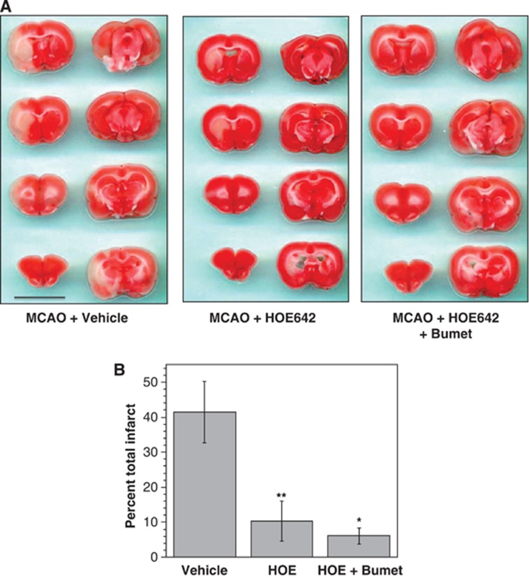
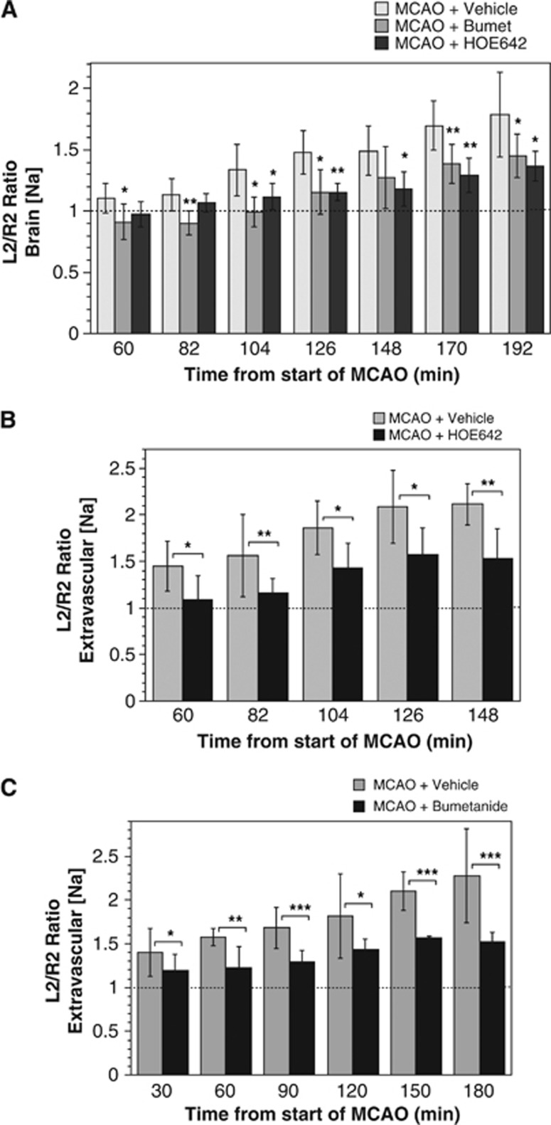
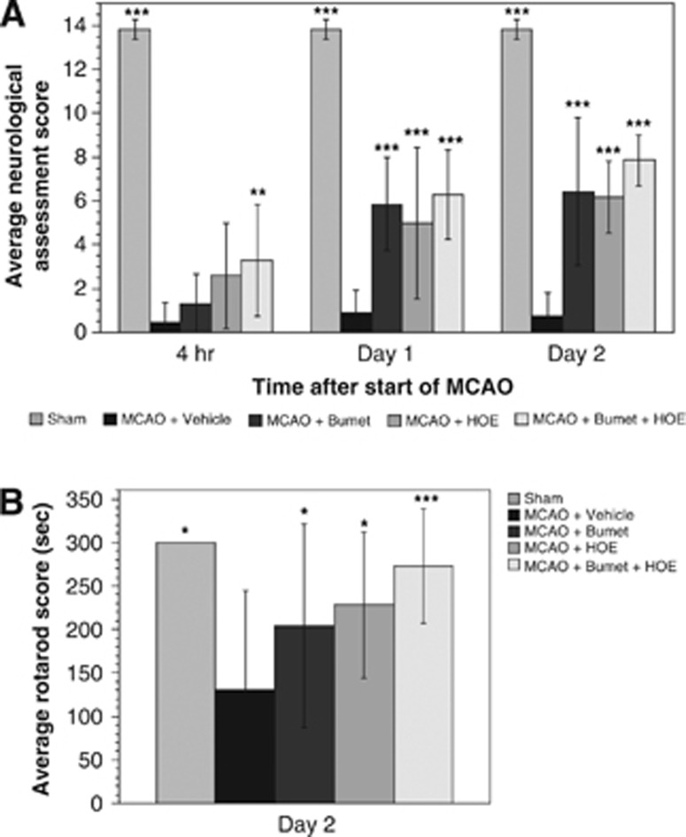
Cerebral edema forms in the early hours of ischemic stroke by processes involving increased transport of Na and Cl from blood into brain across an intact blood-brain barrier (BBB). Our previous studies provided evidence that the BBB Na-K-Cl cotransporter is stimulated by the ischemic factors hypoxia, aglycemia, and arginine vasopressin (AVP), and that inhibition of the cotransporter by intravenous bumetanide greatly reduces edema and infarct in rats subjected to permanent middle cerebral artery occlusion (pMCAO). More recently, we showed that BBB Na/H exchanger activity is also stimulated by hypoxia, aglycemia, and AVP. The present study was conducted to further investigate the possibility that a BBB Na/H exchanger also participates in edema formation during ischemic stroke. Sprague-Dawley rats were subjected to pMCAO and then brain edema and Na content assessed by magnetic resonance imaging diffusion-weighed imaging and magnetic resonance spectroscopy Na spectroscopy, respectively, for up to 210 minutes. We found that intravenous administration of the specific Na/H exchange inhibitor HOE-642 significantly decreased brain Na uptake and reduced cerebral edema, brain swelling, and infarct volume. These findings support the hypothesis that edema formation and brain Na uptake during the early hours of cerebral ischemia involve BBB Na/H exchanger activity as well as Na-K-Cl cotransporter activity.


Figures

Figure 1
Figure 1
Immunoelectron microscopy localization of NHE1 and NHE2 proteins in microvascular endothelial cell membranes of ischemic rat brain. (A, B) Rats were subjected to 90 minutes of permanent middle cerebral artery occlusion (pMCAO), then brains perfusion fixed and prepared for immunoelectron microscopy as described in Materials and Methods. The brain sections were labeled with NHE1 or NHE2 antibodies at dilutions of 1:2,000 and 1:1,000 (NHE1 and NHE2, respectively). Sections were then labeled with gold particle-conjugated secondary antibody. ImmunoEM images shown are representative micrographs. Vessel lumens are at the top of each image with the basal lamina underlying the endothelium and separating the cells from perivascular astrocytes and neurons. Locations of gold particles are indicated with arrowheads. (C, D) NHE1 and NHE2 distribution between luminal (L) and abluminal (A) microvascular endothelial cell membranes of ischemic (ipsilateral core and penumbra) and control (contralateral) frontoparietal cortex of perfusion-fixed rat brains as determined by quantitation of gold particles in immunoelectron micrographs. Values shown are mean values±s.d. of 23 to 32 microvessels for NHE1 core and penumbra 1:1,000 and 1:2,000 antibody dilutions and 22 to 37 microvessels for NHE2 core and penumbra 1:500 and 1:1,000 antibody dilutions. *Significantly different from contralateral penumbra by analysis of variance (ANOVA) with Bonferroni Dunn post hoc test (P<0.0001) p value for NHE2 ANOVA comparing all eight groups is 0.947. It should be noted that the fixation methods used here, required to retain good antigenicity of the tissue, sacrifice some structural detail in the images. Also, although NHE transporter proteins are abundant in the tissue, the section thickness and size of the field of view result in few gold particles visible in each image. Quantitation of microvessels from >20 micrographs for each antibody and dilution revealed that NHE1 and NHE2 are distributed 66% to 75% and 75% to 80%, respectively, in the luminal membrane. Qualitatively, the immunoEM images reveal that NHE1 and NHE2 proteins are found predominantly in the luminal blood–brain barrier (BBB) membrane.
Figure 2
Figure 2
Brain edema formation in permanent middle cerebral artery occlusion (MCAO): inhibition by intravenous HOE-642. (A) Magnetic resonance diffusion-weighted brain images of rats subjected to left pMCAO illustrating relative hyperintensity (edema) on left side. Regions of interest for ipsilateral and contralateral cortex (regions of interest (ROIs) L1–L3 and R1–R3, respectively) and striatum (ROI L4 and R4, respectively) are shown. Left to right panels show images of rats treated with vehicle, HOE-642 and HOE-642+Bumetanide, respectively. (B, C) Rats were administered vehicle, HOE-642 (15 mg/kg) or HOE-642 plus bumetanide (15 mg/kg each) as described in Materials and Methods and then apparent diffusion coefficient (ADC) values were determined for ROIs 1 to 4 for up to 210 minutes following induction of pMCAO. L/R ADC ratios are shown for ROIs 1 and 2 in (B, C), respectively. Values shown are mean values±s.d. for 4, 6, and 3 rats subjected to pMCAO with vehicle, HOE-642, and HOE-642+Bumet, respectively. ADC ratios for HOE-642 and HOE-642+Bumet are significantly different than Vehicle by analysis of variance (ANOVA) with Newman–Keuls post hoc test; *P<0.05, **P<0.01, ***P<0.001.
Figure 3
Figure 3
2,3,5-Triphenyltetrazolium chloride (TTC) assessment of infarct size: effects of HOE-642 and bumetanide. (A) At the conclusion of diffusion-weighted imaging experiments, TTC staining of brain slices was done as described in Materials and Methods. Images are from three representative experiments and show brain slices from rats given intravenous injection of vehicle, HOE-642 (15 mg/kg) or HOE-642 plus bumetanide (15 mg/kg each) then subjected to 210 minutes of permanent middle cerebral artery occlusion (pMCAO). In all three panels, the brain slices start at 2 mm from the frontal pole and continue in 2 mm increments to 16 mm from the frontal pole. (B) The TTC-defined total hemispheric infarct volume was determined by summing infarct volumes of all 2 mm brain slices for each rat (2- to 16-mm slices from the frontal pole). Data represent infarct volume as percent of the total ipsilateral hemisphere volume, corrected for brain swelling, and are mean values±s.d. of 4, 6, and 3 rats subjected to MCAO+Vehicle, MCAO+HOE-642, and MCAO+HOE-642+Bumet, respectively. Values for HOE-642 and HOE-642+Bumet are significantly different from Vehicle by analysis of variance (ANOVA) with Newman–Keuls post hoc test; *P<0.05, **P<0.01.
Figure 4
Figure 4
HOE-642 and bumetanide reduction of brain Na uptake in middle cerebral artery occlusion (MCAO). (A) Rats were administered intravenous HOE-642 (30 mg/kg), bumetanide (30 mg/kg), or vehicle immediately before initiation of pMCAO. Localized 23Na chemical shift imaging (CSI) magnetic resonance spectroscopy (MRS) was used to determine brain [Na] of specified regions of interest (ROI) as described in Materials and Methods. ROIs are as depicted in Figure 2A. Values are mean values±s.d. for 6, 9, and 6 rats for vehicle, bumetanide, and HOE-642, respectively. Sham-operated rat Na L/R ratios, depicted in the figure as the dashed line, averaged 1.0±0.02 (mean±s.d., n=6) over the 192 minute time course following induction of pMCAO. Brain [Na] values for bumetanide and HOE-642-treated rats are significantly different than vehicle-treated rats by analysis of variance (ANOVA) with Newman–Keuls post hoc test at 104, 126, 170, and 192 minutes for bumetanide (P<0.01, P<0.05, P<0.05, and P<0.01, respectively) and at 126, 148, 170, and 192 minutes for HOE-642 (P<0.05, P<0.05, P<0.01, and P<0.001, respectively). Asterisks indicate values that are significantly different from MCAO+Vehicle by two-tailed t-test; *P<0.05, **P<0.01). (B, C) Rats were administered intravenous HOE-642 (30 mg/kg) or bumetanide (30 mg/kg) immediately before pMCAO. Localized 23Na CSI MRS of DyTTHA-infused rats was used to determine extravascular brain [Na] of specified ROIs as described in Materials and Methods. Values are mean values±s.d. of eight rats each for vehicle and HOE-642 (B) five and four rats for vehicle and bumetanide, respectively (C). Extravascular brain [Na] values for HOE are significantly different than vehicle by ANOVA with Newman–Keuls post hoc test at 126 and 148 minutes (P<0.05 for both). Extravascular brain [Na] values for bumetanide are significantly different than vehicle by ANOVA with Newman–Keuls post hoc test at 120, 150, and 180 minutes (P<0.05, P<0.001, and P<0.001, respectively). For (B, C), asterisks indicate values that are different from MCAO+Vehicle by two-tailed t-test; *P<0.05, **P<0.01, ***P<0.001.
Figure 5
Figure 5
Neurologic assessment of rats subjected to middle cerebral artery occlusion (MCAO) and treated with HOE-642 and/or bumetanide. (A) Rats were administered intraperitoneal bumetanide (30 mg/kg), HOE-642 (30 mg/kg), Bumetanide+HOE-642 (30 m/kg each) or Vehicle then subjected to pMCAO. The rats were allowed to awaken from anesthesia and then tested for sensorimotor function at 4 hours, 1 day, and 2 days after induction of MCAO by the 14-score test, as described in Materials and Methods. Values are mean±s.d. with n values of 5, 11, 7, 5, and 7 for Sham, MCAO+Vehicle, MCAO+Bumet, MCAO+HOE, and MCAO+Bumet+HOE, respectively. Values are significantly different than MCAO vehicle scores by analysis of variance (ANOVA) with Newman–Keuls post hoc test; **P<0.01, ***P<0.001. MCAO+HOE score at 4 hours is significantly different from MCAO+vehicle score by two-tailed t-test. (B) Rotarod test. Values are mean±s.d. with n values of 5, 24, 20, 10, and 18 for Sham, MCAO+Vehicle, MCAO+Bumet, MCAO+HOE, and MCAO+Bumet+HOE, respectively. Values are significantly different than MCAO vehicle scores by ANOVA with Newman–Keuls post hoc test; *P<0.05, **P<0.01, ***P<0.001. In some experiments, rats were given bumetanide and/or HOE-642 by intravenous rather than intraperitoneal injection. No differences in outcome between the two routes of administration were observed (data not shown).

Similar articles

Cited by

References

    1. Betz AL. Alterations in cerebral endothelial cell function in ischemia. Adv Neurol. 1996;71:301–313. - PubMed
    1. Foroutan S, Brillault J, Forbush B, O'Donnell ME. Moderate-to-severe ischemic conditions increase activity and phosphorylation of the cerebral microvascular endothelial cell Na+-K+-Cl- cotransporter. Am J Physiol Cell Physiol. 2005;289:C1492–C1501. - PubMed
    1. O'Donnell ME, Tran L, Lam TI, Liu XB, Anderson SE. Bumetanide inhibition of the blood-brain barrier Na-K-Cl cotransporter reduces edema formation in the rat middle cerebral artery occlusion model of stroke. J Cereb Blood Flow Metab. 2004;24:1046–1056. - PubMed
    1. Brillault J, Lam TI, Rutkowsky JM, Foroutan S, O'Donnell ME. Hypoxia effects on cell volume and ion uptake of cerebral microvascular endothelial cells. Am J Physiol Cell Physiol. 2008;294:C88–C96. - PubMed
    1. O'Donnell ME, Lam TI, Tran LQ, Foroutan S, Anderson SE. Estradiol reduces activity of the blood-brain barrier Na-K-Cl cotransporter and decreases edema formation in permanent middle cerebral artery occlusion. J Cereb Blood Flow Metab. 2006;26:1234–1249. - PubMed

Publication types

MeSH terms