. 2012:3:1185.
doi: 10.1038/ncomms2171.

A peptide derived from laminin-γ3 reversibly impairs spermatogenesis in rats

Affiliations

A peptide derived from laminin-γ3 reversibly impairs spermatogenesis in rats

Linlin Su et al. Nat Commun. 2012.

Abstract

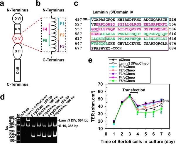
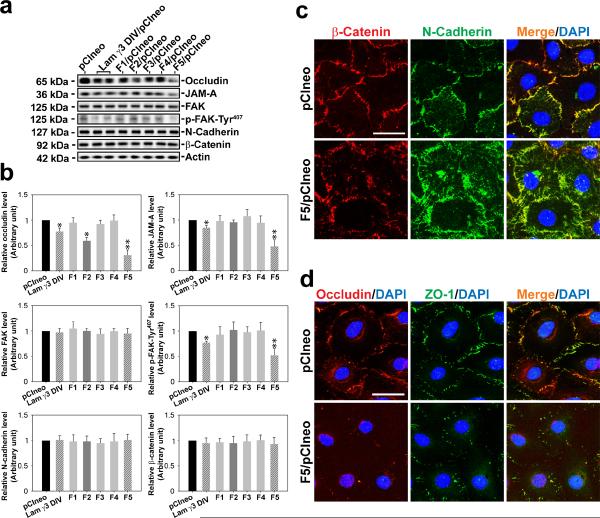
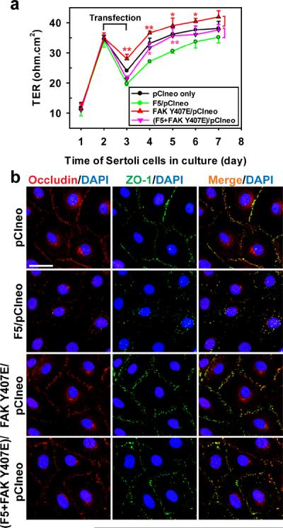
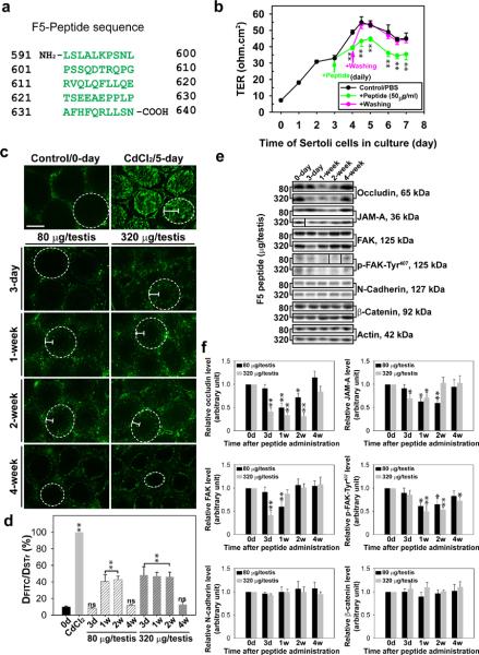
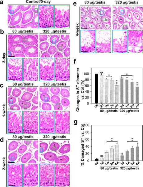
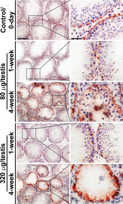
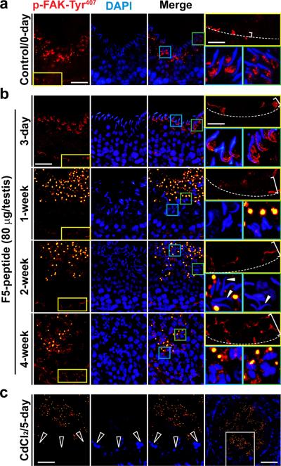
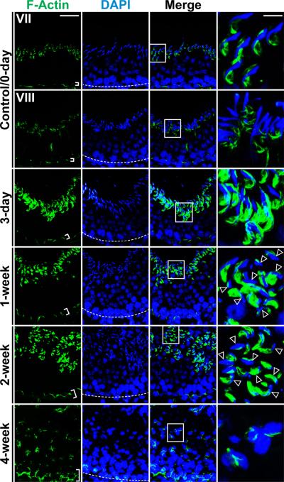
Cellular events that occur across the seminiferous epithelium in the mammalian testis during spermatogenesis are tightly coordinated by biologically active peptides released from laminin chains. Laminin-γ3 domain IV is released at the apical ectoplasmic specialization during spermiation and mediates restructuring of the blood-testis barrier, which facilitates the transit of preleptotene spermatocytes. Here we determine the biologically active domain in laminin-γ3 domain IV, which we designate F5 peptide, and show that the overexpression of this domain, or the use of a synthetic F5 peptide, in Sertoli cells with an established functional blood-testis barrier reversibly perturbs blood-testis barrier integrity in vitro and in the rat testis in vivo. This effect is mediated via changes in protein distribution at the Sertoli and Sertoli-germ-cell cell interface and by phosphorylation of focal adhesion kinase at Tyr(407). The consequences are perturbed organization of actin filaments in Sertoli cells, disruption of the blood-testis barrier and spermatid loss. The impairment of spermatogenesis suggests that this laminin peptide fragment may serve as a contraceptive in male rats.


Figures

Figure 1
Figure 1. Preparation of laminin-γ3 domain IV cDNA constructs to assess their biological activity
(a) Schematic drawing illustrating different functional domains of laminin-γ3 chain. Laminin-γ3 domain IV (DIV, in red) was selected based on earlier studies that this domain is biologically active in regulating BTB dynamics, and it was further divided into 5 fragments of F1–F5 as shown in (b) to assess their effects on BTB function. (c) cDNA-deduced amino acid sequence of laminin-γ3 DIV (GenBank accession code XM_231139) is shown, illustrating the sequences of F1 (blue boxed area), F2 (orange boxed area), F3 (purple boxed area), F4 (pink colored text) and F5 (green colored text). (d) RT-PCR illustrating the expression of laminin-γ3 DIV and the five different fragments (see a) in Sertoli cells after transfection with corresponding vectors versus pCIneo alone for 3 days. Cells transfected with pCIneo alone served as a negative control since Sertoli cells per se did not express laminin-γ3 chains, which are exclusively expressed by elongating/elongated spermatids. S-16 served as an internal PCR control. Identity of the PCR product was confirmed by direct nucleotide sequencing at GeneWiz Inc (South Plainfield, NJ). M, DNA markers; bp, base-pair. (e) Changes in the Sertoli cell TJ-permeability barrier function following transient expression of different fragments of laminin-γ3 DIV versus DIV (positive control) and pCIneo alone (negative control) on day 3 for 24-hr by quantifying TER across the Sertoli cell epithelium in bicameral units (n = 3). This experiment was repeated three times during different batches of Sertoli cells and yielded similar results. *, P<0.05; **, P<0.01.
Figure 2
Figure 2. Laminin-γ3 fragments affect protein expression and localization at the Sertoli cell BTB
(a) On day 3, Sertoli cells with a functional BTB were transfected with different expression vectors for 24 hr and terminated 48 hr thereafter for immunoblotting, illustrating changes in the levels of integral membrane proteins (e.g., occludin, JAM-A, N-cadherin), adaptor protein (e.g., β-catenin) and regulatory proteins (e.g., FAK, p-FAK-Tyr407). (b) Bar graphs summarize results shown in (a). Each bar is a mean±SD of n = 5 experiments, and data were normalized against actin. The protein levels in cells transfected with pCIneo alone were arbitrarily set at 1, against which one-way ANOVA and Dunnett's test were performed. *, P < 0.05; **, P < 0.01. (c) Overexpression of F5-peptide in Sertoli cells caused considerable re-distribution of basal ES-proteins at the BTB: β-catenin (red) and N-cadherin (green) at the Sertoli cell-cell interface, moving from cell surface into cell cytosol on day 3 after transfection. Overexpression of F5-peptide also changed localization and/or distribution of TJ proteins at the BTB: occludin (red) and ZO-1 (green) at the Sertoli cell-cell interface. Co-localized proteins appeared as “orange” in merged images. Nuclei were visualized by DAPI (blue). Scale bar = 10 μm in c and d, which applies to all images in both panels.
Figure 3
Figure 3. Laminin F5-peptide perturbs Sertoli cell TJ function via p-FAK-Tyr407
(a) To examine the mechanism by which the F5-peptide perturbs the Sertoli cell TJ-permeability barrier function as shown in Figs. 1 and 2, p-FAK-Tyr407, a molecular “switch” in the apical ES-BTB axis to regulate BTB restructuring and apical ES adhesion, was expressed either alone as a phosphomimetic mutant (FAK Y407E/pCIneo) or co-expressed with F5 [(F5+FAK Y407E)/pCIneo]. Co-expression of FAK phosphomimetic mutant FAK Y407E with F5-peptide in Sertoli cells blocked the F5-induced TJ-barrier disruption, which is likely mediated by the ability of p-FAK-Tyr407 to promote the distribution of occludin and ZO-1 at the Sertoli cell-cell interface shown in (b), thereby stabilizing the Sertoli TJ-barrier. Each data point is a mean±SD of n = 4 of a representative experiment, and this experiment was repeated 3 times which yielded similar results. *, P < 0.05; **, P < 0.01 Scale bar = 10 μm, which applies to all remaining micrographs.
Figure 4
Figure 4. Laminin F5-peptide reversibly perturbs BTB function in vitro and in vivo
(a) Amino acid residues (green) of the synthetic F5-peptide based on laminin-γ3 DIV. Numerals in “black” denote the sequence of laminin-γ3 chain (see Fig. 1a–c). (b) Effects of the synthetic F5-peptide on the Sertoli cell TJ-permeability barrier. Peptide at 50 μg/ml (~10 μM) was added on day 3 (“green” arrow), which was either included in the daily replacement medium (“green”) (n = 3) or removed by washing on day 4 (“pink” arrow) in medium without peptide (“pink”) versus PBS control. When F5-peptide was removed, the disrupted TJ-barrier was “resealed”, illustrating the disruptive effect was reversible. Each data point is a mean±SD of n = 3 of a representative experiment, and this experiment was repeated 3 times which yielded similar results. **, P < 0.01. (c) BTB integrity assay was performed to assess the ability of an intact BTB to block the movement of FITC-inulin across the BTB from the basal compartment near the basement membrane (annotated by “white” dotted line) to the adluminal compartment. Results of the BTB integrity assay are shown in normal testes (control) versus rats treated with CdCl2 and different peptide treatment groups: 80 μg per testis (or ~10 μM) and 320 μg per testis (~40 μM). Scale bar = 150 μm in c, which also applies to all remaining micrographs. (d) Histograms summarizing data based on findings shown in c by comparing the distance of FITC-inulin diffused into the epithelium (DFITC) (annotated by the “white bracket”) vs. the radius of a seminiferous tubule (DSTr) (average of the longest and shortest axes for sections of oval-shaped tubules) (n = 200 tubules from testes of 3 rats in each group). **, P < 0.01; ns, not significantly different. (e) Immunoblot analysis of different target proteins at the BTB in adult rat testes at different time points after peptide administration. (f) Histograms summarizing data shown in e with n = 3 rats for each time point. *, P<0.05; **, P<0.01.
Figure 5
Figure 5. Laminin F5-peptide impairs spermatogenesis and induces germ cell loss from the testis
Peptide or vehicle control (0.89% NaCl, see a) was injected into each testis of a group of adult rats (~300–350 gm b.w.) with n = 3 rats per time point in each treatment group versus controls Rats were terminated at specific time points at 3-day and 1-, 2- or 4-wk thereafter. (b–e) Representative photographs of paraffin sections of testes stained with hematoxylin and eosin from rats treated with the synthetic F5-peptide at either 80 or 320 μg/testis. Vehicle control is shown in a. Morphometric changes [e.g., shrinkiage of tubule, and % of damaged seminiferous tubules (ST) manifested by germ cell exfoliation – annotated by asterisks in tubules] of the tubules (n = 200 tubules from 3 rat testes) are summarized in the bar graphs shown in f and g. Bar = 150 μm in a, and the micrographs on the left panels in b–e, which applies to the micrographs on the right panels b–e. Bar = 50 μm in micrograph encircled in “blue” which applies to all micrographs encirculed in and “green” in a–e. The “blue” and “green” encircled micrographs are magnified images of the corresponding boxed areas of the lower magnification. ns, not significantly different by ANOVA; *, P < 0.05; and **, P < 0.01 when compared to normal control rats at time 0.
Figure 6
Figure 6. Laminin F5-peptide perturbs occludin disruption at the BTB
Normal rat testis (control, at time 0-day), displaying normal distribution of occludin at the BTB. versus rats received either 80 μg (~10 μM) or 320 μg (~40 μM) F5-peptide per testis by 1- and 4-week with noticeable germ cell loss in the tubules. Micrographs on the right panel are the corresponding magnified images of the boxed area shown on the left panel. Bar = 150 μm in the first micrograph on the left panel and scale bar = 50 μm in the first micrograph on the right panel, which apply to remaining micrographs in the same panel.
Figure 7
Figure 7. Laminin F5-peptide disrupts BTB and spermatid adhesion via p-FAK-Tyr407
Testes received vehicle (control) at time 0 (a) versus synthetic F5-peptide at 80 μg/testis (~10 μM), and terminated at 3-day, and 1-, 2- and 4-week (b), and positive control where rats were treated with CdCl2 (5 mg/kg b.w., i.p., for 5 days) (c). In control testes, p-FAK-Tyr407 was localized near the basement membrane consistent with its localization at the BTB [see “yellow” boxed area in the micrograph on the left, which was magnified in the right panel (top) in each row with the relative BTB location annotated by the “broken white-line”]; p-FAK-Tyr407 was also found in the adluminal compartment, restricted almost exclusively to the concave side of the spermatid head at the apical ES [see “blue” and “green” boxed areas in the micrograph on the third column, which were magnified in the right panel (bottom) in each row]. Following F5-peptide treatment, p-FAK-Tyr407 was down-regulated and mis-localized at the BTB, since it no longer restricted to the BTB, but diffused away (see “white” bracket on the top right panel of each column in treatment groups vs. control rats, which was widened over time in treatment groups). p-FAK-Tyr407 was also down-regulated and its localization at the apical ES also shifted from the concave to cover the convex side of the spermatid head by 3-day, and p-FAK-Tyr407 no longer associated with the apical ES in most elongated spermatids by 1- and 2-week, and by 4-week as elongated spermatids were not found in most tubules. In CdCl2-treated rat testes (c), p-FAK-Tyr407 was no longer detectable at the BTB (see “open” white arrowheads), which was the magnified image of the “white” boxed area of the testis in the right column in (c), and p-FAK-Tyr407 was also considerably diminished. p-FAK-Tyr407 appeared as vesicle-like structures near the tubule lumen in rats after treatment (and also in CdCl2-treated rats), representing cytoplasmic droplets from p-FAK-Tyr407-positive materials engulfed by Sertoli cells. Scale bar = 50 μm in (a–c), which applies to all micrographs in the first three columns in (a–c), ; scale bar in the “yellow” boxed micrograph = 20 μm, which applies to all “blue” and “green” boxed micrographs in (a, b); scale bar in the last micrograph in (c) = 80 μm.
Figure 8
Figure 8. Laminin F5-peptide induces spermatid loss via changes in F-actin distribution
Rat testes received synthetic F5-peptide at 80 μg/testis (~10 μM) at time 0, and terminated at 3-day, and 1-, 2- and 4-week versus control (normal rats). Frozen sections were used to visualize the distribution F-actin in the seminiferous epithelium. Relative location of the BTB was annotated by the “white” broken line in the panel stained for cell nuclei with DAPI. Actin filaments were most abundant at the BTB and the apical ES at the Sertoli-spermatid interface as shown in a stage VII and VIII tubule in control rats at time 0. By 3-day, 1-, 2- and 4-week after F5 peptide treatment, in tubules of stage ~VII–VIII, actin filaments were redistributed, progressively moved away from the BTB site (see the “white” brackets that illustrate the relatively “tightly” packed actin filaments, such as those found in controls, were “unbundled” and the “white” brackets were widened in treat rat testes). While F-actin was also restricted to the apical ES at these time-points, the actin filaments no longer tightly restricted to the Sertoli-spermatid interface at the apical ES as seen in control testes (see “magnified” images in the last column of the “boxed” areas in the third column) and the actin filaments no longer restricted to the elongated spermatids as shown in control testes, but were also found in early elongating spermatids (see “open” arrowheads). This mis-localization of F-actin network at the apical ES no longer supported spermatid adhesion, causing premature “spermiation”. Scale bar = 50 μm in the micrograph in the first column, which applies to all micrographs in the first three columns, scale bar = 20 μm in the micrograph in the fourth column, which applies to all micrographs in this column.

Similar articles

Cited by

References

    1. de Kretser DM, Kerr JB. The cytology of the testis. In: Knobil E, et al., editors. The Physiology of Reproduction. Vol. 1. Raven Press; New York: 1988. pp. 837–932.
    1. Cheng CY, Mruk DD. The blood-testis barrier and its implication in male contraception. Pharmacol Rev. 2012;64:16–64. - PMC - PubMed
    1. Sharpe RM. Regulation of spermatogenesis. In: Knobil E, Neill JD, editors. The Physiology of Reproduction. Raven Press; New York: 1994. pp. 1363–1434.
    1. Parvinen M. Regulation of the seminiferous epithelium. Endocr Rev. 1982;3:404–417. - PubMed
    1. Hess RA, de Franca LR. Spermatogenesis and cycle of the seminiferous epithelium. Adv Exp Med Biol. 2008;636:1–15. - PubMed

Publication types

MeSH terms