. 2012:3:1186.
doi: 10.1038/ncomms2184.

Structural modelling and mutant cycle analysis predict pharmacoresponsiveness of a Na(V)1.7 mutant channel

Affiliations

Structural modelling and mutant cycle analysis predict pharmacoresponsiveness of a Na(V)1.7 mutant channel

Yang Yang et al. Nat Commun. 2012.

Abstract

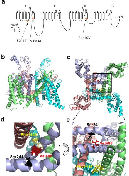
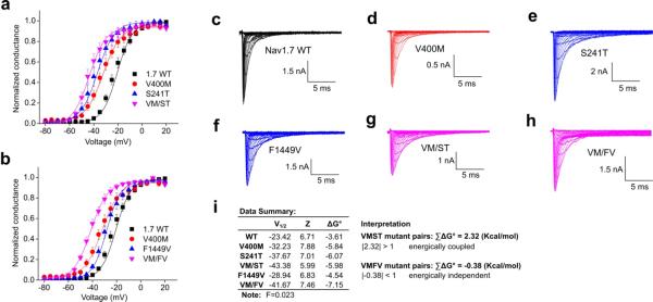
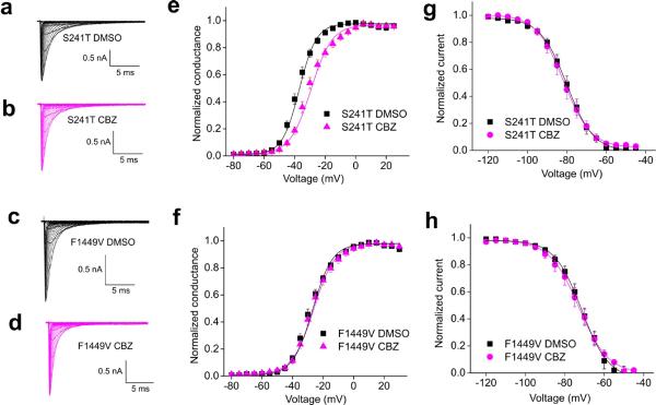
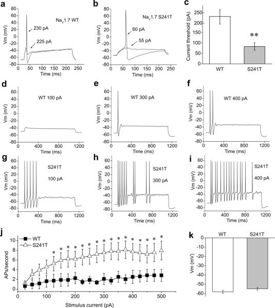
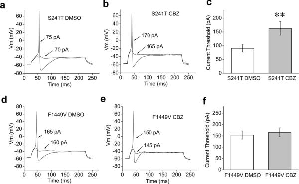
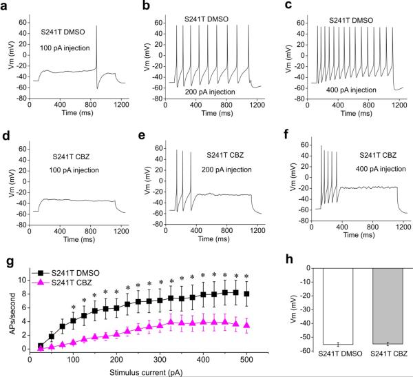
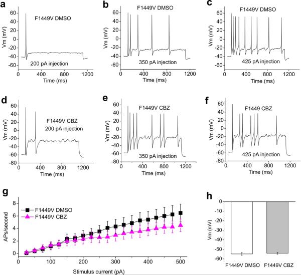
Sodium channel Na(V)1.7 is critical for human pain signalling. Gain-of-function mutations produce pain syndromes including inherited erythromelalgia, which is usually resistant to pharmacotherapy, but carbamazepine normalizes activation of Na(V)1.7-V400M mutant channels from a family with carbamazepine-responsive inherited erythromelalgia. Here we show that structural modelling and thermodynamic analysis predict pharmacoresponsiveness of another mutant channel (S241T) that is located 159 amino acids distant from V400M. Structural modelling reveals that Na(v)1.7-S241T is ~2.4 Å apart from V400M in the folded channel, and thermodynamic analysis demonstrates energetic coupling of V400M and S241T during activation. Atomic proximity and energetic coupling are paralleled by pharmacological coupling, as carbamazepine (30 μM) depolarizes S214T activation, as previously reported for V400M. Pharmacoresponsiveness of S241T to carbamazepine was further evident at a cellular level, where carbamazepine normalized the hyperexcitability of dorsal root ganglion neurons expressing S241T. We suggest that this approach might identify variants that confer enhanced pharmacoresponsiveness on a variety of channels.


Figures

Figure 1
Figure 1. Structural modeling of transmembrane domains of human NaV1.7 channel
(a) Linear schematic of the human NaV1.7 channel topology showing the mutations S241T, V400M and F1449V. (b) Intra-membrane view of structural model of NaV1.7 channel transmembrane domains. Domain I, lightblue; Domain II, salmon; Domain III, cyan; Domain IV, lime. (c) Cyotsolic view of the structural model of NaV1.7 channel transmembrane domains. Boxed area containing S241, V400 and F1449 residues was enlarged in (e). (d) Close-up intra-membrane view of the area containing V400, S241 and F1449 residues. (e) Close-up cytosolic view of the boxed area of (c). V400, S241 and F1449 were shown as stick and colored red, black, and yellow, respectively. Waxman
Figure 2
Figure 2. Mutant cycle analysis of voltage-dependence of activation of NaV1.7 mutations
(a) Voltage-dependence of activation curves of NaV1.7 WT, V400M, S241T, and VM/ST double mutant channels. Curves were Boltzmann fits of the data. (b) Voltage-dependence of activation curves of NaV1.7 WT, V400M, F1449V, and VM/FV double mutant channels. (c–h) Representative traces of current families recorded from HEK293 cells expressing WT (c), V400M (d), S241T (e), F1449V (f), VM/ST double mutant (g), and VM/FV double mutant (h) channels. (i) Data summary for the mutant cycle analysis and interpretation (n=4–5). ΣΔG° was used to determine the level of additivity and was calculated based on the equation described in methods. Waxman
Figure 3
Figure 3. Comparison of voltage-dependence of activation and fast inactivation of S241T and F1449V mutant channels with or without CBZ exposure
(a–d) Representative traces of current families recorded using the activation protocol from HEK293 cells expressing S241T mutant channel treated with DMSO (a), or with CBZ (b); F1449V mutant channel treated with DMSO (c), or with CBZ (d). (e) The averaged voltage-dependence of activation of S241T mutant channel treated with DMSO or CBZ (30 μM) was plotted and fitted with Boltzmann equation. A depolarizing shift of activation of 7.0 mV was observed when S241T mutant channel was treated with CBZ. (f) The averaged voltage-dependence of activation of F1449V mutant channel treated with DMSO or CBZ was plotted and fitted with Boltzmann equation. No notable shift in activation curve of F1449V mutant channel was observed. (g–h) The voltage dependence of fast inactivation of S241T (g) or F1449V (h) mutant channel treated with DMSO or CBZ was plotted and fitted with Bolzmann equation. No notable shift was observed. Waxman
Figure 4
Figure 4. Current-clamp analysis of DRG neurons expressing WT or S241T mutant channel
(a) Representative DRG neuron expressing NaV1.7 WT channel showed sub-threshold response to 225 pA current injection and subsequent action potential evoked by injection of 230 pA, which was the current threshold for this neuron. (b) Representative DRG neuron expressing NaV1.7-S241T mutant channel showed sub-threshold response to 55 pA current injection and subsequent action potential evoked by injection of 60 pA. (c) Comparison of current threshold for DRG neurons expressing WT and S241T mutant channels. Expression of S241T channel reduced current threshold significantly (P<0.01). Current threshold for WT: (227.6±36.7 pA, n= 19); for S241T: (83.5±18.2 pA, n=20). (d–f) responses of a representative DRG neuron expressing WT channel to 1s long depolarization current steps at 100 (d), 300 (e) and 400 (f) pA current injection. (g–i) Responses of a representative DRG neuron expressing S241T mutant channel to 1s long depolarization current steps at 100 (g), 300 (h) and 400 (i) pA current injection. The difference in responses is apparent across this range. (j) The averaged number of action potentials between DRG neurons expressing WT and S241T mutant channel was compared. The response of DRG neurons expressing WT channel to current injection was significantly different compared with DRG neurons expressing S241T mutant channel across a range (125–500 pA) of step current injections (P<0.05). (k) Averaged membrane potentials between DRG neurons expressing WT or S241T mutant channel were not statistically different. Waxman
Figure 5
Figure 5. Comparison of current threshold of CBZ or DMSO treated DRG neurons expressing S241T or F1449V mutant channels
(a–b) The sub- and supra-threshold responses of representative DRG neurons expressing S241T mutant channel treated with DMSO (a) or 30 μM CBZ (b) are shown. (c) Comparison of current threshold for DRG neurons expressing S241T mutant channel treated with DMSO or 30 μM CBZ. The CBZ treatment increased the current threshold significantly (P<0.01). Current threshold for DMSO treated DRG neurons: 90.4±13.2 pA (n=27); for CBZ treated DRG neurons: 162.7±24.4 pA (n=28). (d–e) The sub- and supra-threshold of DRG neurons expressing F1449V mutant channel treated with DMSO (d) or 30 μM CBZ (e) are shown. (f) Comparison of current threshold for DRG neurons expressing F1449V mutant channel with the treatment of DMSO (153.5±17.9 pA, n=29) or 30 μM CBZ (165.5±19.7 pA, n=28). No significant difference was found (P>0.05). Waxman
Figure 6
Figure 6. Comparison of firing frequency and membrane potential of CBZ or DMSO treated DRG neurons expressing S241T mutant channel
(a–c) Responses of a representative DRG neuron expressing S241T mutant channel treated with DMSO to 1sec long depolarization current steps at 100 (a), 200 (b) and 400 (c) pA current injection. (d–f) Similar recording from a representative DRG neuron expressing S241T mutant channel treated with 30 μM CBZ at 100 (d), 300 (e), 400 (f) pA current injection. (g) The averaged response for DRG neurons expressing S241T mutant channel treated with DMSO or CBZ were summarized. CBZ statistically reduced firing frequency starting from 100 pA current injection (P<0.05). (h) Averaged resting membrane potential between DRG neurons expressing S241T mutant channel treated with DMSO or CBZ were not statistically different (DMSO, −55.3±1.4 mV, n=27; CBZ, −54.9±1.3 mV, n=28, P>0.05). Waxman
Figure 7
Figure 7. Comparison of firing frequency and membrane potential of CBZ or DMSO treated DRG Neurons expressing F1449V mutant channel
(a–c) Responses of a representative DRG neurons expressing F1449V mutant channel treated with DMSO to 1s long depolarization current steps at 200 (a), 350 (b) and 425 (c) pA current injection. (d–f) Similar recording from a representative DRG neuron expressing F1449V mutant channel treated with CBZ at 200 (d), 350 (e), and 425 (f) pA current injection. (g) The averaged response of the firing frequency for DRG neurons expressing F1449V mutant channel treated with DMSO or CBZ were compared and no statistical difference was found across the entire range (P>0.05). (h) Averaged resting membrane potentials between DRG neurons expressing F1449V mutant channel treated with DMSO or CBZ were not statistically different (DMSO, −54.8±1.5 mV, n=29; CBZ, −54.1±1.0 mV, n=28, P>0.05). Waxman

Similar articles

Cited by

References

    1. Relieving Pain in America: A Blueprint for Transforming Prevention, Care, Education, and Research. Washington (DC): 2011. - PubMed
    1. Waxman SG. Neurobiology: a channel sets the gain on pain. Nature. 2006;444:831–832. - PubMed
    1. Dib-Hajj SD, Cummins TR, Black JA, Waxman SG. From genes to pain: Na v 1.7 and human pain disorders. Trends Neurosci. 2007;30:555–563. - PubMed
    1. Dib-Hajj SD, Cummins TR, Black JA, Waxman SG. Sodium channels in normal and pathological pain. Annual review of neuroscience. 2010;33:325–347. - PubMed
    1. Minett MS, et al. Distinct Nav1.7-dependent pain sensations require different sets of sensory and sympathetic neurons. Nature communications. 2012;3:791. - PMC - PubMed

Publication types

MeSH terms