. 2013 Jan;33(2):429-43.
doi: 10.1128/MCB.00850-12. Epub 2012 Nov 12.

A quantitative signaling screen identifies CARD11 mutations in the CARD and LATCH domains that induce Bcl10 ubiquitination and human lymphoma cell survival

Affiliations

A quantitative signaling screen identifies CARD11 mutations in the CARD and LATCH domains that induce Bcl10 ubiquitination and human lymphoma cell survival

Waipan Chan et al. Mol Cell Biol. 2013 Jan.

Abstract

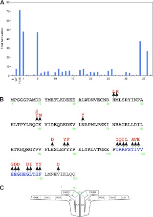
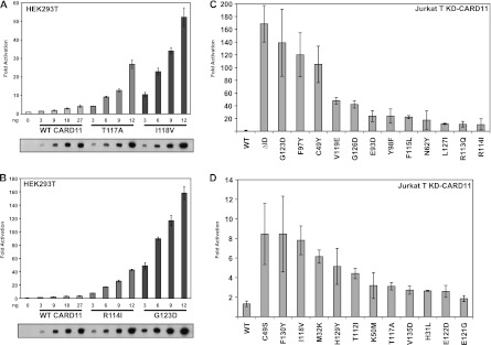
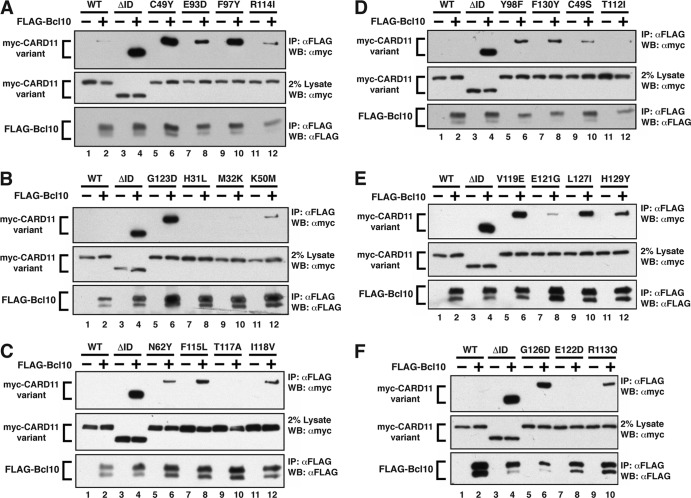
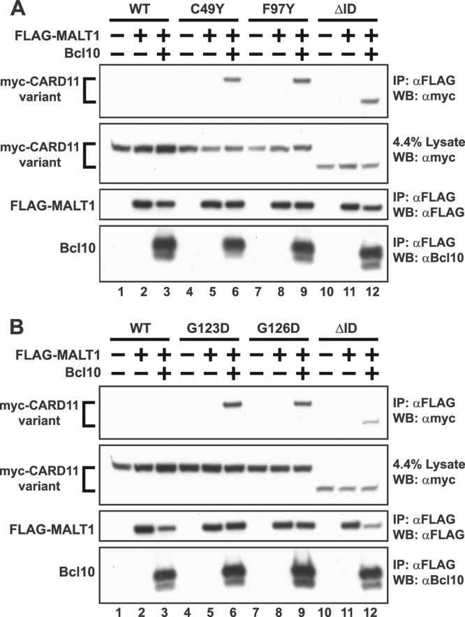
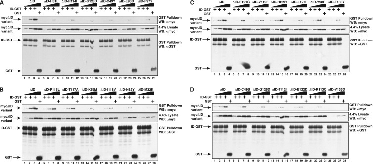
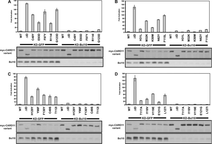
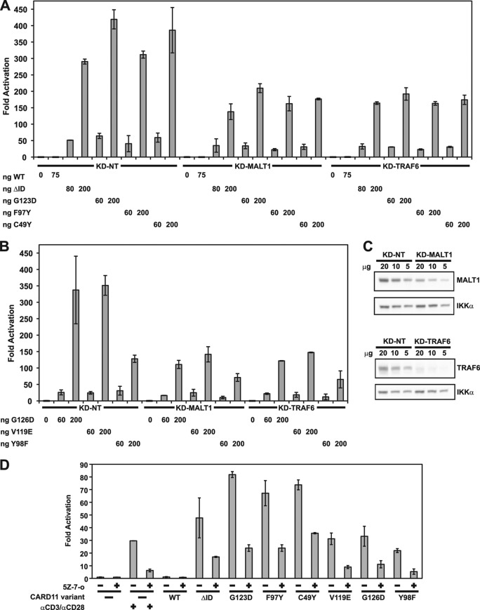
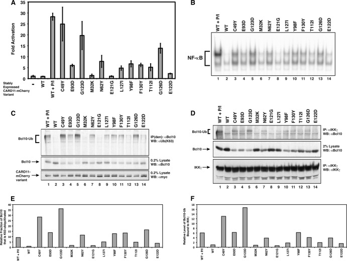
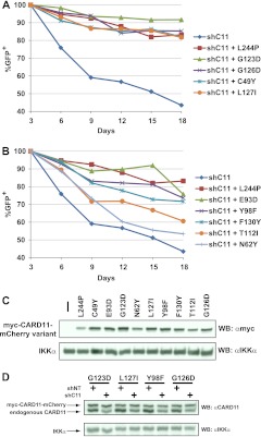
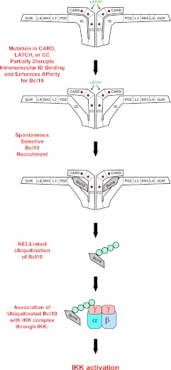
Antigen receptor signaling to NF-κB, essential for normal lymphocyte activation, is dysregulated in several types of lymphoma. During normal signaling, the multidomain adapter CARD11 transitions from a closed, inactive state to an open, active scaffold that assembles a multiprotein complex, leading to NF-κB activation. The regulation of CARD11 scaffold function is bypassed by lymphoma-associated oncogenic CARD11 mutations that induce spontaneous signaling. We report an unbiased high-throughput quantitative signaling screen that identifies new CARD11 hyperactive variants and defines a LATCH domain that functions with the CARD to promote CARD11 autoinhibition. Gain-of-function mutations in the LATCH or CARD disrupt inhibitory domain binding, promote Bcl10 association, and induce Bcl10 ubiquitination, NF-κB activation, and human lymphoma cell survival. Our results identify CARD11 mutations with oncogenic potential, provide a mechanistic explanation for their signaling potency, and offer a straightforward method for the discovery of variants that promote the tumorigenesis of NF-κB-dependent lymphomas.


Figures

Fig 1
Fig 1
A quantitative signaling screen identifies gain-of-function mutations in the CARD and LATCH domains of CARD11. (A) Example of primary screening in which four clones scored as positives. Clones 1, 5, 34, and 36 encoded C49Y, V119E, H129Y, and T117A mutations, respectively. (B) Summary of all mutations identified in the primary sequence at the N terminus of CARD11. CARD domain residues are depicted in black, LATCH domain residues in blue, and the N-terminal end of the coiled-coil domain in gray. (C) Model for CARD11 in the closed inactive state. CARD11 is depicted as a dimer for simplicity, but the oligomerization status has not been determined.
Fig 2
Fig 2
Relative specific activity of CARD11 variants in HEK293T and Jurkat T cells. (A and B) HEK293T cells were transfected with 20 ng of Igκ2-IFN-LUC and 6 ng of CSK-LacZ in the presence of the indicated amounts (in nanograms) of expression vectors for the indicated myc-tagged CARD11 variants. The bottom panel displays Western blots of corresponding lysates probed with anti-myc primary antibody to indicate the relative expression level of each variant. β-Galactosidase activity, driven by CSK-LacZ, was used to normalize luciferase activity and to calculate equivalent amounts of transfected cell lysate for Western analysis. (C and D) Jurkat T cells in which CARD11 was stably knocked down (KD-CARD11) were transfected with 200 ng of CSK-LacZ and 1,500 ng of Igκ2-IFN-LUC in the presence of 50 to 90 ng of expression vectors for the indicated variants. Vector quantities were adjusted to achieve equivalent protein expression levels for all variants, using the relative levels of expression per nanogram in HEK293T cells as a guide.
Fig 3
Fig 3
Mutations in the CARD and LATCH domains enhance the ability of CARD11 to associate with Bcl10. (A to F) HEK293T cells were transfected with 70 to 240 ng of expression vectors for the indicated myc-CARD11 variants and 70 to 225 ng of FLAG-Bcl10, as indicated. Anti-FLAG immunoprecipitations (IP) were performed as described in Materials and Methods and analyzed by Western blotting (WB) with the indicated primary antibodies.
Fig 4
Fig 4
Gain-of-function mutations enhance Bcl10-mediated association of MALT1 with CARD11. (A and B) HEK293T cells were transfected with 400 to 700 ng of expression vectors for the indicated myc-CARD11 variants, 500 to 800 ng of FLAG-MALT1, and 300 to 500 ng of untagged Bcl10, as indicated. Anti-FLAG immunoprecipitations were performed as described in Materials and Methods and analyzed by Western blotting with the indicated primary antibodies.
Fig 5
Fig 5
Mutations in the CARD and LATCH domains disrupt ID binding in trans. (A to D) HEK293T cells were transfected with 800 to 2,000 ng of expression vectors for the indicated CARD11 ΔID variants or with 2,000 ng or 1,500 ng of expression vectors for ID-GST or GST, respectively. Lysates were mixed as indicated and incubated, and glutathione-Sepharose pulldowns were performed as described in Materials and Methods and analyzed by Western blotting with the indicated primary antibodies.
Fig 6
Fig 6
Hyperactive CARD11 variants require Bcl10 for NF-κB activation. (A to D) HEK293T cells stably expressing a shRNA that targets either Bcl10 (KD-Bcl10) or GFP (KD-GFP) as a control were transfected with 20 ng of Igκ2-IFN-LUC, 6 ng of CSK-LacZ, and 8 to 100 ng of expression vectors for the indicated myc-tagged CARD11 variants. Western blots of corresponding lysates probed with anti-myc or anti-Bcl10 primary antibody to indicate the relative expression levels of each variant and Bcl10 in each sample are displayed at the bottom of each panel. β-Galactosidase activity, driven by CSK-LacZ, was used to normalize luciferase activity and to calculate equivalent amounts of transfected cell lysate for Western blot analysis.
Fig 7
Fig 7
Hyperactive variants require MALT1, TRAF6, and TAK1 for NF-κB activation. (A and B) Jurkat T cells stably expressing hairpins that target MALT1 (KD-MALT1) or TRAF6 (KD-TRAF6), or a control hairpin (KD-NT), were transfected with 200 ng of CSK-LacZ and 1,500 ng of Igκ2-IFN-LUC in the presence of the indicated amounts of expression vectors for the indicated CARD11 variants and assayed as described in Materials and Methods. (C) Lysates from KD-MALT1 and KD-TRAF6 Jurkat T cell lines were assayed by Western blot analysis with the indicated primary antibodies. (D) Jurkat T cells were transfected with 200 ng of CSK-LacZ and 1,500 ng of Igκ2-IFN-LUC in the presence of 100 ng of expression vectors for the indicated CARD11 variants. Either DMSO vehicle (−) or 500 nM (5Z)-7-oxozeaenol (5Z-7-o) (+) was added to each sample as indicated every 12 h after transfection for a total of 3 doses. The indicated samples were stimulated with 1 μg/ml anti-CD3/anti-CD28 cross-linking in the absence or presence of (5Z)-7-oxozeaenol for 4 h prior to harvest.
Fig 8
Fig 8
Hyperactive CARD11 variants induce K63-linked ubiquitination of Bcl10 and the association of Ubn(K63)-Bcl10 with IKKγ. (A) Jurkat T cell lines stably expressing the indicated CARD11 variants fused to mCherry were generated by retrovirus infection, transiently transfected with 200 ng of CSK-LacZ and 1,500 ng of Igκ2-IFN-LUC, and assayed as described in Materials and Methods. The line expressing wild-type CARD11 was treated with 50 ng/ml PMA and 1 uM ionomycin (P/I) for 4 h as indicated. (B) Electrophoretic mobility shift assays were performed using 3 μg of nuclear extracts from the stably expressing Jurkat T cell lines described for panel A and a 32P-labeled DNA fragment containing a consensus κB site. The line expressing wild-type CARD11 was treated with 50 ng/ml PMA and 1 μM ionomycin for 30 min as indicated. (C) Immunoprecipitations were performed using denatured lysates [IP(den)] from the stably expressing Jurkat T cell lines described for panel A using anti-Bcl10 antibodies and analyzed by Western blotting with antiubiquitin (K63 linkage specific) [αUb(K63)], anti-Bcl10, or anti-myc primary antibodies as indicated. The line expressing wild-type CARD11 was treated with 50 ng/ml PMA and 1 μM ionomocyin for 20 min as indicated. (D) Immunoprecipitations were performed using lysates from the stably expressing Jurkat T cell lines described for panel A using anti-IKKγ antibodies and analyzed by Western blotting with anti-Bcl10 or anti-IKKγ primary antibodies as indicated. The line expressing wild-type CARD11 was treated with 50 ng/ml PMA and 1 μM ionomocyin for 20 min as indicated. The relative expression level of CARD11 variants in this experiment is also indicated in the anti-myc Western blot in panel C, since the same cell lines were used in panels C and D. (E) Quantitation of the levels of Bcl10-Ub detected in each sample in panel C, divided by the levels of unconjugated Bcl10, normalized to the ratio observed in the WT sample. (F) Quantitation of the levels of Bcl10-Ub detected in the IP with IKKγ in panel D, divided by the levels of unconjugated Bcl10, normalized to the ratio observed in the WT sample.
Fig 9
Fig 9
CARD11 variants containing gain-of-function mutations in the CARD and LATCH domains can promote the survival of OCI-Ly3 human DLBCL-derived cells. (A and B) OCI-Ly3 cells stably expressing the indicated CARD11 variants were generated by retrovirus infection as described in Materials and Methods. Each line was superinfected with a retrovirus that coexpresses GFP and a shRNA that targets the endogenous human CARD11 mRNA (shC11). The percentages of GFP+ cells were normalized to the percentage observed on day 3 after infection. The survival curves for the parental OCI-Ly3 line and the line rescued with the L244P positive control are depicted in each panel for ease of comparison. (C) Lysates from the OCI-Ly3 lines stably expressing the indicated CARD11 variants were analyzed by Western blotting using anti-myc and anti-IKKα antibodies as indicated to reveal the relative expression level of each variant. The leftmost lane represents the parental mCAT-1-expressing OCI-Ly3 line. (D) OCI-Ly3 cells stably expressing the indicated myc-CARD11-mCherry variants were infected with a retrovirus coexpressing GFP with either a hairpin targeting endogenous human CARD11 (shC11) or a control hairpin (shNT). Seven days after infection, GFP+ cells were isolated by FACS sorting and lysates were analyzed by Western blotting with anti-CARD11 or anti-IKKα antibodies.
Fig 10
Fig 10
Model depicting how gain-of-function mutations in the CARD, LATCH, and coiled-coil domains spontaneously induce NF-κB activation.

Similar articles

Cited by

References

    1. Schulze-Luehrmann J, Ghosh S. 2006. Antigen-receptor signaling to nuclear factor kappa B. Immunity 25: 701–715 - PubMed
    1. Jiang C, Lin X. 2012. Regulation of NF-kappaB by the CARD proteins. Immunol. Rev. 246: 141–153 - PMC - PubMed
    1. Kaileh M, Sen R. 2012. NF-kappaB function in B lymphocytes. Immunol. Rev. 246: 254–271 - PubMed
    1. Lim KH, Yang Y, Staudt LM. 2012. Pathogenetic importance and therapeutic implications of NF-kappaB in lymphoid malignancies. Immunol. Rev. 246: 359–378 - PMC - PubMed
    1. Staudt LM. 2010. Oncogenic activation of NF-kappaB. Cold Spring Harb. Perspect. Biol. 2: a000109 doi:10.1101/cshperspect.a000109 - DOI - PMC - PubMed

Publication types

MeSH terms