

Structural brain changes in migraine
- PMID: 23150008
- PMCID: PMC3633206
- DOI: 10.1001/jama.2012.14276
Structural brain changes in migraine
Abstract
Context: A previous cross-sectional study showed an association of migraine with a higher prevalence of magnetic resonance imaging (MRI)-measured ischemic lesions in the brain.
Objective: To determine whether women or men with migraine (with and without aura) have a higher incidence of brain lesions 9 years after initial MRI, whether migraine frequency was associated with progression of brain lesions, and whether progression of brain lesions was associated with cognitive decline.
Design, setting, and participants: In a follow-up of the 2000 Cerebral Abnormalities in Migraine, an Epidemiological Risk Analysis cohort, a prospective population-based observational study of Dutch participants with migraine and an age- and sex-matched control group, 203 of the 295 baseline participants in the migraine group and 83 of 140 in the control group underwent MRI scan in 2009 to identify progression of MRI-measured brain lesions. Comparisons were adjusted for age, sex, hypertension, diabetes, and educational level. The participants in the migraine group were a mean 57 years (range, 43-72 years), and 71% were women. Those in the control group were a mean 55 years (range, 44-71 years), and 69% were women. MAIN OUTCOME MEASURES Progression of MRI-measured cerebral deep white matter hyperintensities, infratentorial hyperintensities, and posterior circulation territory infarctlike lesions. Change in cognition was also measured.
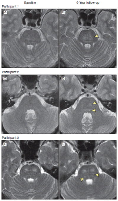
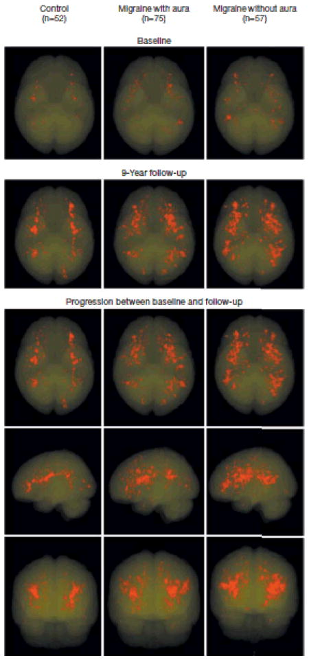
Results: Of the 145 women in the migraine group, 112 (77%) vs 33 of 55 women (60%) in the control group had progression of deep white matter hyperintensities (adjusted odds ratio [OR], 2.1; 95%CI, 1.0-4.1; P = .04). There were no significant associations of migraine with progression of infratentorial hyperintensities: 21 participants (15%) in the migraine group and 1 of 57 participants (2%) in the control group showed progression (adjusted OR, 7.7; 95% CI, 1.0-59.5; P = .05) or new posterior circulation territory infarctlike lesions: 10 of 203 participants (5%) in the migraine group but none of 83 in the control group (P = .07). There was no association of number or frequency of migraine headaches with progression of lesions. There was no significant association of high vs nonhigh deep white matter hyperintensity load with change in cognitive scores (-3.7 in the migraine group vs 1.4 in the control group; 95% CI, -4.4 to 0.2; adjusted P = .07).
Conclusions: In a community-based cohort followed up after 9 years, women with migraine had a higher incidence of deep white matter hyperintensities but did not have significantly higher progression of other MRI-measured brain changes. There was no association of migraine with progression of any MRI-measured brain lesions in men.
Conflict of interest statement
Conflict of Interest Disclosures: All authors have completed and submitted the ICMJE Form for Disclosure of Potential Conflicts of Interest. Dr Ferrari reported receiving grants and consultancy or industry support from Almirall, Coherex, Colucid, Eisai, GlaxoSmithKline, Linde, MAP, Medtronic, Menarini, Merck, Minster, Pfizer, and St Jude, and independent support from the Netherlands Organisation for Scientific Research (NOW). Dr Terwindt reported receiving consultancy support from Merck, Janssen-Cilag, Almirall, and Menarini. Dr Koppen reported consultancy or industry support from Allergan, Benecke congres, Pfizer, and In circulation website. The other authors reported no financial disclosures.
Figures
Comment in
-
White matter hyperintensities in migraine: reason for optimism.JAMA. 2012 Nov 14;308(18):1920-1. doi: 10.1001/jama.2012.36530. JAMA. 2012. PMID: 23150012 No abstract available.
-
[Migraine: Structural brain changes in MRI].Rofo. 2013 Dec;185(12):1130. doi: 10.1055/s-0033-1346802. Rofo. 2013. PMID: 24432396 German. No abstract available.
References
-
- Launer LJ, Terwindt GM, Ferrari MD. The prevalence and characteristics of migraine in a populationbased cohort: the GEM study. Neurology. 1999;53(3):537–542. - PubMed
-
- Ferrari MD. Migraine. Lancet. 1998;351(9108):1043–1051. - PubMed
-
- Headache Classification Committee of the International Headache Society. Classification and diagnostic criteria for headache disorders, cranial neuralgias and facial pain. Cephalalgia. 1988;8(suppl 7):1–96. - PubMed
-
- Kruit MC, van Buchem MA, Hofman PA, et al. Migraine as a risk factor for subclinical brain lesions. JAMA. 2004;291(4):427–434. - PubMed
-
- Kruit MC, Launer LJ, Ferrari MD, van Buchem MA. Infarcts in the posterior circulation territory in migraine: the population-based MRI CAMERA study. Brain. 2005;128(Pt 9):2068–2077. - PubMed
Publication types
MeSH terms
Grants and funding
LinkOut - more resources
Full Text Sources
Medical
Miscellaneous
