

doi: 10.1007/978-1-62703-080-9_2.
Mouse models for studies of retinal degeneration and diseases
Affiliations
- PMID: 23150358
- PMCID: PMC3856760
- DOI: 10.1007/978-1-62703-080-9_2
Item in Clipboard
Mouse models for studies of retinal degeneration and diseases
Methods Mol Biol.
2013.
Abstract
Mouse models, with their well-developed genetics and similarity to human physiology and anatomy, serve as powerful tools with which to investigate the etiology of human retinal degeneration. Mutant mice also provide reproducible, experimental systems for elucidating pathways of normal development and function. Here, I describe the tools used in the discoveries of many retinal degeneration models, including indirect ophthalmoscopy (to look at the fundus appearance), fundus photography and fluorescein angiography (to document the fundus appearance), electroretinography (to check retinal function), as well as the heritability test (for genetic characterization).
Figures

Ophthalmic instruments used for mouse fundus examination.

The major components of the electroretinogram system used in our laboratory.

An eye prior to dilation in pigmented mice (A) and albino mice (B) and the pupil of the same eye in its dilated state (C, D).
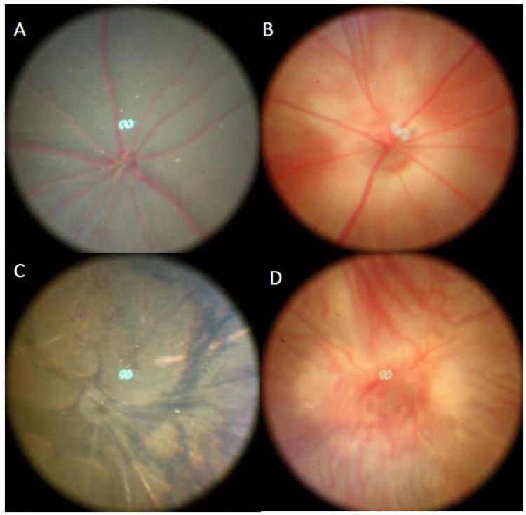
Normal mouse fundus in pigmented mice (A) and albino mice (B) as well as retinal degeneration fundus in pigmented mice (C) and albino mice (D).

Normal mouse fundus prior to the fluorescein injection (A) and the same eye after the fluorescein injection (C). Mouse fundus with neovascular depigmented spots (B) prior to the fluorescein injection and the same eye after the fluorescein injection (D).

Representative ERG responses to a bright flash obtained from a mouse with normal retinal function (A) and a mouse with abnormal retinal function (B).
References
-
- Chang B, Hawes NL, Hurd RE, Wang J, Howell D, Davisson MT, Roderick TH, Nusinowitz S, Heckenlively JR. Mouse models of ocular diseases. Vis Neurosci. 2005;22:587–593. - PubMed
-
- Samardzija M, Neuhauss SCF, Joly S, Kurz-Levin M, Grimm C. Animal Models for Retinal Degeneration. In: Pang I, Clark AF, editors. Animal Models for Retinal Diseases. The Human Press, Inc.; 2010. pp. 51–79.
-
- Redmond TM, Yu S, Lee E, Bok D, Hamasaki D, Chen N, Goletz P, Ma JX, Crouch RK, Pfeifer K. Rpe65 is necessary for production of 11-cis-vitamin A in the retinal visual cycle. Nat Genet. 1998;20(4):344–51. - PubMed
-
- Samardzija M, von Lintig J, Tanimoto N, Oberhauser V, Thiersch M, Reme CE, Seeliger M, Grimm C, Wenzel A. R91W mutation in Rpe65 leads to milder early-onset retinal dystrophy due to the generation of low levels of 11-cis-retinal. Hum Mol Genet. 2008;17(2):281–92. - PubMed
Publication types
MeSH terms
Grants and funding
LinkOut - more resources
Full Text Sources
Other Literature Sources
