. 2013 Feb;27(2):832-42.
doi: 10.1096/fj.12-213900. Epub 2012 Nov 12.

Substrate specificity of Pasteurella multocida toxin for α subunits of heterotrimeric G proteins

Affiliations

Substrate specificity of Pasteurella multocida toxin for α subunits of heterotrimeric G proteins

Joachim H C Orth et al. FASEB J. 2013 Feb.

Abstract

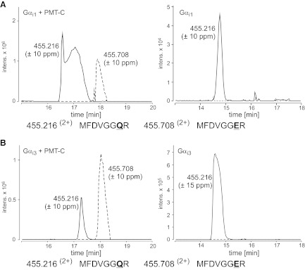
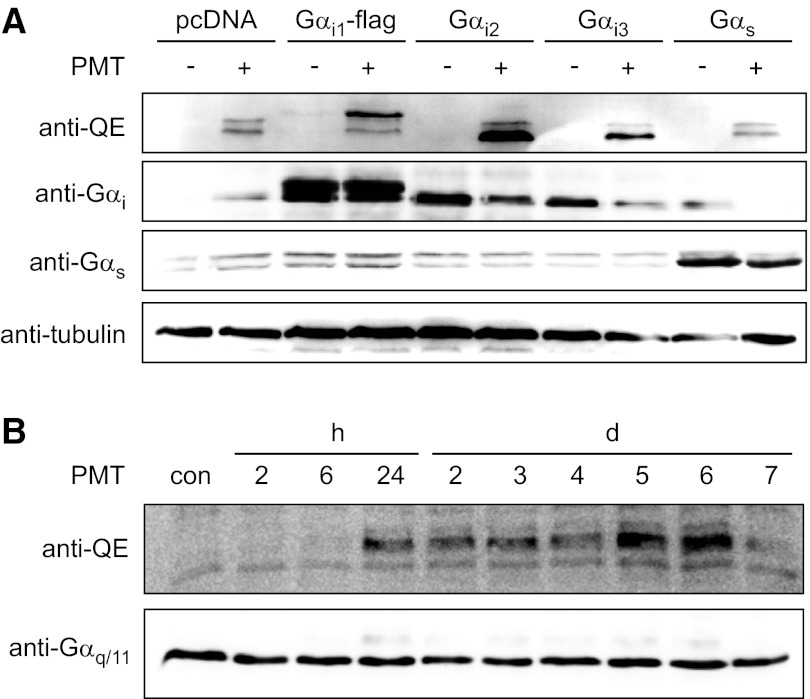
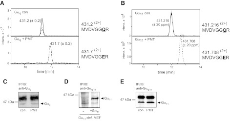
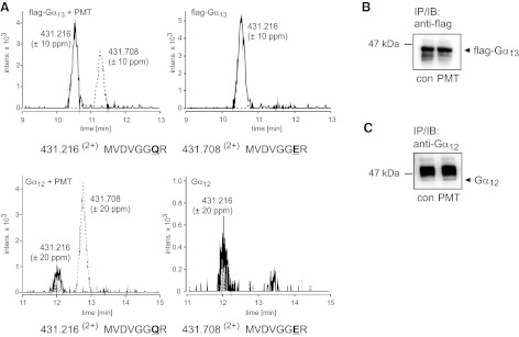
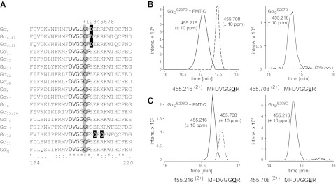
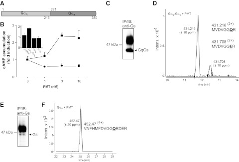
Pasteurella multocida is the causative agent of a number of epizootic and zoonotic diseases. Its major virulence factor associated with atrophic rhinitis in animals and dermonecrosis in bite wounds is P. multocida toxin (PMT). PMT stimulates signal transduction pathways downstream of heterotrimeric G proteins, leading to effects such as mitogenicity, blockade of apoptosis, or inhibition of osteoblast differentiation. On the basis of Gα(i2), it was demonstrated that the toxin deamidates an essential glutamine residue of the Gα(i2) subunit, leading to constitutive activation of the G protein. Here, we studied the specificity of PMT for its G-protein targets by mass spectrometric analyses and by utilizing a monoclonal antibody, which recognizes specifically G proteins deamidated by PMT. The studies revealed deamidation of 3 of 4 families of heterotrimeric G proteins (Gα(q/11), Gα(i1,2,3), and Gα(12/13) of mouse or human origin) by PMT but not by a catalytic inactive toxin mutant. With the use of G-protein fragments and chimeras of responsive or unresponsive G proteins, the structural basis for the discrimination of heterotrimeric G proteins was studied. Our results elucidate substrate specificity of PMT on the molecular level and provide evidence for the underlying structural reasons of substrate discrimination.


Figures

Figure 1.
Figure 1.
i family members are substrates of PMT. Gαi proteins were coexpressed with an active PMT fragment and subjected to MS analysis. Left panels: combined extracted ion chromatograms (Q-TOF data) for m/z 455.216 (±10 ppm; solid line) and 455.708 (±10 ppm; dashed line), corresponding to the tryptic peptides MFDVGGQR and MFDVGGER (aa 198–205) of Gαi1 (A, left panel) and Gαi3 (B, left panel). The presence of the tryptic peptide MFDVGGER indicates deamidation of Gαi1 and Gαi3 at the essential Gln-204 by PMT. Right panels: no relevant deamidation product was detectable when Gαi1 (A) and Gαi3 (B) were expressed without PMT.
Figure 2.
Figure 2.
Determination of the substrate specificity of PMT using a monoclonal anti-QE antibody that detects toxin-deamidated Gα proteins. A) HEK-293 cells were transfected with pcDNA3-based plasmids expressing the indicated Gα subunits. After 24 h of incubation, the cells were treated with or without PMT (1 nM) for a further 24 h. Cells were lysed and subjected to SDS-PAGE, followed by immunoblotting with monoclonal rat anti-Gαq Q209E (3G3; anti-QE), polyclonal rabbit anti-Gαi, polyclonal rabbit anti-Gαs, and monoclonal mouse anti-tubulin antibody as described in Materials and Methods. Note that control HEK-293 cells (pcDNA) and recombinant Gαs-expressing cells contain small amounts of endogenous G proteins, which are deamidated by PMT and labeled with anti-QE (double bands in top panel, lanes 2 and 10). The double bands labeled by anti-Gαs indicate the two isoforms of Gαs. B) CaCo-2 cells were treated with PMT (1 nM) for 1 h. Toxin-containing medium was discarded, and cells were washed with medium and incubated for indicated times. Cells were lysed and subjected to SDS-PAGE, followed by immunoblotting with monoclonal rat anti-Gαq Q209E (3G3; anti-QE) and polyclonal rabbit anti-Gαq/11 antibody.
Figure 3.
Figure 3.
PMT substrate specificity with respect to Gαq and Gα11. A) Combined extracted ion chromatograms (ion trap data) for m/z 431.2 ± 0.2 (solid line) and 431.7 ± 0.2 (dashed line), corresponding to the doubly protonated precursors of the tryptic peptides MVDVGGQR and MFDVGGER (aa 203–210) of Gαq. Top panel: endogenous Gαq from untreated WT-MEFs. Bottom panel: endogenous Gαq from PMT-treated WT-MEFs. B) Combined extracted ion chromatograms (Q-TOF data) for m/z 431.216 ± 20 ppm (solid line) and 431.708 ± 20 ppm (dashed line), corresponding to the doubly protonated precursors of the tryptic peptides MVDVGGQR and MFDVGGER (aa 203–210) of Gα11. Gα11 was precipitated from Gαq/11-deficient MEFs, which were transduced with retrovirus encoding for Gα11.Top panel: Gα11 from untreated cells. Bottom panel: Gα11 from PMT-treated cells. Deamidation of Gln-209 of Gαq and Gα11 is complete, and MVDVGGQR is no longer detectable after PMT treatment. C) Immunoblot of endogenous Gαq after immunoprecipitation with a specific Gαq antibody. Precipitations were performed from untreated MEF (con) or from PMT-treated MEF (1 nM, 18 h). D) Detection of Gα11 in Gαq/11-deficient MEFs (−) or in Gαq/11-deficient MEFs transduced with Gα11-encoding retrovirus (+Gα11). E) Immunoblot of Gα11 after immunoprecipitation with anti-Gαq/11 antibody from Gαq/11-deficient MEFs transduced with Gα11-encoding retrovirus.
Figure 4.
Figure 4.
PMT substrate specificity with respect to Gα12 and Gα13. A) Combined extracted ion chromatograms for m/z 431.216 ± 10 ppm (solid line) and 431.708 ± 10 ppm (dashed line), corresponding to the doubly protonated precursors of the tryptic peptides MVDVGGQR and MFDVGGER (Gα13: aa 220–227, Gα12: aa 225–232) of Gα13 and Gα12. Top panel: Gα13 from PMT-treated (left) or untreated cells (right; Q-TOF data). Bottom panel: Gα12 from PMT-treated (left) or untreated cells (right; Q-TOF data). The 1-Da shift demonstrates the deamidation of Gln-226 (Gα13) and Gln-231 (Gα12) to Glu. The deamidated form of the tryptic peptide (MVDVGGER) is only detectable when Gα13 or Gα12 is precipitated from PMT-treated cells. In addition, unmodified peptide (MVDVGGQR) is present in precipitates from PMT-treated cells. B) Gα13 cDNA was engineered to harbor an internal flag tag. The construct was ectopically expressed in HEK-293 cells. After treatment with or without PMT (1 nM, 18 h), cells were lysed, and flag-tagged protein was immunoprecipitated, isolated, and subjected to MS analysis. Shown is an immunoblot of Gα13 after immunoprecipitation with anti-flag-tag antibody from PMT-treated or untreated (con) cells. C) Gα12 was ectopically expressed in HEK-293 cells. After treatment with or without PMT (1 nM, 18 h), cells were lysed, and Gα12 protein was immunoprecipitated, using a specific anti-Gα12 antibody, isolated, and subjected to MS analysis. Immunoblot shows Gα12 after immunoprecipitation with anti-Gα12 antibody from PMT-treated or untreated (con) cells. The strong band above Gα12 is part of the antibody used for immunoprecipitation.
Figure 5.
Figure 5.
Switch II of G-protein α subunits is not involved in PMT substrate specificity. A) Alignment of amino acid sequences of the switch II region in the α subunits of mouse heterotrimeric GTPases. Nucleotide sequences are obtained from the U.S. National Center for Biotechnology Information (NCBI): Gαs (P63094), Gαolf1 (NP_034437), Gαolf2 (NP_796111), Gαi1 (NP_034435), Gαi2 (AAH65159), Gαi3 (NP_034436), GαoA (NP_034438), GαoB (P18873.3), Gαt1 (NP_032166), Gαt2 (NP_032167), Gαz (NP_034441), Gα15 (NP_034434), Gα14 (NP_032163), Gα13 (NP_034433), Gα12 (NP_034432), Gα11 (NP_034431), and Gαq (NP_032165). Numbers below the alignment correspond to the amino acid positions of Gαq. The active site Gln (at position 209 in Gαq) is indicated by a gray box, highly conserved flanking residues by boldface type, and flanking residues that result in notable charge differences by are indicated in reverse type on a black box. Asterisks denote identical amino acid residues; colons denote highly conserved residues; periods denote conserved residues. Numbers in the top row indicate positions downstream of the essential Gln. B, C) Combined extracted ion chromatograms (Q-TOF data) for m/z 455.216 ± 10 and 455.708 ± 10 ppm, corresponding to the tryptic peptides MFDVGGQR and MFDVGGER (aa 199–206) of Gαi2S207D (B) and Gαi2E208Q (C). Gαi2 mutants were coexpressed with an active PMT fragment and subjected to MS analysis. The presence of the tryptic peptide MFDVGGER indicates deamidation of Gαi2S207D and Gαi2E208Q at Gln-205 by PMT. No relevant deamidation product was detectable when Gαi2S207D and Gαi2E208Q were expressed without PMT (right panels).
Figure 6.
Figure 6.
PMT-induced activation of Gαs-containing chimera. A) Scheme of Gαq-Gαs chimera. N-terminal part including the HD and switch II of Gαq (aa 1–216) were fused to the C-terminal effector-binding domain of Gαs (aa 221–380). B) cAMP accumulation was measured after transfection of Gαq-Gαs chimera into HEK-293-cells (squares) or after transfection with an empty pcDNA vector (circles). PMT was added at indicated concentrations for 3 h. Inset: cAMP accumulation of PMT-treated (10 nM, 3 h) cells transfected as indicated. Gαq-and Gαs-overexpressing cells show no increase in cAMP levels after PMT incubation. C) Chimera Gαq-Gαs was immunoprecipitated from PMT-treated cells (1 nM, 18 h) with a Gs antibody against the C-terminal region. Immunoblot shows Gαq-Gαs chimera after immunoprecipitation with anti-Gs antibody. D) Combined extracted ion chromatograms (Q-TOF data) for m/z 431.216 ± 10 and 431.708 ± 10 ppm, corresponding to the doubly protonated precursors of the tryptic peptides MVDVGGQR and MFDVGGER (Gαq-Gαs: aa 203–210) of Gαq-Gαs. The 1-Da shift demonstrates the deamidation of Gln-209 to Glu. E) Endogenous Gαs was immunoprecipitated from PMT-treated MEF (1 nM, 18 h) with a Gs antibody against the C-terminal region. Immunoblot shows Gαs after immunoprecipitation with anti-Gs antibody. F) Combined extracted ion chromatogram (Q-TOF data) for m/z 452.464 ± 20 ppm corresponding to the quadruple protonated precursors of the tryptic peptide VNFHMFDVGGQRDER (Gαs: aa 203–217) of Gαs. No deamidation of Gln-213 was detectable.
Figure 7.
Figure 7.
PMT effect on Gαi2 fragments. A) Scheme of Gαi2 fragments. Gαi2 consists of the GTPase domain and an HD insert. In the case of Gαi2ΔHD, the HD (residues 61–175) was deleted and replaced by a short linker (Ser-Ala-Gly-Ala). The construct Gαi2swII comprises only the region around the switch II (residues 184–218). B) GST-fusion proteins of Gαi2, Gαi2ΔHD, and Gαi2swII were coexpressed with active or inactive PMT-C and subjected to immunoblot analysis with monoclonal rat anti-Gαq Q209E (3G3; anti-QE), Gαpan antibody as described in Materials and Methods. Deamidation of Gαi2 and Gαi2ΔHD was detectable. C) Combined extracted ion chromatograms (Q-TOF data) for m/z 455.216 ± 10 ppm (solid line) and 455.708 ± 10 ppm (dashed line), corresponding to the tryptic peptides MFDVGGQR and MFDVGGER (aa 198–205) of Gαi2ΔHD coexpressed with active PMT-Cwt. D) Combined extracted ion chromatograms (Q-TOF data) for m/z 455.216 ± 10 ppm (solid line) and 455.708 ± 10 ppm (dashed line) of Gαi2swII coexpressed with active PMT-Cwt. Only the tryptic peptide MFDVGGQR (m/z 455.216), but no MFDVGGER (m/z 455.708), was identified.

Similar articles

Cited by

References

    1. Orth J. H. C., Aktories K. (2010) Pasteurella multocida toxin activates various heterotrimeric G proteins by deamidation. Toxins 2, 205–214 - PMC - PubMed
    1. Wilson B. A., Ho M. (2010) Recent insights into Pasteurella multocida toxin and other G-protein-modulating bacterial toxins. Future Microbiol. 5, 1185–1201 - PMC - PubMed
    1. Lax A. J., Grigoriadis A. E. (2001) Pasteurella multocida toxin: the mitogenic toxin that stimulates signalling cascades to regulate growth and differentiation. Int. J. Med. Microbiol. 291, 261–268 - PubMed
    1. Wilson B. A., Zhu X., Ho M., Lu L. (1997) Pasteurella multocida toxin activates the inositol triphosphate signaling pathway in Xenopus oocytes via Gqα-coupled phospholipase C-β1. J. Biol. Chem. 272, 1268–1275 - PubMed
    1. Rozengurt E., Higgins T., Chanter N., Lax A. J., Staddon J. M. (1990) Pasteurella multocida toxin: potent mitogen for cultured fibroblasts. Proc. Natl. Acad. Sci. U. S. A. 87, 123–127 - PMC - PubMed

Publication types

MeSH terms

LinkOut - more resources