. 2012 Nov 27;109(48):19864-9.
doi: 10.1073/pnas.1214996109. Epub 2012 Nov 12.

Epitranscriptional orchestration of genetic reprogramming is an emergent property of stress-regulated cardiac microRNAs

Affiliations

Epitranscriptional orchestration of genetic reprogramming is an emergent property of stress-regulated cardiac microRNAs

Yuanxin Hu et al. Proc Natl Acad Sci U S A. .

Abstract

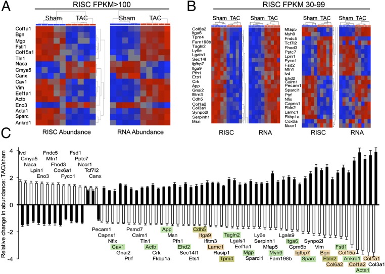
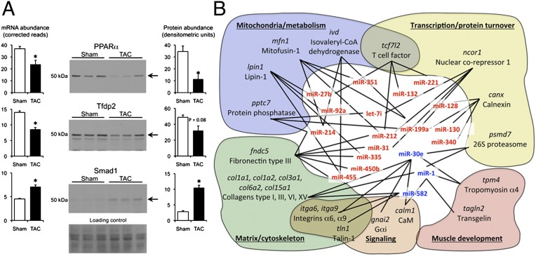
Cardiac stress responses are driven by an evolutionarily conserved gene expression program comprising dozens of microRNAs and hundreds of mRNAs. Functionalities of different individual microRNAs are being studied, but the overall purpose of interactions between stress-regulated microRNAs and mRNAs and potentially distinct roles for microRNA-mediated epigenetic and conventional transcriptional genetic reprogramming of the stressed heart are unknown. Here we used deep sequencing to interrogate microRNA and mRNA regulation in pressure-overloaded mouse hearts, and performed a genome-wide examination of microRNA-mRNA interactions during early cardiac hypertrophy. Based on abundance and regulatory patterns, cardiac microRNAs were categorized as constitutively expressed housekeeping, regulated homeostatic, or dynamic early stress-responsive microRNAs. Regulation of 62 stress-responsive cardiac microRNAs directly affected levels of only 66 mRNAs, but the global impact of microRNA-mediated epigenetic regulation was amplified by preferential targeting of mRNAs encoding transcription factors, kinases, and phosphatases exerting amplified secondary effects. Thus, an emergent cooperative property of stress-regulated microRNAs is orchestration of transcriptional and posttranslational events that help determine the stress-reactive cardiac phenotype. This global functionality explains how large end-organ effects can be induced through modest individual changes in target mRNA and protein content by microRNAs that sense and respond dynamically to a changing physiological milieu.


Conflict of interest statement

The authors declare no conflict of interest.

Figures

Fig. 1.
Fig. 1.
Compensated pressure-overload hypertrophy early after transverse aortic banding. (A) Simultaneous left ventricular (LV, black) and aortic (Ao, gray) pressure tracings 1 wk after transverse aortic banding (TAC). Shaded area is abnormal pressure gradient. (B) LV pressure–volume relations in a sham-operated (gray) and TAC (black) mouse heart; 12 cardiac cycles are shown for each heart. (C) Gravimetric cardiac hypertrophy (n = 9 sham, n = 5 TAC). Ht, height. Data are presented as mean ± SEM. (D) M-mode cardiac echocardiograms. (E) Mean group echo data. EDD, end-diastolic dimension; ESD, end-systolic dimension; NS, not significant.
Fig. 2.
Fig. 2.
MicroRNA signature of active pressure-overload hypertrophy. (A and B) MicroRNA-Seq results for highly expressed (A) and moderately expressed (B) cardiac microRNAs. Volcano plots (Left) show fold-change TAC/sham vs. P value. Bold horizontal and vertical lines show threshold levels [±25% fold change and P value at false discovery rate (FDR) of 0.02]. Heat maps show unsupervised hierarchical clustering of normalized regulated microRNA sequence data; each column is a single mouse heart. (C) Quantitative levels for all 62 hypertrophy-regulated microRNAs. Sham (white) and TAC (black) are shown as reads per sample. Data are presented as mean ± SEM. (D) Correlation of early hypertrophy-induced changes in 21 representative microRNA levels assessed by miR-Seq vs. RT-qPCR.
Fig. 3.
Fig. 3.
Transcriptional signature of the pathologically hypertrophying mouse heart. (A) Volcano plot (Left) and heat maps (Right) of 787 highly abundant cardiac mRNAs. Heat maps show unsupervised hierarchical clustering of raw (Left) and normalized (Right) RNA-sequence data for the 279 regulated mRNAs. (B) As in A for 1,758 moderately abundant mRNAs. (C) Pie charts show functional classification of up-regulated and down-regulated mRNAs.
Fig. 4.
Fig. 4.
Changes in the cardiac proteome 1 wk after surgical pressure overloading. (A) Representative 2D differential in-gel electrophoresis showing Cy 3-labeled sham-operated (Upper) and Cy 5-labeled 1-wk TAC (Lower) cardiac proteomes. (B) Merged Cy 3 (green) and Cy 5 (red) images. Regulated myosin-binding protein C (MyBPc3), transferrin (Trf), protein disulfide isomerase (P4hb), and myosin light-chain isoform 4 (Myl4) are labeled. (CE) Exploded views from a separate experiment of Myl4 (C), P4hb (D), and Trf (E), with accompanying quantifications corresponding to RISC-associated versus total mRNA levels.
Fig. 5.
Fig. 5.
Regulation of mRNA–RISC association during pressure-overload hypertrophy. (A) Normalized heat maps for 17 highly RISC-enriched, significantly regulated cardiac mRNAs showing RISC abundance (Left) and corresponding abundance in the transcriptome (Right). (B) As with A for 45 moderately RISC-abundant cardiac mRNAs. (C) Quantitative group mean data showing fold change after TAC for the 66 mRNAs whose abundance is regulated in the RISC (white bars) together with their respective mRNA levels in the transcriptome (black bars). Data are presented as mean ± SEM. Highlighting indicates mRNAs in gene-ontology categories of cell adhesion (orange) or cardiac contraction/calcium signaling (green) (per Fig. 3C).
Fig. 6.
Fig. 6.
Distinct functions of microRNA-regulated and microRNA-independent cardiac mRNAs. (A) RNA-Seq and immunoblot analyses of hypertrophy-regulated cardiac transcription factors. Ponceau stain is loading control. *P < 0.05. (B) Schematic representation of regulated microRNAs interacting with putative regulated mRNA targets within common functional networks.

References

    1. Dorn GW, II, Robbins J, Sugden PH. Phenotyping hypertrophy: Eschew obfuscation. Circ Res. 2003;92(11):1171–1175. - PubMed
    1. Olson EN. Gene regulatory networks in the evolution and development of the heart. Science. 2006;313(5795):1922–1927. - PMC - PubMed
    1. Bartel DP. MicroRNAs: Target recognition and regulatory functions. Cell. 2009;136(2):215–233. - PMC - PubMed
    1. Small EM, Olson EN. Pervasive roles of microRNAs in cardiovascular biology. Nature. 2011;469(7330):336–342. - PMC - PubMed
    1. van Rooij E, et al. A signature pattern of stress-responsive microRNAs that can evoke cardiac hypertrophy and heart failure. Proc Natl Acad Sci USA. 2006;103(48):18255–18260. - PMC - PubMed

Publication types