. 2012 Nov 27;109(48):19846-51.
doi: 10.1073/pnas.1211517109. Epub 2012 Nov 12.

Role for kisspeptin/neurokinin B/dynorphin (KNDy) neurons in cutaneous vasodilatation and the estrogen modulation of body temperature

Affiliations

Role for kisspeptin/neurokinin B/dynorphin (KNDy) neurons in cutaneous vasodilatation and the estrogen modulation of body temperature

Melinda A Mittelman-Smith et al. Proc Natl Acad Sci U S A. .

Abstract

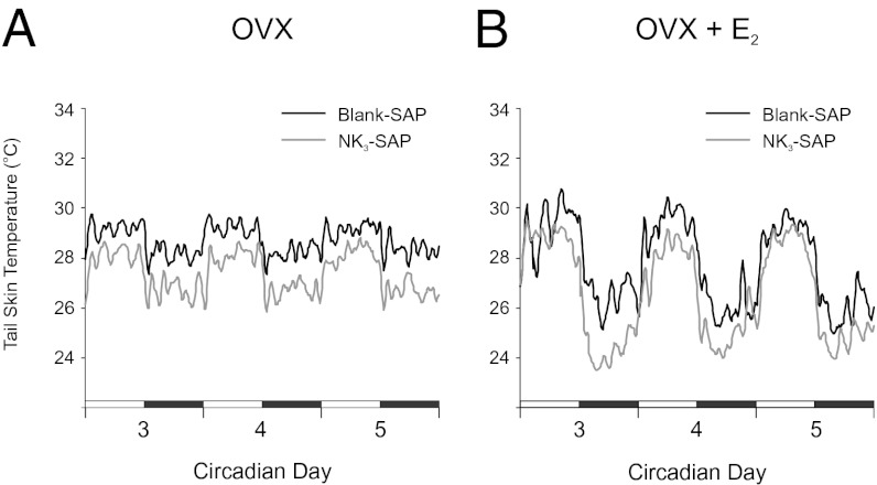
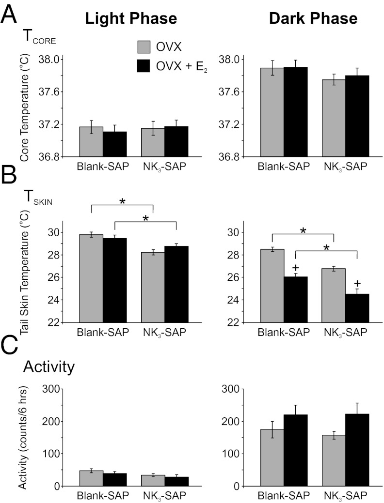
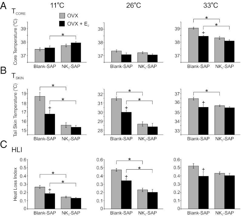
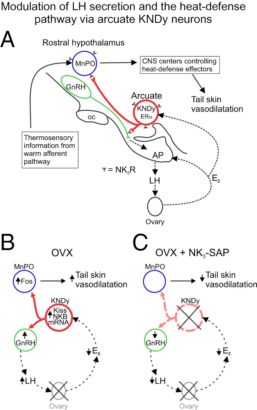
Estrogen withdrawal in menopausal women leads to hot flushes, a syndrome characterized by the episodic activation of heat dissipation effectors. Despite the extraordinary number of individuals affected, the etiology of flushes remains an enigma. Because menopause is accompanied by marked alterations in hypothalamic kisspeptin/neurokinin B/dynorphin (KNDy) neurons, we hypothesized that these neurons could contribute to the generation of flushes. To determine if KNDy neurons participate in the regulation of body temperature, we evaluated the thermoregulatory effects of ablating KNDy neurons by injecting a selective toxin for neurokinin-3 expressing neurons [NK(3)-saporin (SAP)] into the rat arcuate nucleus. Remarkably, KNDy neuron ablation consistently reduced tail-skin temperature (T(SKIN)), indicating that KNDy neurons facilitate cutaneous vasodilatation, an important heat dissipation effector. Moreover, KNDy ablation blocked the reduction of T(SKIN) by 17β-estradiol (E(2)), which occurred in the environmental chamber during the light phase, but did not affect the E(2) suppression of T(SKIN) during the dark phase. At the high ambient temperature of 33 °C, the average core temperature (T(CORE)) of ovariectomized (OVX) control rats was significantly elevated, and this value was reduced by E(2) replacement. In contrast, the average T(CORE) of OVX, KNDy-ablated rats was lower than OVX control rats at 33 °C, and not altered by E(2) replacement. These data provide unique evidence that KNDy neurons promote cutaneous vasodilatation and participate in the E(2) modulation of body temperature. Because cutaneous vasodilatation is a cardinal sign of a hot flush, these results support the hypothesis that KNDy neurons could play a role in the generation of flushes.


Conflict of interest statement

The authors declare no conflict of interest.

Figures

Fig. 1.
Fig. 1.
Experimental protocol. On day 0, the rats were ovariectomized, injected with Blank-SAP or NK3-SAP in the arcuate nucleus, implanted with telemetry devices and returned to their home cages. After a 12- to 15-d recovery period (shown as day 14 in this example), TCORE, TSKIN, and activity were recorded every 10 min over 5 d to evaluate changes over the light/dark cycle (circadian). The next three mornings, TCORE and TSKIN were recorded in rats exposed to TAMBIENT of 26 °C, 11 °C, and 33 °C (in that order) in an environmental chamber. One to 3 d later (shown as day 22 in this example), rats received s.c. E2 capsules. The circadian rhythm recordings were repeated, followed by the TAMBIENT exposures. The rats were killed 11 d after E2 treatment. E2,17β-estradiol; OVX, ovariectomized; SAP, saporin; TAMBIENT, ambient temperature.
Fig. 2.
Fig. 2.
Effects of KNDy neuron ablation on average TSKIN in OVX (A) and OVX + E2 rats (B). (A) Circadian rhythms of TSKIN were not different between NK3-SAP and Blank-SAP rats. However, OVX NK3-SAP rats exhibited consistently lower TSKIN than OVX Blank-SAP rats. (B) After E2 treatment, TSKIN was still consistently lower in NK3-SAP rats than Blank-SAP rats. E2 reduced TSKIN during the dark phase in both Blank-SAP and NK3-SAP rats (compare with A). These waveforms represent the average TSKIN for each group (6–11 rats per group) and were generated using a moving average of five data points. Dark bars represent the dark phase of the 24-h cycle, and numbers refer to circadian recording days 3, 4, and 5. Statistical analyses of these data are shown in Fig. 3B.
Fig. 3.
Fig. 3.
Effects of KNDy neuron ablation and E2 treatment on TCORE (A), TSKIN (B), and activity (C) during the light (Left) and dark (Right) phases. (A) Average TCORE was increased in the dark phase (compared with light) in all groups with no significant effect of NK3-SAP or E2 treatment. (B) Average TSKIN was consistently lower in NK3-SAP rats compared with Blank-SAP rats, indicative of lower levels of cutaneous vasodilatation. In the dark phase, TSKIN was decreased by E2 treatment in both Blank-SAP and NK3-SAP rats. (C) Activity (detected by the DSI telemetry probe) was markedly increased in the dark (active) phase, with no significant differences between Blank-SAP– and NK3-SAP–treated groups. There was a trend (P = 0.06) for E2 to increase activity in the dark phase in both Blank-SAP and NK3-SAP rats. Values represent mean ± SEM, n = 6–11 rats per group. *Significantly different (NK3-SAP vs. Blank-SAP, within OVX or OVX + E2). +Significantly different (OVX vs. OVX + E2, within Blank-SAP or NK3-SAP).
Fig. 4.
Fig. 4.
Average TCORE (A), TSKIN (B), and heat loss index (HLI) (C) in Blank-SAP and NK3-SAP rats exposed to TAMBIENT of 11 °C, 26 °C, or 33 °C (Left to Right). (A) At the TAMBIENT of 33 °C the TCORE was the highest in OVX Blank-SAP rats and reduced in these rats by E2 treatment. At the TAMBIENT of 33 °C, the TCORE of NK3-SAP rats was significantly lower than Blank-SAP animals and was not affected by E2. (B) At the TAMBIENT of 11 °C and 26 °C, the TSKIN of NK3-SAP animals was significantly lower than in Blank-SAP animals. The TSKIN of the Blank-SAP rats was reduced by E2 at all TAMBIENT, but TSKIN was not lowered by E2 treatment in NK3-SAP rats at any TAMBIENT. (C) At the TAMBIENT of 11 °C and 26 °C, the average HLI of NK3-SAP rats was significantly lower than in Blank-SAP rats, indicating lower levels of vasodilatation. HLI was reduced by E2 in Blank-SAP rats but not in NK3-SAP rats. Values represent mean ± SEM, 6–11 rats per group. *Significantly different (NK3-SAP vs. Blank-SAP, within OVX or OVX + E2). +Significantly different (OVX + E2 vs. OVX, within Blank-SAP or NK3-SAP).
Fig. 5.
Fig. 5.
(A) Relationship between KNDy neurons, GnRH neurons, and the heat-defense pathway in the rat. KNDy neurons branch within the arcuate nucleus and project to GnRH terminals in the median eminence (22, 56) and preoptic areas that regulate body temperature, including the MnPO (–24, 42, 45, 48). Secretion of GnRH into portal capillaries stimulates LH secretion from the anterior pituitary gland, which stimulates the secretion of E2 from the ovaries. E2 negative feedback reduces serum LH and decreases NKB and kisspeptin mRNA in KNDy neurons (57, 58). ERα, the isoform required for estrogen negative feedback (59), is expressed in arcuate KNDy neurons (60) but not GnRH neurons (61). NK3R is expressed on arcuate KNDy neurons (60) and GnRH terminals in the median eminence (56). GnRH neurons express kisspeptin receptor mRNA (62), but the distribution of the kisspeptin receptor protein on GnRH neurons has not been described. MnPO neurons express NK3R and pharmacological activation of these neurons reduces body temperature (25). The MnPO receives information from warm-sensitive, cutaneous thermoreceptors and projects to CNS centers to modulate heat-dissipation effectors (24, 42). (B) Effects of ovariectomy on serum LH and tail skin vasodilatation: Loss of E2 after ovariectomy markedly increases NKB and kisspeptin gene expression in KNDy neurons (57, 58), increases GnRH secretion into the portal capillaries (63) and LH secretion from the anterior pituitary gland. In the MnPO, at neutral TAMBIENT, Fos is increased in OVX rats, compared with OVX + E2 rats (37). Tail skin vasodilatation is increased by ovariectomy and decreased in OVX rats by E2 replacement (–31). (C) Effects of KNDy neuron ablation in OVX rats: KNDy neuron ablation lowers serum LH and prevents the rise in LH secretion after ovariectomy (11). Because KNDy neurons do not project to the portal capillary system to directly influence pituitary gonadotrophs (56), this effect is likely mediated via lower levels of GnRH secretion. Ablation of KNDy neurons (with loss of their projections to the rostral hypothalamus) also reduces tail skin vasodilatation. These findings suggest that withdrawal of E2 in OVX rats increases LH secretion and tail skin vasodilatation by increasing the activity of KNDy neurons. AP, anterior pituitary gland; ERα, estrogen receptor α; E2, estradiol-17β; GnRH, gonadotropin-releasing hormone; LH, luteinizing hormone; Kiss, kisspeptin; KNDy, kisspeptin, neurokinin B, and dynorphin-expressing neurons; MnPO, median preoptic nucleus; NKB, neurokinin B; NK3R, neurokinin 3 receptors; oc, optic chiasm.

References

    1. Kronenberg F. Hot flashes: Epidemiology and physiology. Ann N Y Acad Sci. 1990;592:52–86, discussion 123–133. - PubMed
    1. Stearns V, et al. Hot flushes. Lancet. 2002;360(9348):1851–1861. - PubMed
    1. Santoro N. Symptoms of menopause: Hot flushes. Clin Obstet Gynecol. 2008;51(3):539–548. - PubMed
    1. Freedman RR. Physiology of hot flashes. Am J Hum Biol. 2001;13(4):453–464. - PubMed
    1. Casper RF, Yen SSC, Wilkes MM. Menopausal flushes: A neuroendocrine link with pulsatile luteninizing hormone secreation. Science. 1979;205(4408):823–825. - PubMed

Publication types

LinkOut - more resources