. 2012 Nov 27;109(48):19798-803.
doi: 10.1073/pnas.1212900109. Epub 2012 Nov 12.

Function of glycoprotein E of herpes simplex virus requires coordinated assembly of three tegument proteins on its cytoplasmic tail

Affiliations

Function of glycoprotein E of herpes simplex virus requires coordinated assembly of three tegument proteins on its cytoplasmic tail

Jun Han et al. Proc Natl Acad Sci U S A. .

Abstract

Glycoprotein E (gE) of HSV plays a key role in cell-to-cell spread and virus-induced cell fusion. Here, we report that this function of gE requires the cooperation of tegument proteins UL11, UL16, and UL21. We found that the four proteins come together with very high efficiency to form a complex in transfected cells and in a manner that is regulated and coordinated. In particular, the inefficient interaction of UL16 with each membrane protein (UL11 and gE) observed in pairwise transfections became efficient when other binding partners were present. The significance of these interactions was revealed in studies of viral mutants, which showed that each of these tegument proteins is critical for processing, transport, and biological activity of gE. These findings provide insights into the mechanisms of how gE executes its function and also have implications in understanding HSV assembly and budding.


Conflict of interest statement

The authors declare no conflict of interest.

Figures

Fig. 1.
Fig. 1.
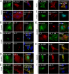
UL11, UL16, UL21 and gE form a complex in transfected cells. (A) Summary of the known interactions among UL11, UL16, UL21, and gE that have been identified by in vitro assays (Left). The dotted lines represent weak or inefficient interactions whereas the solid lines represent efficient interactions in pairwise transfection assays. The four proteins are proposed to assemble into a complex in mammalian cells (Right). (B) Top row: subcellular distribution of UL16-GFP, gE, UL21, or UL11 when each is expressed alone. The lower three rows show the locations of proteins in quadruple cotransfections. (C) Membrane flotation analysis of UL16. Vero cells expressing UL16-GFP alone or in different combinations with UL11, UL21, and gE were osmotically disrupted, and the ability of the proteins to float to the upper fractions of sucrose step gradients during centrifugation was examined. Representative immunoblots are shown. The tops and bottoms of the gradients are indicated, along with the direction of flotation (arrows). (D) Densitometry was used to quantitate immunoblots from three different experiments, and the results are shown as the percentage of floating proteins (top three fractions) relative to the total proteins (all fractions).
Fig. 2.
Fig. 2.
UL21 activates the UL11–UL16 interaction. Vero cells were transfected to express the indicated proteins, either alone (A), pairwise (B, D, G, H, and I), or in combinations of three (C, E, and F). At 16 to 18 h after transfection, the cells were fixed, stained with the appropriate antibodies, and examined by confocal microscopy. For double transfections, the merged images are shown in the right panels. For triple transfections, the right panels indicate which proteins were coexpressed and show the images for the two being analyzed.
Fig. 3.
Fig. 3.
UL11 is required to activate the gE–UL16 interaction. Vero cells were transfected to express the indicated proteins alone (A), pairwise (B), or in combinations of three (C and D). At 16 to 18 h after transfection, the cells were fixed, stained with the appropriate antibodies, and examined by confocal microscopy. For triple transfections, the panels show the images of individual proteins being analyzed in the same cell.
Fig. 4.
Fig. 4.
UL11, UL16, and UL21 are needed for fusion of Vero cells by a gBsyn mutant. (A) Vero cells were infected at a multiplicity of infection (MOI) of 0.01 with WT HSV or Syn mutants. Virus-induced cytopathic effect was monitored daily and recorded by an inverted microscope. (B) Vero cells were infected with WT HSV or mutants at an MOI of 5. At 18 to 24 h after infection, the cells were harvested, lysed in sample buffer, subjected to electrophoresis in denaturing gels, and immunoblotted with indicated antibodies. Capsid protein VP5 served as a loading control. The presence (+) and absence (−) of syncytia is indicated.
Fig. 5.
Fig. 5.
gE fails to accumulate on the surface of infected Vero cells in the absence of its binding partners. (A) Vero cells were infected at an MOI of 0.01 with WT or the indicated mutant and revertant viruses. At 18 to 24 h after infection, the cells were fixed and reacted with a mouse monoclonal antibody (clone 3114) to gE before microscopy. (B) Vero cells were infected with WT or mutants at an MOI of 5. At 18 to 24 h after infection, the cells were harvested, lysed in sample buffer, subjected to electrophoresis in denaturing gels, and immunoblotted with indicated antibodies. Capsid protein VP5 was used as the loading control.
Fig. 6.
Fig. 6.
The Syn phenotype is lost in HaCaT cells even though gE accumulates on the cell surface when its binding partners are absent. (A) HaCaT cells grown on coverslips were infected with WT or the indicated mutant viruses at an MOI of 0.01. At 24 h after infection, the cells were fixed and reacted with a mouse monoclonal antibody (clone 3114) to gE before microscopy. (B) HaCaT cells were infected with WT or mutants viruses at an MOI of 5. At 18 to 24 h after infection, the cells were harvested, lysed in sample buffer, subjected to electrophoresis in denaturing gels, and reacted with the indicated antibodies. VP5 was used as a loading control. (C) HaCaT cells grown in six-well plates were infected with WT or Syn mutant viruses at low MOI. Virus-induced cytopathic effect was monitored daily and recorded by an inverted microscope. The presence (+) and absence (−) of syncytia is indicated.

Similar articles

Cited by

References

    1. Johnson DC, Huber MT. Directed egress of animal viruses promotes cell-to-cell spread. J Virol. 2002;76(1):1–8. - PMC - PubMed
    1. Johnson DC, Baines JD. Herpesviruses remodel host membranes for virus egress. Nat Rev Microbiol. 2011;9(5):382–394. - PubMed
    1. Balan P, et al. An analysis of the in vitro and in vivo phenotypes of mutants of herpes simplex virus type 1 lacking glycoproteins gG, gE, gI or the putative gJ. J Gen Virol. 1994;75(Pt 6):1245–1258. - PubMed
    1. Dingwell KS, et al. Herpes simplex virus glycoproteins E and I facilitate cell-to-cell spread in vivo and across junctions of cultured cells. J Virol. 1994;68(2):834–845. - PMC - PubMed
    1. Dingwell KS, Doering LC, Johnson DC. Glycoproteins E and I facilitate neuron-to-neuron spread of herpes simplex virus. J Virol. 1995;69(11):7087–7098. - PMC - PubMed

Publication types

Substances