. 2012 Dec 26;109(52):21336-41.
doi: 10.1073/pnas.1218087109. Epub 2012 Nov 12.

Exploring conformational states of the bacterial voltage-gated sodium channel NavAb via molecular dynamics simulations

Affiliations

Exploring conformational states of the bacterial voltage-gated sodium channel NavAb via molecular dynamics simulations

Cristiano Amaral et al. Proc Natl Acad Sci U S A. .

Abstract

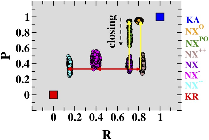
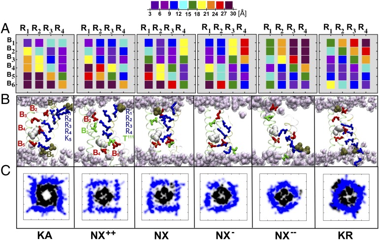
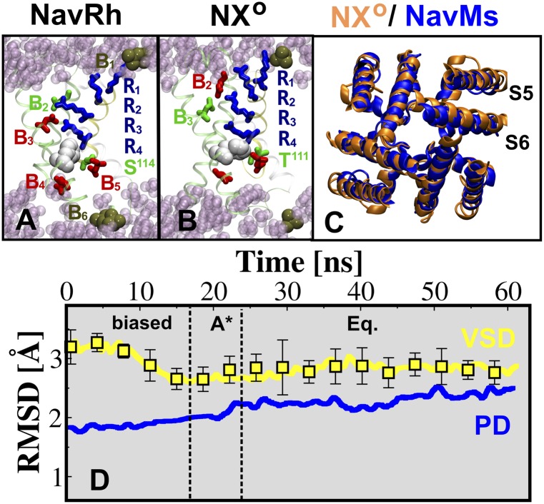
The X-ray structure of the bacterial voltage-gated sodium channel NavAb has been reported in a conformation with a closed conduction pore. Comparison between this structure and the activated-open and resting-closed structures of the voltage-gated Kv1.2 potassium channel suggests that the voltage-sensor domains (VSDs) of the reported structure are not fully activated. Using the aforementioned structures of Kv1.2 as templates, molecular dynamics simulations are used to identify analogous functional conformations of NavAb. Specifically, starting from the NavAb crystal structure, conformations of the membrane-bound channel are sampled along likely pathways for activation of the VSD and opening of the pore domain. Gating charge computations suggest that a structural rearrangement comparable to that occurring between activated-open and resting-closed states is required to explain experimental values of the gating charge, thereby confirming that the reported VSD structure is likely an intermediate along the channel activation pathway. Our observation that the X-ray structure exhibits a low pore domain-opening propensity further supports this notion. The present molecular dynamics study also identifies conformations of NavAb that are seemingly related to the resting-closed and activated-open states. Our findings are consistent with recent structural and functional studies of the orthologous channels NavRh, NaChBac, and NavMs and offer possible structures for the functionally relevant conformations of NavAb.


Conflict of interest statement

The authors declare no conflict of interest.

Figures

Fig. 1.
Fig. 1.
Conformations associated with voltage-sensor domain activation and pore-domain opening of NavAb. The present unconstrained MD trajectories for membrane-equilibrated NavAb conformations NX−−, NX, NX, NX++, NXPO, and NXO are shown as projections onto a conformational distance space, defined by the generalized coordinates R (NavAb, VSD; t) and P (NavAb, PD; t). These six conformations correspond to distinct positions along the VSD-activation (red arrows) and PD-opening (yellow arrows) pathways of NavAb. The conformational distance positions corresponding to the activated-open (blue square) and resting-closed (red square) conformations of Kv1.2 are also indicated. The time series of R (NavAb, VSD; t) and P (NavAb, PD; t), including the biased and equilibrium phases of the simulations, are presented in Fig. S1.
Fig. 2.
Fig. 2.
Voltage-sensor conformations of NavAb. (A) Distance matrices Mij (NavAb, VSD) mapping the domain electrostatic interactions between the S4 basic residues (numbered R1 to R4) and their binding sites [numbered B1 to B6 for (Upper) PO4, E32, N49, E59, and D80 and (Lower) PO4, respectively] in each of the VSD conformations NX++, NX, NX, and NX−−. For the purpose of comparison, the reference, distance matrices Mij (Kv1.2, VSD) for Kv1.2 in the KA and KR conformations are also depicted; Kv1.2 binding sites are numbered B1 to B6 for (Upper) PO4, E183, E226, E236, and D259 and (Lower) PO4, respectively. Distance matrices are computed by averaging over the four independent subunits. (B) Snapshots of the VSD for the NavAb and Kv1.2 conformations, highlighting the position of the S4 basic residues (blue sticks) and the salt bridges/hydrogen bonds they form with the acidic/polar residues (red/green sticks) of other VSD segments or with lipid phosphate moieties (ochre). Note that the NavAb “catalytic center,” formed by B4, B5, and F56 (white; F233 in Kv1.2), is occupied by the S4 residues R2, R3, R4, and T111. (C) Atomic-density plots for the selected NavAb and Kv1.2 conformations, depicting the positions of the S4–S5 linkers (blue) and S6 helices (black) at the membrane plane placed at the intracellular entrance of the channel. The S4–S5 linker constrains the bundle-crossing region of S6 in the conformations NX and NX−−, thereby prohibiting pore opening in these structures.
Fig. 3.
Fig. 3.
(A and B) Snapshots of the VSD of NavAb NXO and NavRh, highlighting the position of the S4 basic residues (blue sticks) and the salt bridges/hydrogen bonds they form with the acidic/polar residues (red/green sticks) of other VSD segments or with the lipid phosphate moieties (ocre). The highly conserved Phe residue (white), forming the catalytic center, is shown for both structures. (C) Structural superposition of the PD of NXO (orange) and NavMs (blue). (D) Rmsd profiles of selected segments of NavAb NXO, VSD (yellow), and PD (blue). The rms deviations of the backbone atoms for the VSD and PD are calculated with respect to the X-ray structure of NavRh and NavMs, respectively; for the VSD, the acidic/polar binding sites within the VSD were superposed and the average over the four subunits is reported, with associated error bars.

Comment in

References

    1. Hille B. Ionic Channels of Excitable Membranes. 2nd Ed. Sunderland, MA: Sinauer; 1992.
    1. Bezanilla F. The voltage-sensor structure in a voltage-gated channel. Trends Biochem Sci. 2005;30(4):166–168. - PubMed
    1. Catterall WA. Ion channel voltage sensors: Structure, function, and pathophysiology. Neuron. 2010;67(6):915–928. - PMC - PubMed
    1. Long SB, Campbell EB, MacKinnon R. Crystal structure of a mammalian voltage-dependent Shaker family K+ channel. Science. 2005;309(5736):897–903. - PubMed
    1. Treptow W, Tarek M. Environment of the gating charges in the Kv1.2 Shaker potassium channel. Biophys J. 2006;90(9):L64–L66. - PMC - PubMed

Publication types

MeSH terms

Substances

LinkOut - more resources