. 2012 Nov 27;109(48):19679-84.
doi: 10.1073/pnas.1211274109. Epub 2012 Nov 13.

Dynamic force sensing of filamin revealed in single-molecule experiments

Affiliations

Dynamic force sensing of filamin revealed in single-molecule experiments

Lorenz Rognoni et al. Proc Natl Acad Sci U S A. .

Abstract

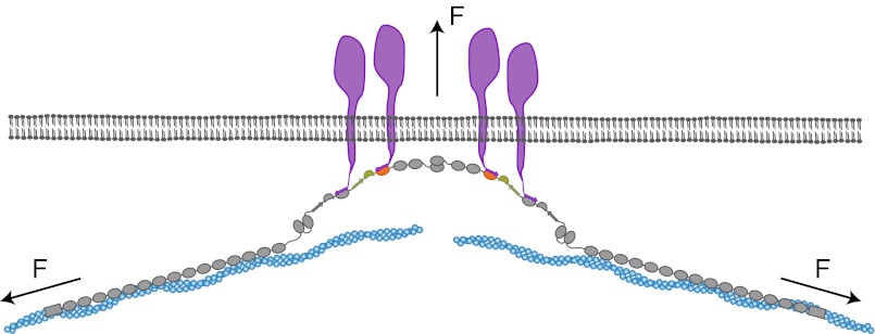
Mechanical forces are important signals for cell response and development, but detailed molecular mechanisms of force sensing are largely unexplored. The cytoskeletal protein filamin is a key connecting element between the cytoskeleton and transmembrane complexes such as integrins or the von Willebrand receptor glycoprotein Ib. Here, we show using single-molecule mechanical measurements that the recently reported Ig domain pair 20-21 of human filamin A acts as an autoinhibited force-activatable mechanosensor. We developed a mechanical single-molecule competition assay that allows online observation of binding events of target peptides in solution to the strained domain pair. We find that filamin force sensing is a highly dynamic process occurring in rapid equilibrium that increases the affinity to the target peptides by up to a factor of 17 between 2 and 5 pN. The equilibrium mechanism we find here can offer a general scheme for cellular force sensing.


Conflict of interest statement

The authors declare no conflict of interest.

Figures

Fig. 1.
Fig. 1.
Interaction of filamin with transmembrane proteins. (A) Schematics of the domain arrangement of filamin and its interaction with the transmembrane proteins integrin and GPIbα. Filamin consists of 24 Ig domains that dimerize at domain 24 and carry an N-terminal actin-binding domain. In its rod 2 region (Ig domains 16–24), domains 16–21 (FLNa16-21) arrange in pairs that contain binding sites for integrins (dark green) and GPIbα (purple). The domain pair FLNa20-21 is highlighted in orange and green. (B) Schematics of the autoinhibition mechanism of the domain pair FLNa20-21. The A-strand of FLNa20 binds to FLNa21, thus autoinhibiting its interaction with the transmembrane proteins (upper cartoon). It has been hypothesized that mechanical stress releases the autoinhibition and promotes interaction with the targets (lower cartoon). (C) Schematics of the single-molecule assay using a double-beam optical trap (see text and SI Materials and Methods).
Fig. 2.
Fig. 2.
Interaction of filamin with tethered peptides. (A) Force extension trace of the interaction between the target peptide of GPIbα and FLNa21 (GPIbα-FLNa21). At forces around 10 pN, the target peptide rapidly fluctuates between bound and unbound conformations and stays permanently unbound at higher loads (>12 pN). (B) Equilibrium traces obtained at three different biasing forces. At high loads (top trace), the probability (black histogram) is shifted to the unbound state, whereas at decreasing loads, the bound state becomes more and more populated (bottom two traces). (C) Opening (circles) and closing (triangles) rates as a function of force. The solid line is an extrapolation of the rates to zero-load taking into account the compliance of all mechanical elements in the construct (SI Materials and Methods). (D) Force extension trace of the interaction between the target peptide of β7-integrin and FLNa21 (ITβ7-FLNa21). (E) Equilibrium traces of ITβ7-FLNa21 obtained at three different biasing forces (compare B). (F) Opening (circles) and closing (triangles) rates as a function of force (compare C). (G) Force extension trace of the interaction between the target peptide of migfilin and FLNa21 (Mig-FLNa21). (H) Equilibrium traces of Mig-FLNa21 obtained at three different biasing forces (compare B and E). (I) Opening (circles) and closing (triangles) rates as a function of force (compare C and F).
Fig. 3.
Fig. 3.
Single-molecule mechanical competition assay to study peptide binding from solution. (A) Time traces of opening and closing of the GPIbα-FLNa21 construct held at a force bias of 8.5 pN in the presence of 2.3 μM GPIbα peptide in solution. The colored traces correspond to the 20-kHz data (gray), moving average filtered with 2.5 ms (black) and 50 ms (white) time window. Apparent high-SD regions with lifetimes formula image 
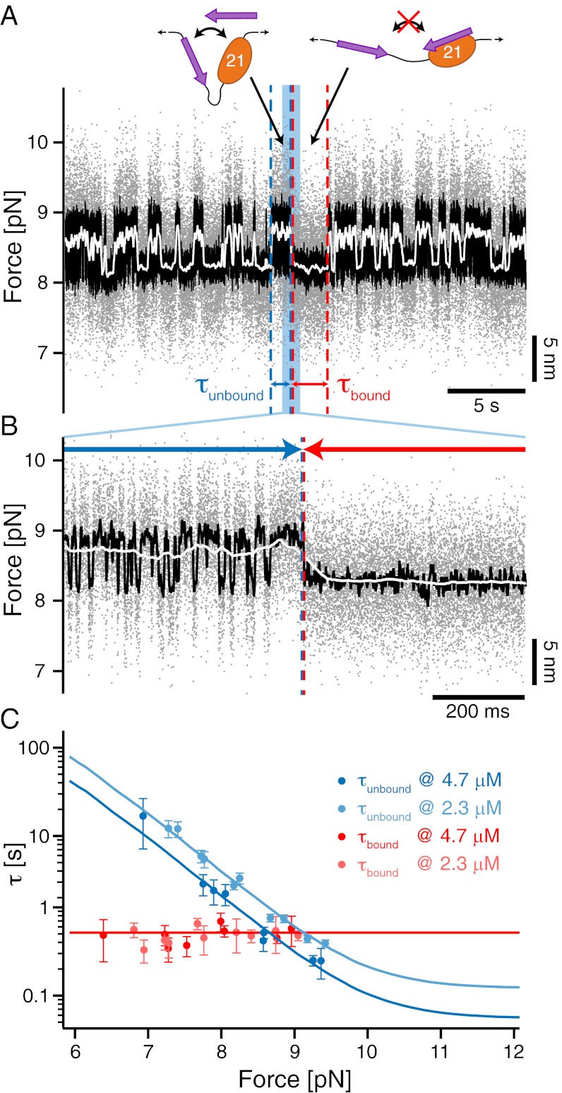
in the black and gray traces are interrupted by low-SD regions with lifetimes formula image. (B) A zoom into the blue region shows the transition between a high-SD (rapid opening and closing cycles of the tethered construct) and low-SD region (blocked fluctuations due to competitive peptide binding from solution). (C) Dependence of the bound and unbound lifetimes as a function of applied force and solution concentration. As expected for binding from solution, formula image depends on the opening probability of the tethered construct and hence the applied force, as well as the solution concentration, whereas formula image is independent (see text and SI Materials and Methods).
Fig. 4.
Fig. 4.
Filamin domain pairs act as a precisely tuned mechano-sensor. (A) Low-resolution stretch (blue) and relax (gray) trace of FLNa20-21. At forces around 15 pN, FLNa20 unfolds, and at higher loads exceeding 30 pN, FLNa21 unfolds. Opening of the domain pair and release of the autoinhibition is not visible at this experimental resolution. (B) (Upper) Competition assay of domain pair opening in the presence of 2.7 μM GPIbα peptide in solution observed at loads of 4.5 pN (color scheme as in Fig. 3A). High-SD regions (opening–closing fluctuations) are interrupted with low-SD regions where bound peptide blocks fluctuations. (Lower) Zoom into a transition region. (C) Force-dependent binding of peptide from solution observed at biasing forces from 2.3 to 4.0 pN. From low to high loads, the binding probability (black histograms) increases constantly. (D) Force-dependent gating characteristics of the force-sensing domain pair as obtained from the force and concentration-dependent dwell times of Fig. S4C (blue data points; black solid line shows global fit). The three symbols (triangle, square, and circle) denote three different solution concentrations. The dashed black line is an independent measure of the force-dependent opening probability as obtained from the HMM analysis of the fluctuating state where no ligand from solution is bound (Fig. S6B).
Fig. 5.
Fig. 5.
Model of the force-dependent binding and clustering of membrane receptors to filamin. Mechanical force in a strained cytoskeleton will lead to domain pair opening in the rod 2 segment of filamin, allowing the binding to membrane receptors. The simultaneous interaction with many domain pairs will significantly stabilize the cytoskeleton–membrane interaction and potentially induce clustering of receptors.

Similar articles

Cited by

References

    1. Engler AJ, Sen S, Sweeney HL, Discher DE. Matrix elasticity directs stem cell lineage specification. Cell. 2006;126(4):677–689. - PubMed
    1. Hoshijima M. Mechanical stress-strain sensors embedded in cardiac cytoskeleton: Z disk, titin, and associated structures. Am J Physiol Heart Circ Physiol. 2006;290(4):H1313–H1325. - PMC - PubMed
    1. Nakamura F, Stossel TP, Hartwig JH. The filamins: Organizers of cell structure and function. Cell Adhes Migr. 2011;5(2):160–169. - PMC - PubMed
    1. Glogauer M, et al. The role of actin-binding protein 280 in integrin-dependent mechanoprotection. J Biol Chem. 1998;273(3):1689–1698. - PubMed
    1. Gehler S, et al. Filamin A-beta1 integrin complex tunes epithelial cell response to matrix tension. Mol Biol Cell. 2009;20(14):3224–3238. - PMC - PubMed

Publication types