. 2013 Jan;193(1):125-41.
doi: 10.1534/genetics.112.146100. Epub 2012 Nov 12.

Genetic dissection of peroxisome-associated matrix protein degradation in Arabidopsis thaliana

Affiliations

Genetic dissection of peroxisome-associated matrix protein degradation in Arabidopsis thaliana

Sarah E Burkhart et al. Genetics. 2013 Jan.

Abstract

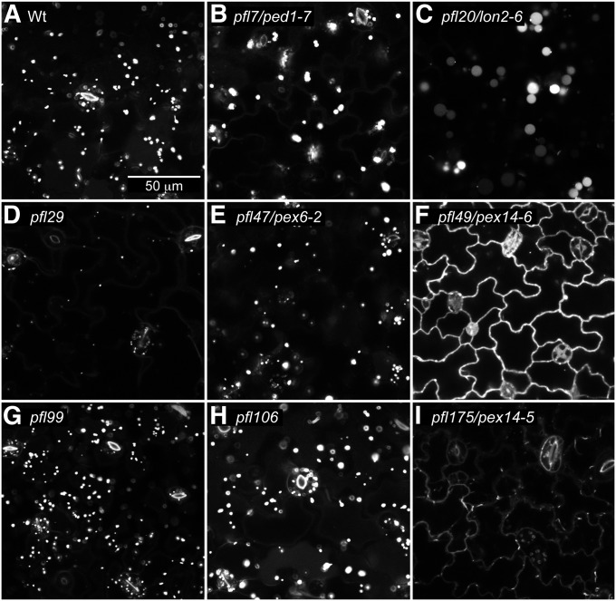
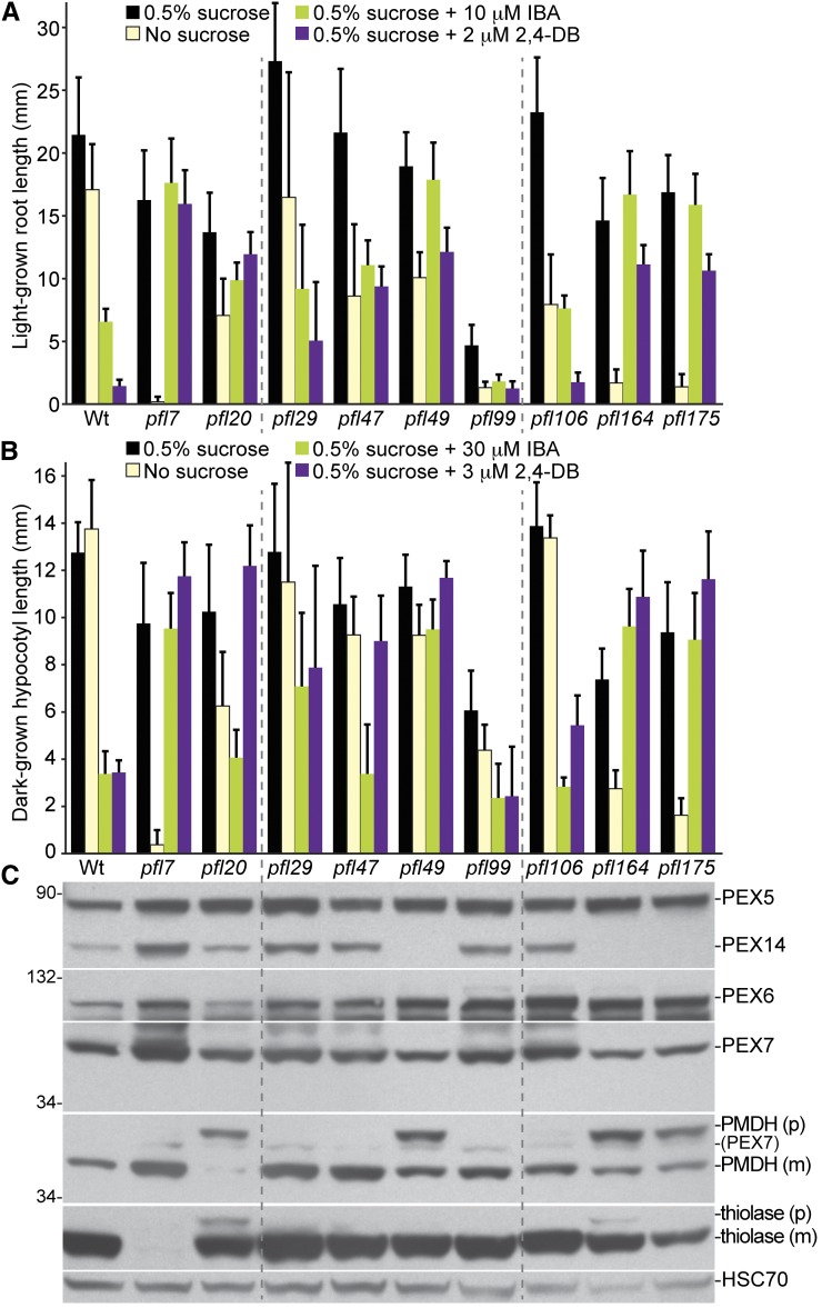
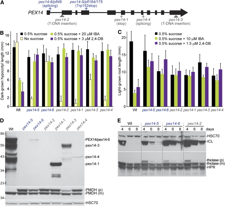
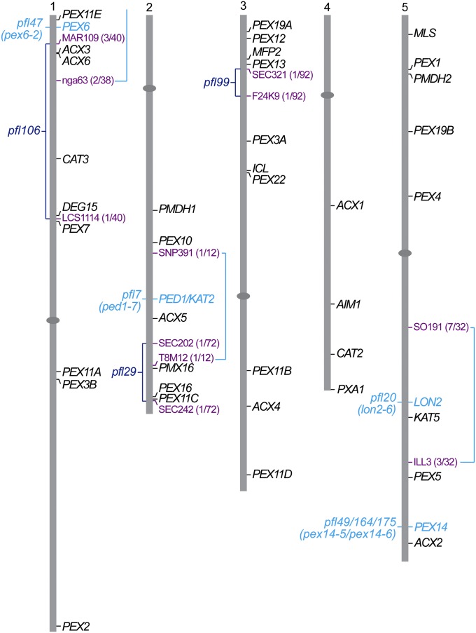
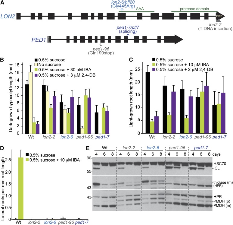
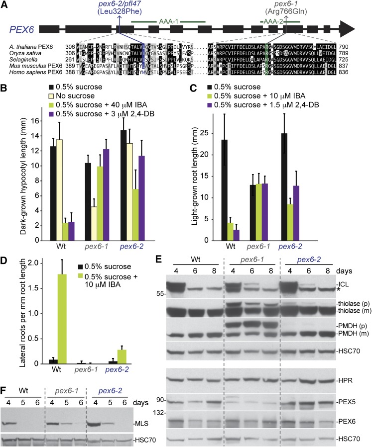
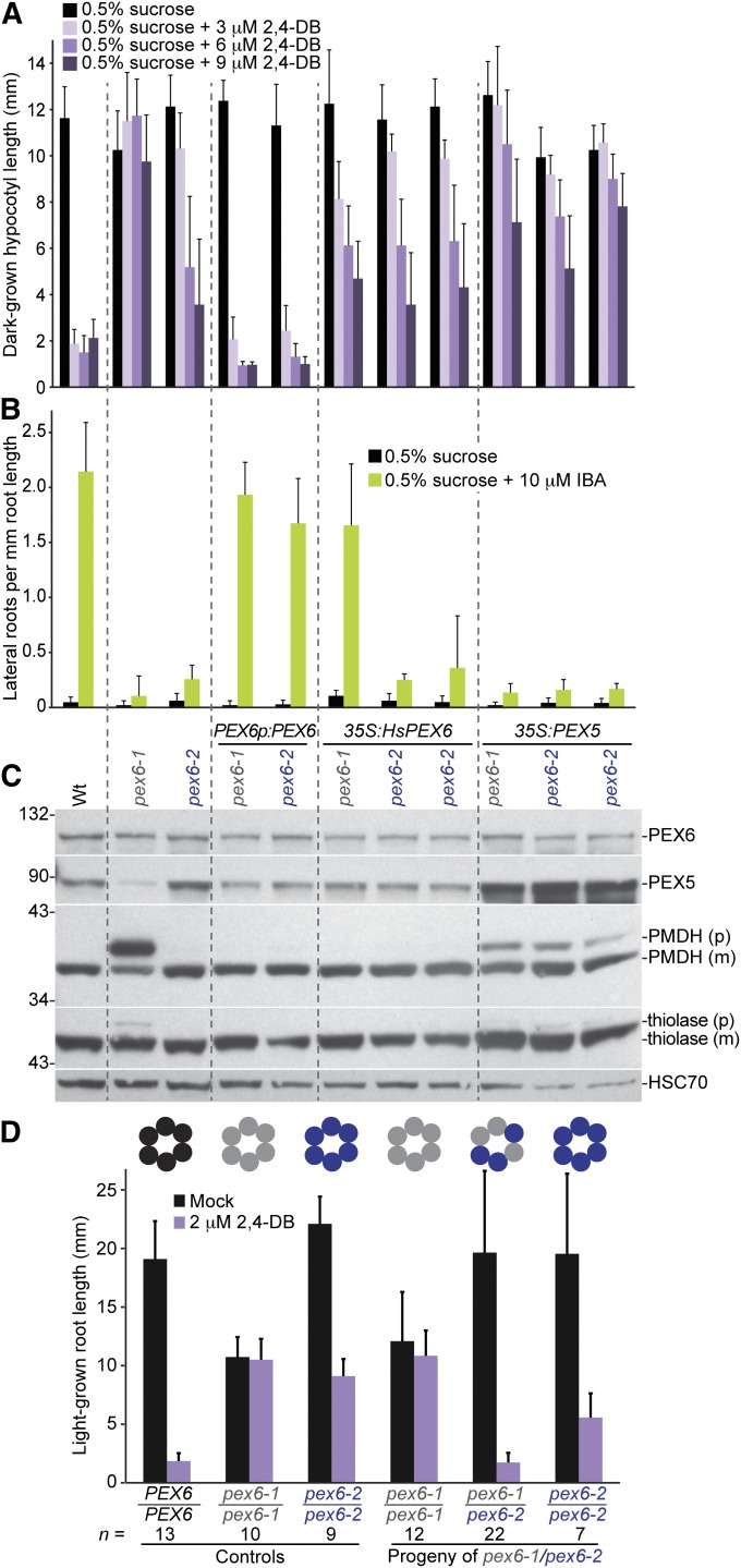
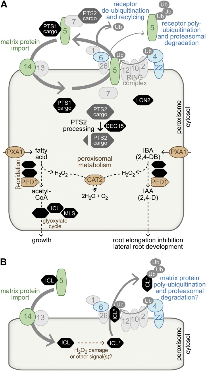
Peroxisomes are organelles that sequester certain metabolic pathways; many of these pathways generate H(2)O(2), which can damage proteins. However, little is known about how damaged or obsolete peroxisomal proteins are degraded. We exploit developmentally timed peroxisomal content remodeling in Arabidopsis thaliana to elucidate peroxisome-associated protein degradation. Isocitrate lyase (ICL) is a peroxisomal glyoxylate cycle enzyme necessary for early seedling development. A few days after germination, photosynthesis begins and ICL is degraded. We previously found that ICL is stabilized when a peroxisome-associated ubiquitin-conjugating enzyme and its membrane anchor are both mutated, suggesting that matrix proteins might exit the peroxisome for ubiquitin-dependent cytosolic degradation. To identify additional components needed for peroxisome-associated matrix protein degradation, we mutagenized a line expressing GFP-ICL, which is degraded similarly to endogenous ICL, and identified persistent GFP-ICL fluorescence (pfl) mutants. We found three pfl mutants that were defective in PEROXIN14 (PEX14/At5g62810), which encodes a peroxisomal membrane protein that assists in importing proteins into the peroxisome matrix, indicating that proteins must enter the peroxisome for efficient degradation. One pfl mutant was missing the peroxisomal 3-ketoacyl-CoA thiolase encoded by the PEROXISOME DEFECTIVE1 (PED1/At2g33150) gene, suggesting that peroxisomal metabolism influences the rate of matrix protein degradation. Finally, one pfl mutant that displayed normal matrix protein import carried a novel lesion in PEROXIN6 (PEX6/At1g03000), which encodes a peroxisome-tethered ATPase that is involved in recycling matrix protein receptors back to the cytosol. The isolation of pex6-2 as a pfl mutant supports the hypothesis that matrix proteins can exit the peroxisome for cytosolic degradation.


Figures

Figure 1
Figure 1
Localization of GFP–ICL using confocal microscopy separates pfl mutants into three categories: (1) cytosolic, (2) both cytosolic and punctate, and (3) punctate patterns. Cotyledon epidermal cells of 4-day-old light-grown Wt (Col-0 transformed with ICLp:GFP-ICL) (A) or pfl mutant (B–I) seedlings were imaged for GFP using confocal microscopy. pfl20/lon2-6 (C), pfl47/pex6-2 (E), and pfl99 (G) display punctate GFP–ICL fluorescence characteristic of peroxisomal localization. Cytosolic GFP–ICL is visible at the cell margins in pfl49/pex14-6 (F) and pfl175/pex14-5 (I). pfl7/ped1-7 (B), pfl29 (D), and pfl106 (H) display both punctate and cytosolic localization. This experiment was repeated twice with similar results. Scale bar, 50 µm.
Figure 2
Figure 2
Most pfl mutants display physiological and/or molecular defects suggestive of peroxisomal defects. (A) Root lengths of 8-day-old pfl or Wt (Col-0) seedlings grown in yellow light in the presence or absence of sucrose or on sucrose-supplemented medium containing inhibitory concentrations of IBA or 2,4-DB are shown. Error bars show standard deviations of the means (n ≥ 12). (B) Hypocotyl lengths of 6-day-old pfl or Wt (Col-0) seedlings grown in the dark in the presence or absence of sucrose or on sucrose-supplemented medium containing inhibitory concentrations of IBA or 2,4-DB are shown. Error bars show standard deviations of the means (n ≥ 12). (C) Protein extracts from the 8-day-old seedlings grown in the light on 0.5% sucrose (in A) were processed for immunoblotting. The membrane was serially probed with antibodies to the indicated proteins. The positions of molecular mass markers (in kilodaltons) are indicated at the left. PMDH and thiolase (PED1) are synthesized as precursors (p) containing the PTS2 signal that is processed into the mature (m) protein in peroxisome. Residual PEX7 (PEX7) from a previous probing remains visible in the PMDH panel. HSC70 is a loading control. Experiments in A through C were repeated twice with similar results.
Figure 3
Figure 3
pex14 mutants display physiological and molecular peroxisomal defects and stabilize ICL. (A) Positions of newly identified pfl alleles are shown above and characterized alleles are shown below a PEX14 gene model in which exons are shown as boxes and introns as lines. (B) Hypocotyl lengths of 7-day-old pfl or Wt (Col-0 transformed with ICLp:GFP-ICL) seedlings grown in the dark in the presence or absence of sucrose or on sucrose-supplemented medium containing inhibitory concentrations of IBA or 2,4-DB are shown. Error bars show standard deviations of the means (n ≥ 10). (C) Root lengths of 8-day-old seedlings pfl or Wt (Col-0 transformed with ICLp:GFP-ICL) grown under yellow-filtered light on sucrose-supplemented medium containing inhibitory concentrations of IBA or 2,4-DB are shown. Error bars show standard deviations of the means (n ≥ 10). (D) Protein extracts from the 8-day-old seedlings grown in the light on 0.5% sucrose in C were processed for immunoblotting. The membrane was serially probed with antibodies to the indicated proteins. The positions of molecular mass markers (in kilodaltons) are indicated at the left. An overexposed anti-PEX14 immunoblot revealed PEX14 protein in all pex14 alleles except pex14-5 and pex14-2. PMDH is synthesized as a precursor (p) with a cleavable PTS2 signal that is processed into mature (m) PMDH in the peroxisome; this cleavage is impaired in pex14 mutants. HSC70 is a loading control. (E) ICL is stabilized in pex14 mutants. Protein extracts from 4-, 6-, and 8-day-old light-grown Wt (Col-0) and pex14 seedlings were processed for immunoblotting. The membrane was serially probed with antibodies to the indicated proteins. Thiolase is synthesized as a precursor (p) with a cleavable PTS2 signal that is processed into mature (m) thiolase in the peroxisome. HSC70 is a loading control. Experiments in B through E were repeated twice with similar results.
Figure 4
Figure 4
Map positions of pfl mutants were determined using recombination mapping. Map positions of genes encoding peroxins and selected additional peroxisomal proteins (in black) and bordering mapping markers used (in lavender) are shown to the right of the five Arabidopsis thaliana chromosome. Identified pfl mutants (in turquoise) and unidentified pfl mutants (in dark blue) are shown to the left of the chromosomes with mapping intervals bracketed and the number of recombinants/number of chromosomes scored shown in parentheses (in lavender).
Figure 5
Figure 5
Both lon2 and ped1 mutants display physiological and molecular peroxisomal defects, but only ped1 mutants stabilize ICL. (A) Positions of newly identified pfl alleles are shown above and characterized alleles are shown below gene models of LON2 and PED1. Green lines above the LON2 gene model delineate regions encoding the central AAA domain and the C-terminal protease domain. LON2 and PED1 encode proteins that are targeted to peroxisomes via a C-terminal PTS1 signal or an N-terminal PTS2 signal, respectively. (B) Hypocotyl lengths of 6-day-old pfl or Wt (Col-0) seedlings grown in the dark in the presence or absence of sucrose or on sucrose-supplemented medium containing inhibitory concentrations of IBA or 2,4-DB are shown. Error bars show standard deviations of the means (n ≥ 12). (C) Root lengths of 8-day-old pfl or Wt (Col-0) seedlings grown under yellow-filtered light on sucrose-supplemented medium containing inhibitory concentrations of IBA or 2,4-DB are shown. Error bars show standard deviations of the means (n ≥ 15). (D) Lateral roots per millimeter of root length of 8-day-old pfl or Wt (Col-0) seedlings 4 days after transfer to sucrose-containing medium with or without 10 µM IBA are shown. Error bars show standard deviations of the means (n ≥ 8). (E) ICL is stabilized in ped1 mutants but is degraded similarly to wild type in lon2 mutants. Protein extracts from 4-, 6-, and 8-day-old light-grown Wt (Col-0) and mutant seedlings were processed for immunoblotting. The membrane was serially probed with antibodies to the indicated proteins. The positions of molecular mass markers (in kilodaltons) are indicated at the left. PMDH and thiolase are synthesized as precursors (p) with a cleavable PTS2 signal that are processed into mature (m) versions in the peroxisome. Residual HPR (HPR) from a previous probing remains visible in the thiolase panel. HSC70 is a loading control. Experiments in B, C, and E were repeated twice with similar results.
Figure 6
Figure 6
pex6-2 and pex6-1 display partially overlapping physiological and molecular peroxisomal defects and stabilize ICL and MLS. (A) The positions of the newly identified pfl47/pex6-2 allele and the characterized pex6-1 allele are shown above a gene model of PEX6. Green lines above the gene model delineate regions encoding the two PEX6 AAA domains. Arabidopsis PEX6 regions containing the pex6-2 and pex6-1 lesions are shown below the gene model aligned with orthologs from Oryza sativa (NP_001053886), Selaginella moellendorffii (XP_002979987), Mus musculus (NP_663463), and Homo sapiens (NP_000278). (B) Hypocotyl lengths of 6-day-old pfl or Wt (Col-0 transformed with ICLp:GFP-ICL) seedlings grown in the dark in the presence or absence of sucrose or on sucrose-supplemented medium containing inhibitory concentrations of IBA or 2,4-DB are shown. Error bars show standard deviations of the means (n ≥ 10). (C) Root lengths of 8-day-old pfl or Wt (Col-0 transformed with ICLp:GFP-ICL) seedlings grown under yellow-filtered light on sucrose-supplemented medium containing inhibitory concentrations of IBA or 2,4-DB are shown. Error bars show standard deviations of the means (n ≥ 8). (D) Lateral roots per millimeter root length of 8-day-old pfl or Wt (Col-0) seedlings 4 days after transfer to sucrose-containing medium with or without 10 µM IBA are shown. Error bars show standard deviations of the means (n ≥ 8). (E) Both pex6 alleles stabilize ICL, whereas only pex6-1 displays reduced PEX5 levels or severe PTS2 processing defects. Protein extracts from 4-, 6-, and 8-day-old light-grown Wt (Col-0 transformed with ICLp:GFP-ICL) or mutant seedlings were processed for immunoblotting. Membranes from duplicate gels were serially probed with antibodies to the indicated proteins to obtain the top four panels and the bottom four panels. The positions of molecular mass markers (in kilodaltons) are indicated at the left. PMDH and thiolase are synthesized as precursors (p) with a cleavable PTS2 signal that is processed into mature (m) versions in the peroxisome. An asterisk marks a cross-reacting band detected by the ICL antibody that is not present in an icl null mutant (Lingard et al. 2009). HSC70 is a loading control. (F) Both pex6 alleles stabilize MLS. Protein extracts from 4-, 5-, and 6-day-old Wt (Col-0 transformed with ICLp:GFP-ICL) or mutant light-grown seedlings were processed for immunoblotting with antibodies to MLS and HSC70, a loading control. Experiments in B through F were repeated at least twice with similar results.
Figure 7
Figure 7
pex6 complementation analysis. (A) The 2,4-DB resistance of pex6-1 is fully rescued by the pBINPEX6 genomic Arabidopsis PEX6 construct (PEX6p:PEX6) and partially rescued by expression of a human PEX6 cDNA (35S:HsPEX6) or Arabidopsis PEX5 overexpression (35S:PEX5), whereas pex6-2 2,4-DB resistance is rescued by the genomic PEX6 construct, unaffected by expression of human PEX6 (two transformants shown), and enhanced by Arabidopsis PEX5 overexpression (two transformants shown). Hypocotyl lengths of 6-day-old Wt (Col-0) or mutant seedlings grown in the dark on sucrose-supplemented medium containing increasing concentrations of 2,4-DB are shown. Error bars show standard deviations of the means (n ≥ 15). (B) The IBA resistance of both pex6-1 and pex6-2 lateral root production is fully rescued by a genomic Arabidopsis PEX6 construct but not by Arabidopsis PEX5 overexpression. Human PEX6 expression restores IBA-responsive lateral rooting to pex6-1 but not to pex6-2 (two transformants shown). Lateral roots per millimeter root length of 8-day-old Wt (Col-0) or mutant seedlings 4 days after transfer to sucrose-containing medium with or without 10 µM IBA are shown. Error bars show standard deviations of the means (n ≥ 8). (C) The PTS2 processing defect and reduced PEX5 levels of pex6-1 are rescued by a genomic Arabidopsis PEX6 construct and by expression of human PEX6 and are partially rescued by Arabidopsis PEX5 overexpression; pex6-2 acquires PTS2 processing defects when Arabidopsis PEX5 is overexpressed. Protein extracts from the 8-day-old light-grown control seedlings from B were processed for immunoblotting. The membrane was serially probed with antibodies to the indicated proteins. The positions of molecular mass markers (in kilodaltons) are indicated at the left. PMDH and thiolase are synthesized as precursors (p) with a cleavable PTS2 signal that are processed into mature (m) proteins in the peroxisome. HSC70 is a loading control. (D) pex6-1 and pex6-2 exhibit intragenic complementation of 2,4-DB resistant root elongation. Control and F2 progeny were plated on media without and with 2,4-DB and root lengths of 8-day-old seedlings were measured. The genotype of each seedling was then determined. The number of seedlings (n) of each genotype is indicated. This intragenic complementation suggests that the pex6-1 and pex6-2 missense lesions affect different PEX6 functions and that mixed oligomers with both pex6-1 (gray circles) and pex6-2 (purple circles) can carry out PEX6 (black circles) functions. Experiments in A, C, and D were repeated at least twice with similar results.
Figure 8
Figure 8
Arabidopsis peroxisomal matrix protein degradation is influenced by proteins implicated in matrix protein import, receptor recycling, and peroxisomal metabolism. (A) Likely functions of Arabidopsis peroxins (numbered ovals) in peroxisome matrix protein import based on data from Arabidopsis and other systems (reviewed in Hu et al. 2012). Matrix proteins are targeted to the peroxisome via a C-terminal PTS1 or an N-terminal PTS2, which are recognized in the cytosol by the PEX5 and PEX7 receptors, respectively. Receptors dock with the membrane peroxins PEX13 and PEX14, deliver cargo, and are recycled. PEX5 recycling requires the ubiquitin-conjugating enzyme PEX4 and a RING-finger complex composed of PEX2, PEX10, and PEX12. The PEX6 and PEX1 AAA–ATPases promote retrotranslocation of ubiquitinated PEX5 out of the peroxisome; in the absence of efficient recycling, PEX5 can be multi-ubiquitinated and degraded in the proteasome. Once in the peroxisome, PTS2 proteins are processed by the peroxisomal protease DEG15 (Helm et al. 2007; Schumann et al. 2008). Both PTS2 and PTS1 proteins contribute to peroxisome metabolism, including fatty acid and IBA β-oxidation, exemplified by PED1 (Hayashi et al. 1998; Zolman et al. 2000), the glyoxylate cycle, exemplified by ICL (Eastmond et al. 2000a) and MLS (Cornah et al. 2004), and H2O2 decomposition by catalases including CAT2. PXA1 is a membrane protein that likely transports fatty acids and IBA into the peroxisome (Zolman et al. 2001). Mutants defective in proteins shown in color alter the degradation rate of glyoxylate cycle enzymes, including proteins involved in matrix protein import (green), receptor recycling components (blue), and proteins involved in peroxisomal metabolism (brown). (B) A model for peroxisomal matrix protein degradation. Efficient ICL degradation requires PEX5 (Lingard et al. 2009) and PEX14 (this work), implying that ICL import into the peroxisome precedes ICL degradation. Once in the peroxisome, peroxisome metabolism influences the ICL degradation rate, perhaps by modulating the extent of H2O2 damage. For example, ICL degradation is slowed in ped1 (this work) and pxa1 (Lingard et al. 2009) and is enhanced in a cat2 mutant (Lingard et al. 2009). The stabilization of ICL (and MLS) in the pex4-1 pex22-1 mutant (Lingard et al. 2009) and pex6 mutants (this work) is consistent with the possibility that ICL may exit the peroxisome for cytosolic degradation in the proteasome.

References

    1. Adham A. R., Zolman B. K., Millius A., Bartel B., 2005. Mutations in Arabidopsis acyl-CoA oxidase genes reveal distinct and overlapping roles in β-oxidation. Plant J. 41: 859–874 - PubMed
    1. Azevedo J. E., Schliebs W., 2006. Pex14p, more than just a docking protein. Biochim. Biophys. Acta 1763: 1574–1584 - PubMed
    1. Bartoszewska M., Williams C., Kikhney A., Opalinski L., Van Roermund C. W., et al. , 2012. Peroxisomal proteostasis involves a Lon family protein that functions as protease and chaperone. J. Biol. Chem. 287: 27380–27395 - PMC - PubMed
    1. Bell C. J., Ecker J. R., 1994. Assignment of 30 microsatellite loci to the linkage map of Arabidopsis. Genomics 19: 137–144 - PubMed
    1. Boisson-Dernier A., Frietsch S., Kim T.-H., Dizon M. B., Schroeder J. I., 2008. The peroxin loss-of-function mutation abstinence by mutual consent disrupts recognition between male and female gametophytes. Curr. Biol. 18: 63–68 - PMC - PubMed

Publication types

Substances