. 2012 Dec 13;53(13):8222-31.
doi: 10.1167/iovs.12-10076.

Immunoproteomic analysis of potential serum biomarker candidates in human glaucoma

Affiliations

Immunoproteomic analysis of potential serum biomarker candidates in human glaucoma

Gülgün Tezel et al. Invest Ophthalmol Vis Sci. .

Abstract

Purpose: Evidence supporting the immune system involvement in glaucoma includes increased titers of serum antibodies to retina and optic nerve proteins, although their pathogenic importance remains unclear. This study using an antibody-based proteomics approach aimed to identify disease-related antigens as candidate biomarkers of glaucoma.

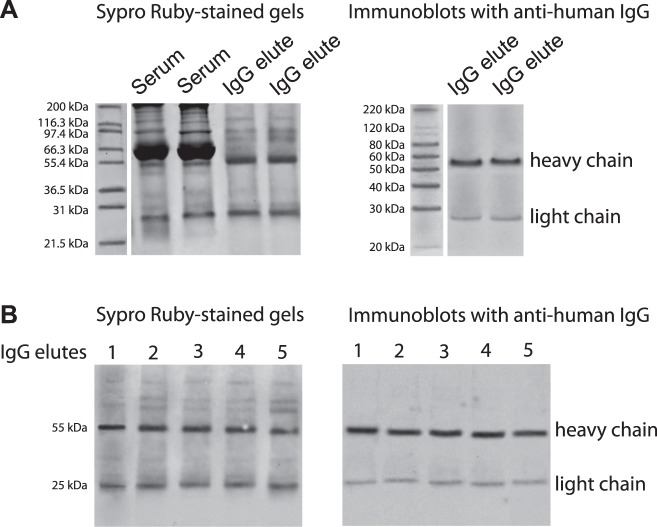
Methods: Serum samples were collected from 111 patients with primary open-angle glaucoma and an age-matched control group of 49 healthy subjects without glaucoma. For high-throughput characterization of antigens, serum IgG was eluted from five randomly selected glaucomatous samples and analyzed by linear ion trap mass spectrometry (LC-MS/MS). Serum titers of selected biomarker candidates were then measured by specific ELISAs in the whole sample pool (including an additional control group of diabetic retinopathy).

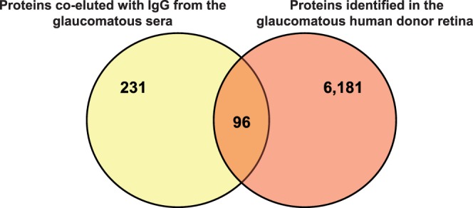
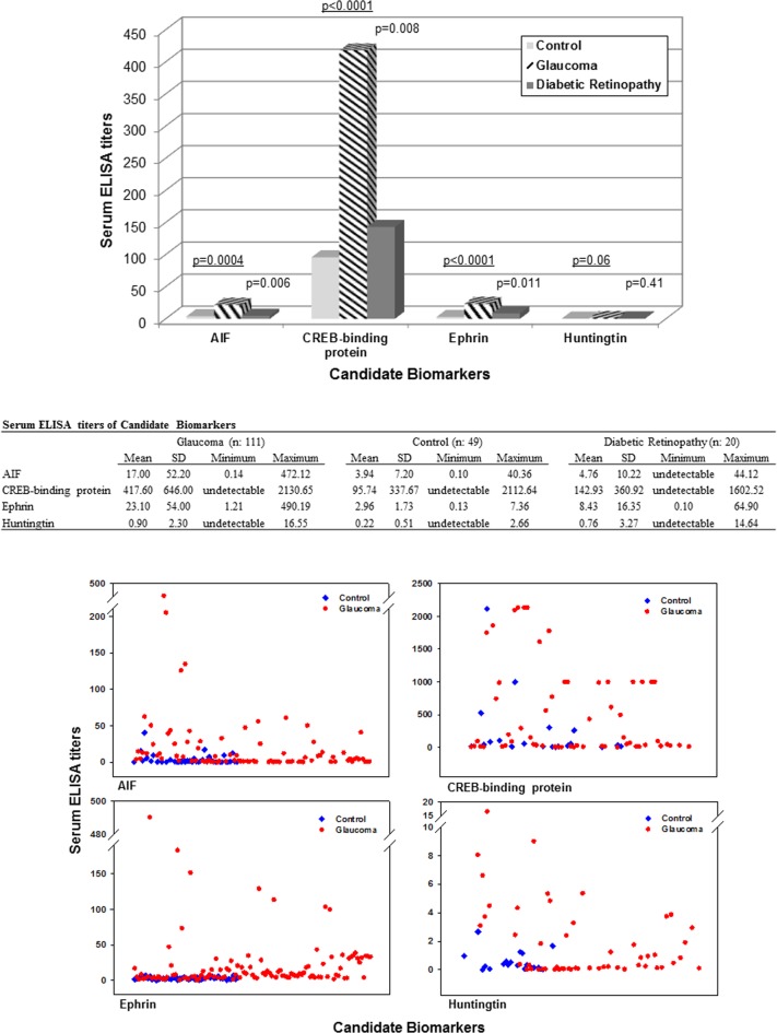
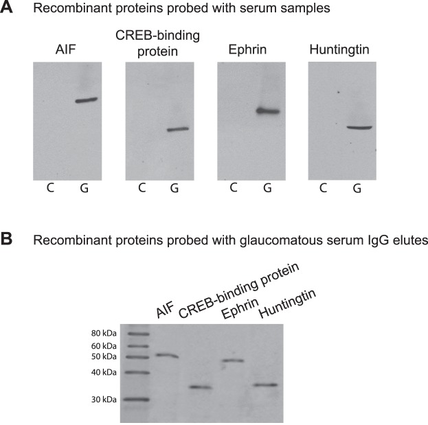
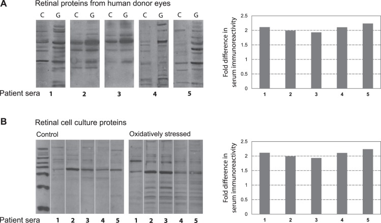
Results: LC-MS/MS analysis of IgG elutes revealed a complex panel of proteins, including those detectable only in glaucomatous samples. Interestingly, many of these antigens corresponded to upregulated retinal proteins previously identified in glaucomatous donors (or that exhibited increased methionine oxidation). Moreover, additional analysis detected a greater immunoreactivity of the patient sera to glaucomatous retinal proteins (or to oxidatively stressed cell culture proteins), thereby suggesting the importance of disease-related protein modifications in autoantibody production/reactivity. As a narrowing-down strategy for selection of initial biomarker candidates, we determined the serum proteins overlapping with the retinal proteins known to be up-regulated in glaucoma. Four of the selected 10 candidates (AIF, cyclic AMP-responsive element binding protein, ephrin type-A receptor, and huntingtin) exhibited higher ELISA titers in the glaucomatous sera.

Conclusions: A number of serum proteins identified by this immunoproteomic study of human glaucoma may represent diseased tissue-related antigens and serve as candidate biomarkers of glaucoma.


Conflict of interest statement

Disclosure: G. Tezel, None; I.L. Thornton, None; M.G. Tong, None; C. Luo, None; X. Yang, None; J. Cai, None; D.W. Powell, None; J.B. Soltau, None; J.M. Liebmann, None; R. Ritch, None

Figures

Figure 1.
Figure 1.
Serum IgG-containing protein mixtures. To identify antigenic targets of serum antibodies for high-throughput characterization, serum proteins co-eluted with IgG were analyzed by LC-MS/MS. (A) SDS-PAGE was performed to verify serum IgG elutes prior to further analysis. Shown Sypro Ruby-stained gels include samples of the elutes whole serum and the IgG elutes isolated using two alternative methods: Melon gel spin column purification and protein G-Dynabeads-based purification, respectively. Immunoblots of these elutes with anti-human IgG antibody detected IgG heavy (approximately 55 kDa) and light (approximately 25 kDa) chains. Since two alternative methods for serum IgG isolation using Melon gel spin columns or protein G-Dynabeads indicated consistent results, the following analysis used the samples eluted by spin column purification. (B) Sypro Ruby-stained gels of IgG elutes and corresponding immunoblots with anti-human IgG antibody represent randomly selected five glaucomatous sera that were subjected to following LC-MS/MS analysis.
Figure 2.
Figure 2.
Candidate biomarkers of glaucoma. As a narrowing-down strategy, we determined whether the MS/MS data obtained from the glaucomatous serum IgG elutes overlapped with our previous high-throughput proteomic data obtained from the glaucomatous human retina. Ninety-six proteins overlapped between the serum and retinal proteomic data.
Figure 3.
Figure 3.
Serum ELISA titers of candidate biomarkers. Serum levels of 10 candidate biomarkers were measured in glaucomatous and nonglaucomatous samples by specific ELISAs. In addition to healthy controls, nonglaucomatous samples also included those collected from an additional group of patients with diabetic retinopathy. As shown in the bar graph of mean values and the Table summarizing the serum ELISA data, glaucomatous samples exhibited higher ELISA titers (ng/mL) for four biomarker candidates. The P values underlined indicate the statistical difference between glaucomatous samples versus samples from healthy controls; other P values indicate the statistical difference between glaucomatous samples versus samples from patients with diabetic retinopathy (Mann-Whitney rank sum test). The scatter graphs of serum ELISA titers in glaucomatous and control samples present individual variability.
Figure 4.
Figure 4.
Serum immunoreactivity to candidate biomarkers. (A) To test serum immunoreactivity by Western blot analysis, recombinant proteins were probed with glaucomatous (G) and nonglaucomatous control (C) sera (1:500), IgG elutes of which were analyzed by LC-MS/MS. Western blots indicated prominent immunoreactivity of the glaucomatous serum to studied proteins, including AIF (approximately 57 kDa), CREB-binding protein (approximately 37 kDa), ephrin (approximately 46 kDa), and huntingtin (approximately 38 kDa). (B) When similarly tested against recombinant proteins, isolated IgG elutes of the glaucomatous sera (15 μg) also exhibited reactivity to these proteins. Presented are the most representative immunoreactivities. The secondary antibody incubation used anti-human IgG conjugated with horse-radish peroxidase (1:2000).
Figure 5.
Figure 5.
Serum immunoreactivity to retinal proteins. To test serum immunoreactivity by Western blot analysis, retinal proteins were separated by SDS-PAGE, and membranes were cut into strips to probe with different serum samples. (A) Glaucomatous serum samples (1–5) exhibited a greater immunoreactivity to retinal proteins obtained from glaucomatous human donors (G) relative to retinal proteins obtained from non-glaucomatous controls (C). (B) We also tested serum immunoreactivity to retinal protein samples obtained from cell cultures. Immunoreactivity of the glaucomatous patient sera (1–5) against oxidatively stressed retinal cell culture proteins was greater than their immunoreactivity to controls. Bar graphs show fold increase in serum immunoreactivity to glaucomatous human retinal proteins (relative to nonglaucomatous retinal proteins), or fold increase in serum immunoreactivity to oxidatively stressed retinal proteins (relative to control proteins) as calculated based on band intensities. Serum dilution was 1:500, and the secondary antibody incubation used anti-human IgG conjugated with horse-radish peroxidase (1:2000).

Similar articles

Cited by

References

    1. Tezel G. Immune regulation towards immunomodulation for neuroprotection in glaucoma. Curr Opin Pharmacol. In press - PMC - PubMed
    1. Maruyama I, Ohguro H, Ikeda Y. Retinal ganglion cells recognized by serum autoantibody against gamma-enolase found in glaucoma patients. Invest Ophthalmol Vis Sci. 2000;41:1657–1665 - PubMed
    1. Tezel G, Wax MB. Glaucoma. Chem Immunol Allergy. 2007;92:221–227 - PMC - PubMed
    1. Reichelt J, Joachim SC, Pfeiffer N, Grus FH. Analysis of autoantibodies against human retinal antigens in sera of patients with glaucoma and ocular hypertension. Curr Eye Res. 2008;33:253–261 - PubMed
    1. Dervan EW, Chen H, Ho SL, et al. Protein macroarray profiling of serum autoantibodies in pseudoexfoliation glaucoma. Invest Ophthalmol Vis Sci. 2010;51:2968–2975 - PubMed

Publication types

MeSH terms