. 2013 Jan 11;288(2):848-58.
doi: 10.1074/jbc.M112.428573. Epub 2012 Nov 13.

Identification of the components of a glycolytic enzyme metabolon on the human red blood cell membrane

Affiliations

Identification of the components of a glycolytic enzyme metabolon on the human red blood cell membrane

Estela Puchulu-Campanella et al. J Biol Chem. .

Abstract

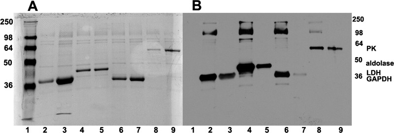
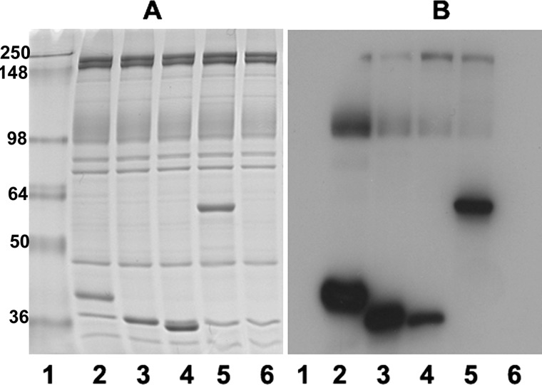
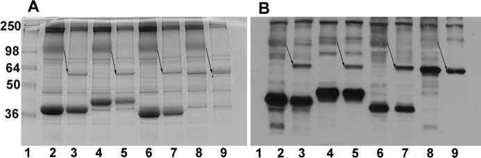
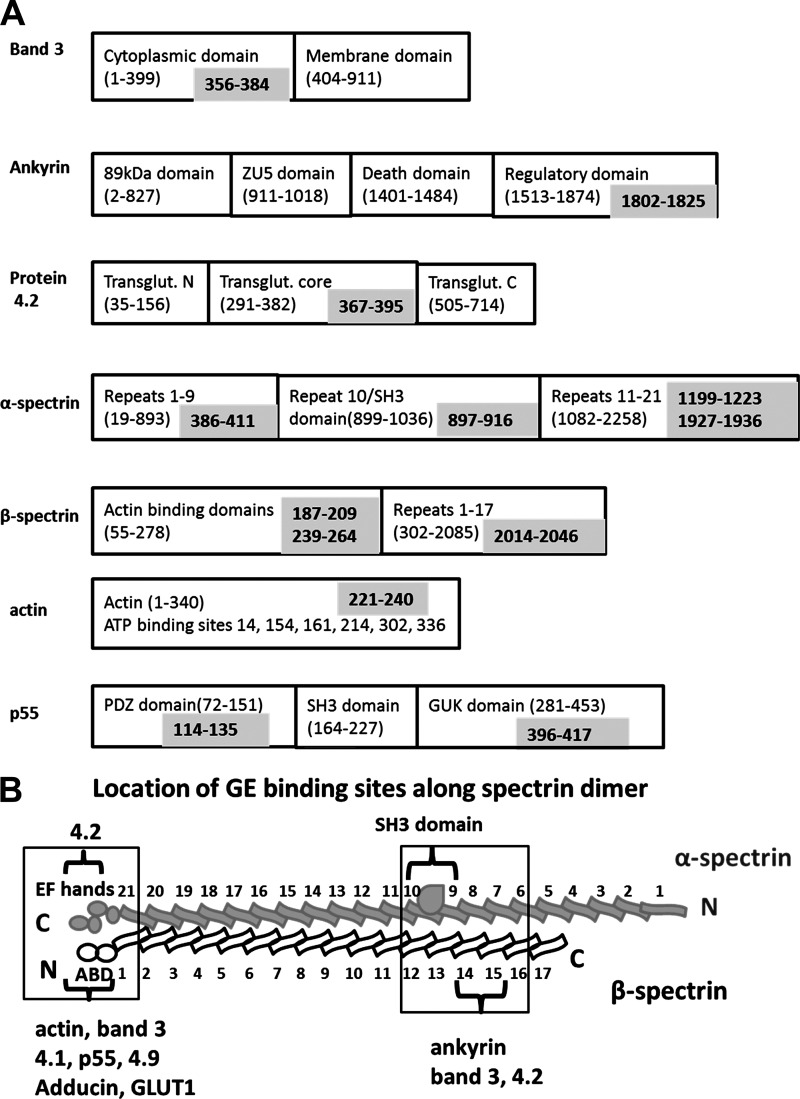
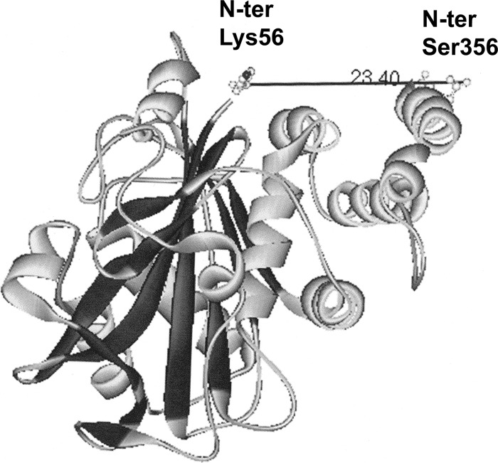
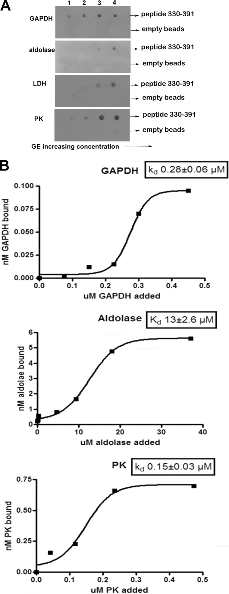
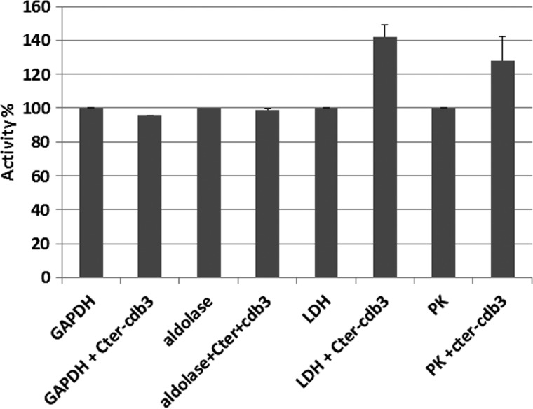
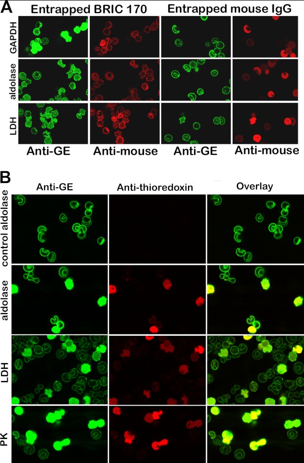
Glycolytic enzymes (GEs) have been shown to exist in multienzyme complexes on the inner surface of the human erythrocyte membrane. Because no protein other than band 3 has been found to interact with GEs, and because several GEs do not bind band 3, we decided to identify the additional membrane proteins that serve as docking sites for GE on the membrane. For this purpose, a method known as "label transfer" that employs a photoactivatable trifunctional cross-linking reagent to deliver a biotin from a derivatized GE to its binding partner on the membrane was used. Mass spectrometry analysis of membrane proteins that were biotinylated following rebinding and photoactivation of labeled GAPDH, aldolase, lactate dehydrogenase, and pyruvate kinase revealed not only the anticipated binding partner, band 3, but also the association of GEs with specific peptides in α- and β-spectrin, ankyrin, actin, p55, and protein 4.2. More importantly, the labeled GEs were also found to transfer biotin to other GEs in the complex, demonstrating for the first time that GEs also associate with each other in their membrane complexes. Surprisingly, a new GE binding site was repeatedly identified near the junction of the membrane-spanning and cytoplasmic domains of band 3, and this binding site was confirmed by direct binding studies. These results not only identify new components of the membrane-associated GE complexes but also provide molecular details on the specific peptides that form the interfacial contacts within each interaction.


Figures

FIGURE 1.
FIGURE 1.
Identification of membrane proteins labeled by photoactivated biotin transfer from sulfo-SBED-derivatized glycolytic enzymes. Sulfo-SBED-labeled enzymes were incubated with fresh erythrocyte ghosts as described under “Experimental Procedures.” Samples were separated on 10% SDS-polyacrylamide gels, transferred to nitrocellulose, and stained first with Ponceau S stain (A) to reveal all transferred proteins and, after removal of Ponceau stain, with streptavidin HRP (B) to selectively reveal only biotinylated proteins. Lane 1, molecular weight markers; lane 2, sulfo-SBED-GAPDH incubated with ghosts; lane 3, same as lane 2 but containing a 10-fold excess of unlabeled GAPDH; lane 4, sulfo-SBED-aldolase incubated with ghosts; lane 5, same as lane 4 but containing a 6-fold excess of unlabeled aldolase; lane 6, sulfo-SBED-LDH incubated with ghosts; lane 7, same as lane 6 but containing a 10-fold excess of unlabeled LDH; lane 8, sulfo-SBED-PK incubated with ghosts; lane 9, same as lane 7 but containing a 10-fold excess of unlabeled PK. Molecular weights are indicated in the right and left margins. Positions of the enzymes are labeled.
FIGURE 2.
FIGURE 2.
Binding of aldolase, GAPDH, LDH, PK, and ovalbumin to fresh porous ghosts. Sulfo-SBED-labeled aldolase, GAPDH, LDH, PK, and ovalbumin were incubated with fresh ghosts, as described under “Experimental Procedures,” and after washing and photoactivating, bound proteins were separated by SDS-PAGE and stained with Coomassie Blue (A) or transferred to nitrocellulose and incubated with streptavidin-HRP to reveal biotinylated proteins (B). Because ovalbumin does not bind to erythrocyte membranes, washing to remove unbound protein prevents labeling of any membrane proteins with sulfo-SBED-ovalbumin. Lane 1, molecular weight markers; lanes 2–6, ghosts that were incubated with sulfo-SBED-aldolase (lane 2), sulfo-SBED-GAPDH (lane 3), sulfo-SBED-LDH (lane 4), sulfo-SBED-PK (lane 5), and sulfo-SBED-ovalbumin (lane 6).
FIGURE 3.
FIGURE 3.
Treatment of intact erythrocytes with chymotrypsin shifts the ∼100 kDa band to ∼55 kDa, as expected for band 3. Ghosts prepared from control cells (lanes 2, 4, 6, and 8) or chymotrypsin-digested whole cells (lanes 3, 5, 7, and 9) were incubated with sulfo-SBED-labeled enzymes and then processed for SDS-PAGE as described under “Experimental Procedures.” Proteins were separated in 10% SDS-polyacrylamide gels and stained with Coomassie (A) or transferred to nitrocellulose and visualized with streptavidin HRP (B). The NH2-terminal half of band 3 containing the full cytoplasmic domain and part of the membrane domain migrates at ∼55 kDa following chymotrypsin digestion of intact erythrocytes (see arrows). This new 55 kDa band is labeled by each of the GE lanes (3, 5, 7, and 9). GAPDH (lanes 2 and 3), aldolase (lanes 4 and 5), LDH (lanes 6 and 7), and PK (lanes 8 and 9).
FIGURE 4.
FIGURE 4.
Schematic representation of the prominent labeling sites of GEs on major membrane proteins. A, the domain structure of each major membrane protein is shown in the black boxes, with the component amino acid sequences provided in parentheses. The specific peptide in each protein that is photolabeled during the biotin transfer reaction is indicated in the gray box. B, a more detailed map of the labeling of spectrin.
FIGURE 5.
FIGURE 5.
Crystal structure of CDB3 (Protein Data Bank entry 1HYN). The distance in angstroms from the first resolved amino acid at the NH2 terminus of band 3 (Lys-56) to the last resolved amino acid near the COOH terminus of CDB3 (Ser-356) is shown.
FIGURE 6.
FIGURE 6.
Effect of increasing GE concentration on the binding of GEs to the peptide comprising residues 330–391 of band 3. A, evaluation of the affinity of GEs for the His-tagged fusion protein containing residues 330–391 of band 3 by nickel bead pull-down assay. Fusion protein (0.17 μmol) was incubated with increasing amounts (5–50 nmol) of GE in a total volume of 100 μl overnight at 4 °C. Nickel beads blocked with binding buffer containing 5% BSA were added, and the His-tagged fragment with bound GE was collected and washed before analysis. A similar sample containing only GE was run in parallel and dot-blotted as a control for nonspecific binding to the nickel beads. The concentrations of GE tetramers applied to the blot in each lane were as follows. Lane 1, GAPDH, 54 nm; aldolase, 51 nm; LDH, 50 nm; PK, 51 nm. Lane 2, GAPDH, 134 nm; aldolase, 126 nm; LDH, 126 nm; PK, 123 nm. Lane 3, GAPDH, 268 nm; aldolase, 252 nm; LDH, 246 nm; PK, 249 nm. Lane 4, GAPDH, 536 nm; aldolase, 504 nm; LDH, 492 nm; PK, 497 nm. B, determination of the Kd of each GE for the peptide 330–391 of band 3. Data are plotted using GraphPad Prism 4 and fitted to a Boltzman sigmoidal equation.
FIGURE 7.
FIGURE 7.
Effect of the band 3 peptide comprising residues 330–391 on the catalytic activities of GEs. GEs were incubated without or with the above band 3 peptide for 5 min prior to assaying the activities of the indicated enzymes as described by Chu and Low (17). Error bars, S.E.
FIGURE 8.
FIGURE 8.
Displacement of GEs from erythrocyte membranes by entrapment of a monoclonal antibody to residues 368–382 of band 3 (BRIC 170) (A) or a fusion protein comprising residues 330–391 linked to thioredoxin (B). A, the first and second columns show the location of endogenous GEs (green stain) and the entrapment of BRIC 170 (red stain). Parallel control experiments following entrapment of a nonspecific mouse IgG are shown in columns 3 and 4. Quantitation of the intensity of GE staining in the cytosol using an Olympus Fluo View microscope version 2.1 reveals that the cytosolic GE staining for GAPDH, aldolase, and LDH is 20, 4, and 8 times greater on average in BRIC 170-entrapped cells than in isotype control-entrapped cells. B, a fusion protein containing peptide 330–391 linked to thioredoxin was entrapped into resealed ghosts, and its effect on GE binding was evaluated by confocal microscopy. A control in which no peptide fusion protein was entrapped during the incubation step with leaky ghosts was run in parallel and is shown in the first row (labeled control aldolase). The first column shows the staining of the GEs; the second column identifies the “ghosts” that have successfully entrapped the fusion protein (red stain of the anti-thioredoxin antibody); and the third column shows the overlay of both stains.

References

    1. Vonck J., Schäfer E. (2009) Supramolecular organization of protein complexes in the mitochondrial inner membrane. Biochim. Biophys. Acta 1793, 117–124 - PubMed
    1. Dai S., Hall D. D., Hell J. W. (2009) Supramolecular assemblies and localized regulation of voltage-gated ion channels. Physiol. Rev. 89, 411–452 - PMC - PubMed
    1. Sterling D., Reithmeier R. A., Casey J. R. (2001) A transport metabolon. J. Biol. Chem. 276, 47886–47894 - PubMed
    1. Sowah D., Casey J. R. (2011) An intramolecular transport metabolon. Fusion of carbonic anhydrase II to the COOH terminus of the Cl−/HCO3− exchanger, AE1. Am. J. Physiol. Cell Physiol. 301, C336–C346 - PubMed
    1. Endeward V., Cartron J.-P., Ripoche P., Gros G. (2008) RhAG protein of the Rhesus complex is a CO2 channel in the human red cell membrane. FASEB J. 22, 64–73 - PubMed

Publication types

LinkOut - more resources