. 2013 Jan 4;288(1):654-65.
doi: 10.1074/jbc.M112.400168. Epub 2012 Nov 13.

C1q-induced LRP1B and GPR6 proteins expressed early in Alzheimer disease mouse models, are essential for the C1q-mediated protection against amyloid-β neurotoxicity

Affiliations

C1q-induced LRP1B and GPR6 proteins expressed early in Alzheimer disease mouse models, are essential for the C1q-mediated protection against amyloid-β neurotoxicity

Marie E Benoit et al. J Biol Chem. .

Abstract

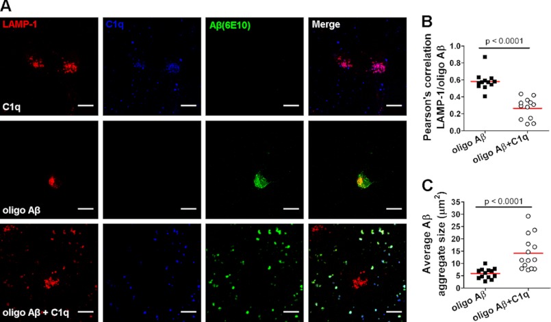
Complement protein C1q is induced in the brain in response to a variety of neuronal injuries, including Alzheimer disease (AD), and blocks fibrillar amyloid-β (fAβ) neurotoxicity in vitro. Here, we show that C1q protects immature and mature primary neurons against fAβ toxicity, and we report for the first time that C1q prevents toxicity induced by oligomeric forms of amyloid-β (Aβ). Gene expression analysis reveals C1q-activated phosphorylated cAMP-response element-binding protein and AP-1, two transcription factors associated with neuronal survival and neurite outgrowth, and increased LRP1B and G protein-coupled receptor 6(GPR6) expression in fAβ-injured neurons. Silencing of cAMP-response element-binding protein, LRP1B or GPR6 expression inhibited C1q-mediated neuroprotection from fAβ-induced injury. In addition, C1q altered the association of oligomeric Aβ and fAβ with neurons. In vivo, increased hippocampal expression of C1q, LRP1B, and GPR6 is observed as early as 2 months of age in the 3 × Tg mouse model of AD, whereas no such induction of LRP1B and GPR6 was seen in C1q-deficient AD mice. In contrast, expression of C1r and C1s, proteases required to activate the classical complement pathway, and C3 showed a significant age-dependent increase only after 10-13 months of age when Aβ plaques start to accumulate in this AD model. Thus, our results identify pathways by which C1q, up-regulated in vivo early in response to injury without the coordinate induction of other complement components, can induce a program of gene expression that promotes neuroprotection and thus may provide protection against Aβ in preclinical stages of AD and other neurodegenerative processes.


Figures

FIGURE 1.
FIGURE 1.
C1q protects immature and mature neurons against fibrillar and oligomeric Aβ toxicity. A and B, mouse primary mature neurons were treated with 15 μm fAβ and/or 20 nm C1q for 24 h. C–F, immature neurons isolated from wild-type (C, E, and F) or C1q−/− (D) mice were treated with 5 μm fAβ and/or 10 nm C1q (C and D) or 1 μm oligomeric (oligo) Aβ and/or 10 nm C1q (E) for 24 h or 5 μm fAβ for 24 h with 10 nm C1q added for the last 8 h of incubation (F). Neuronal integrity was assessed by MAP-2 immunocytochemistry (A, images representative of three independent experiments) and image analysis (B–F). Scale bar, 100 μm. Results represent mean ± S.D. (n = 3, five fields per condition) and were compared using one-way ANOVA (Kruskall-Wallis test). *, p < 0.05; **, p < 0.01. and ***, p < 0.001.
FIGURE 2.
FIGURE 2.
C1q modulates gene expression profiles of fAβ-injured neurons. A, Pearson correlation coefficient-based heat map (complete linkage method) representation of the log2 fold-change of significantly modulated genes 3 h after treatment of rat primary immature neurons with fAβ (5 μm) and/or C1q (10 nm) over untreated neurons shown with a color gradient from blue (down-regulated) to red (up-regulated). Neurons were from the same pool of primary rat cortical neurons with each condition (untreated, Aβ, Aβ+C1q) performed in separate triplicate wells, and each replicate was run on a separate array. B, gene ontology-based functional classification of genes modulated by C1q in fAβ-treated neurons. C, C1q-dependent modulation of LRP1B, GPR6, WASF2, and IER3 mRNA levels assessed by qRT-PCR in fAβ-injured rat or mouse primary immature neurons (n = 3). Dotted lines represent a 2-fold increase and decrease by C1q over fAβ alone-treated neurons. D, GPR6 and actin protein levels in neurons stimulated with fAβ (5 μm) and/or C1q (10 nm) for 24 h by Western blot. E, heat map of significantly over-represented transcription factor-binding sites in the promoter sequences of genes up-regulated by C1q using PAINT and MatInspector. Each red square indicates the presence of the transcription factor-binding site (for example AP-1) in a gene up-regulated by C1q (gene tree indicated by lines at the bottom).
FIGURE 3.
FIGURE 3.
CREB is a central transcription factor in C1q-mediated neuroprotection. A and B, mouse primary immature neurons were stimulated with 5 μm fAβ and/or 10 nm C1q for 30, 60, and 180 min, stained for MAP-2 (red) and pc-Jun (green), mounted in DAPI (blue), and analyzed by confocal microscopy (A) and image quantification (B) to determine pc-Jun nuclear translocation (pc-Jun area over DAPI). Images are representative of three independent experiments. C and D, mouse primary immature neurons were stimulated with 5 μm fAβ and/or 10 nm C1q for 15 and 30 min, and phosphorylation of JNK was determined by Western blot (C) and band intensity analysis (D). Blots are representative of two independent experiments. E–H, mouse primary immature neurons were stimulated with 5 μm fAβ and/or 10 nm C1q for 15, 30, 60, and 180 min. E and F, neurons were stained for MAP-2 (red), pCREB (green), mounted in DAPI (blue), and analyzed by confocal microscopy (E, images representative of three independent experiments) and image quantification (F) to determine pCREB nuclear translocation (pCREB area over DAPI). G and H, phosphorylation of CREB determined by Western blot (G) and band intensity analysis (H). Blots are representative of three independent experiments. I, neurons were transfected with 10 nm scrambled siRNA (scr siRNA) or siRNA targeting CREB 24 h before treatment with 5 μm fAβ ± 10 nm C1q. CREB inhibition was determined by WB 24 h post-transfection (inset). Neuronal integrity was assessed by MAP-2 staining and quantitative image analysis after 24 h of treatment, n = 2 (5 fields per condition). All results represent means ± S.E. and are compared using two-way ANOVA test, *, p < 0.05; **, p < 0.01 and ***, p < 0.001. Scale bar, 10 μm. NT, untransfected.
FIGURE 4.
FIGURE 4.
LRP1B and GPR6 are central effectors of C1q-induced neuroprotection in fAβ-injured neurons. Mouse primary immature neurons were transfected with 10 nm scr siRNA or siRNA targeting LRP1B, GPR6, or GAPDH 24 h before treatment with 5 μm fAβ ± 10 nm C1q. A, LRP1B, GAPDH, and GPR6 mRNA levels in neurons transfected with LRP1B, GPR6, or GAPDH siRNA normalized to levels in neurons transfected with scr siRNA were assessed by qRT-PCR at 24 and 48 h post-transfection. B, neuronal integrity was assessed by MAP-2 staining and quantitative image analysis after 24 h of treatment, n = 3 (five fields per condition). Results represent means ± S.E. and are compared using two-way ANOVA test; *, p < 0.05; **, p < 0.01, and ***, p < 0.001. C, GPR6 and actin protein levels identified by Western blot in neurons transfected with scr, LRP1B, or GPR6 siRNA for 24 h and then stimulated with 5 μm fAβ and/or 10 nm C1q for 24 h. Blots are representative of two independent experiments. UT, untreated.
FIGURE 5.
FIGURE 5.
Increased hippocampal expression of C1q, LRP1B, and GPR6 at early ages in AD mice. LRP1B (A), GPR6 (B), C1q (C), C1r (D), C1s (E), and C3 (F) mRNA levels were assessed by qRT-PCR in the hippocampus of 3×TgBUB (● and ■, solid lines) or nontransgenic BUB (○ and □, dotted lines) mice sufficient (● and ○) or deficient (■ and□) for C1q at 2 (n = 4–5), 4 (n = 8), 10 (n = 6), 13 (n = 6), and 18 (n = 6) months of age (performed in duplicate). Results represent means ± S.E. of mRNA levels relative to GAPDH (2−ΔCt, ΔCt = (CtTargetCtGAPDH)) and are compared using two-way ANOVA test. *, p < 0.05; **, p < 0.01, and ***, p < 0.001.
FIGURE 6.
FIGURE 6.
C1q alters fAβ association with neurons in an LRP1B-dependent manner. Mouse primary immature neurons were treated with 5 μm fAβ and/or 10 nm C1q for 24 h (A and B) or transfected with 10 nm scr siRNA or siRNA targeting LRP1B or GPR6 24 h before treatment with 5 μm fAβ ± 10 nm C1q (C), stained for MAP-2 (red), C1q (blue), and Aβ (green), and analyzed by confocal microscopy. A, images representative of three different experiments. Scale bar, 10 μm. B and C, scatter dot plot of Pearson's correlation coefficient for co-localization between Aβ and MAP-2 (n = 3, 5 fields per condition); red line, mean values. Results were compared using two-tailed non parametric Mann-Whitney U test (B) and one-way ANOVA (Kruskall-Wallis test) (C).
FIGURE 7.
FIGURE 7.
C1q decreases internalization of Aβ oligomers through enhanced Aβ aggregation. Mouse primary immature neurons were treated with 1 μm oligomeric Aβ and/or 10 nm C1q for 8 h, stained for LAMP1 (red), C1q (blue), and Aβ (green), and analyzed by confocal microscopy. A, images representative of three different experiments. Scale bar, 10 μm. B and C, scatter dot plot of Pearson's correlation coefficient for co-localization between LAMP1 and Aβ (B) and average Aβ aggregate size in μm2 (C) (n = 3, 5 fields per condition); red line, mean values. Results were compared using two-tailed nonparametric Mann-Whitney U test.
FIGURE 8.
FIGURE 8.
Model of C1q-induced neuroprotective pathways against Aβ. C1q induces CREB phosphorylation and AP-1 activation followed by up-regulation of the expression of LRP1B, which is involved in C1q-dependent GPR6 expression and C1q-mediated neuroprotection. LRP1B can also bind Aβ and reduces its internalization. In addition, C1q directly binds Aβ and increases its aggregation that sequesters Aβ away from the neurons and thus limits its toxic association with neurons.

References

    1. Alexander J. J., Anderson A. J., Barnum S. R., Stevens B., Tenner A. J. (2008) The complement cascade. Yin-Yang in neuroinflammation–neuro-protection and -degeneration. J. Neurochem. 107, 1169–1187 - PMC - PubMed
    1. Bohlson S. S., Fraser D. A., Tenner A. J. (2007) Complement proteins C1q and MBL are pattern recognition molecules that signal immediate and long-term protective immune functions. Mol. Immunol. 44, 33–43 - PubMed
    1. Benoit M. E., Clarke E. V., Morgado P., Fraser D. A., Tenner A. J. (2012) Complement protein C1q directs macrophage polarization and limits inflammasome activity during the uptake of apoptotic cells. J. Immunol. 188, 5682–5693 - PMC - PubMed
    1. Fraser D. A., Pisalyaput K., Tenner A. J. (2010) C1q enhances microglial clearance of apoptotic neurons and neuronal blebs, and modulates subsequent inflammatory cytokine production. J. Neurochem. 112, 733–743 - PMC - PubMed
    1. Melchior B., Garcia A. E., Hsiung B. K., Lo K. M., Doose J. M., Thrash J. C., Stalder A. K., Staufenbiel M., Neumann H., Carson M. J. (2010) Dual induction of TREM2 and tolerance-related transcript, Tmem176b, in amyloid transgenic mice. Implications for vaccine-based therapies for Alzheimer's disease. ASN Neuro. 2, e00037. - PMC - PubMed

Publication types

MeSH terms