

MicroRNA expression array identifies novel diagnostic markers for conventional and oncocytic follicular thyroid carcinomas
- PMID: 23150679
- PMCID: PMC3537083
- DOI: 10.1210/jc.2012-2694
MicroRNA expression array identifies novel diagnostic markers for conventional and oncocytic follicular thyroid carcinomas
Abstract
Objective: The most difficult thyroid tumors to be diagnosed by cytology and histology are conventional follicular carcinomas (cFTCs) and oncocytic follicular carcinomas (oFTCs). Several microRNAs (miRNAs) have been previously found to be consistently deregulated in papillary thyroid carcinomas; however, very limited information is available for cFTC and oFTC. The aim of this study was to explore miRNA deregulation and find candidate miRNA markers for follicular carcinomas that can be used diagnostically.
Design: Thirty-eight follicular thyroid carcinomas (21 cFTCs, 17 oFTCs) and 10 normal thyroid tissue samples were studied for expression of 381 miRNAs using human microarray assays. Expression of deregulated miRNAs was confirmed by individual RT-PCR assays in all samples. In addition, 11 follicular adenomas, two hyperplastic nodules (HNs), and 19 fine-needle aspiration samples were studied for expression of novel miRNA markers detected in this study.
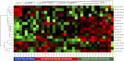
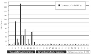
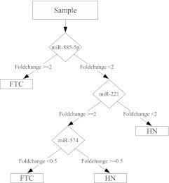
Results: The unsupervised hierarchical clustering analysis demonstrated individual clusters for cFTC and oFTC, indicating the difference in miRNA expression between these tumor types. Both cFTCs and oFTCs showed an up-regulation of miR-182/-183/-221/-222/-125a-3p and a down-regulation of miR-542-5p/-574-3p/-455/-199a. Novel miRNA (miR-885-5p) was found to be strongly up-regulated (>40-fold) in oFTCs but not in cFTCs, follicular adenomas, and HNs. The classification and regression tree algorithm applied to fine-needle aspiration samples demonstrated that three dysregulated miRNAs (miR-885-5p/-221/-574-3p) allowed distinguishing follicular thyroid carcinomas from benign HNs with high accuracy.
Conclusions: In this study we demonstrate that different histopathological types of follicular thyroid carcinomas have distinct miRNA expression profiles. MiR-885-5p is highly up-regulated in oncocytic follicular carcinomas and may serve as a diagnostic marker for these tumors. A small set of deregulated miRNAs allows for an accurate discrimination between follicular carcinomas and hyperplastic nodules and can be used diagnostically in fine-needle aspiration biopsies.
Figures
References
-
- DeLellis R, Lloyd R, Heitz P, Eng C, eds. 2004. Pathology and genetics of tumours of endocrine organs. Lyon, France: IARC Press [World Health Organization classification of tumours]
-
- Nikiforov YE. 2012. Thyroid tumors: classification, staging and general considerations. In: Nikiforov Y, Biddinger PW, Thompson LDR, eds. Diagnostic pathology and molecular genetics of the thyroid. Baltimore: Lippincott Williams &Wilkins; 108–118
-
- Nikiforov YE, Ohori NP, Hodak SP, Carty SE, LeBeau SO, Ferris RL, Yip L, Seethala RR, Tublin ME, Stang MT, Coyne C, Johnson JT, Stewart AF, Nikiforova MN. 2011. Impact of mutational testing on the diagnosis and management of patients with cytologically indeterminate thyroid nodules: a prospective analysis of 1056 FNA samples. J Clin Endocrinol Metab 96:3390–3397 - PMC - PubMed
-
- Nikiforov YE, Steward DL, Robinson-Smith TM, Haugen BR, Klopper JP, Zhu Z, Fagin JA, Falciglia M, Weber K, Nikiforova MN. 2009. Molecular testing for mutations in improving the fine-needle aspiration diagnosis of thyroid nodules. J Clin Endocrinol Metab 94:2092–2098 - PubMed
-
- Cantara S, Capezzone M, Marchisotta S, Capuano S, Busonero G, Toti P, Di Santo A, Caruso G, Carli AF, Brilli L, Montanaro A, Pacini F. 2010. Impact of proto-oncogene mutation detection in cytological specimens from thyroid nodules improves the diagnostic accuracy of cytology. J Clin Endocrinol Metab 95:1365–1369 - PubMed
Publication types
MeSH terms
Substances
Supplementary concepts
Grants and funding
LinkOut - more resources
Full Text Sources
Other Literature Sources
Medical
