

Mitochondrial Pathway of α-Tocopheryl Succinate-Induced Apoptosis in Human Epidermoid Carcinoma A431 Cells
- PMID: 23150807
- PMCID: PMC3491895
Mitochondrial Pathway of α-Tocopheryl Succinate-Induced Apoptosis in Human Epidermoid Carcinoma A431 Cells
Abstract
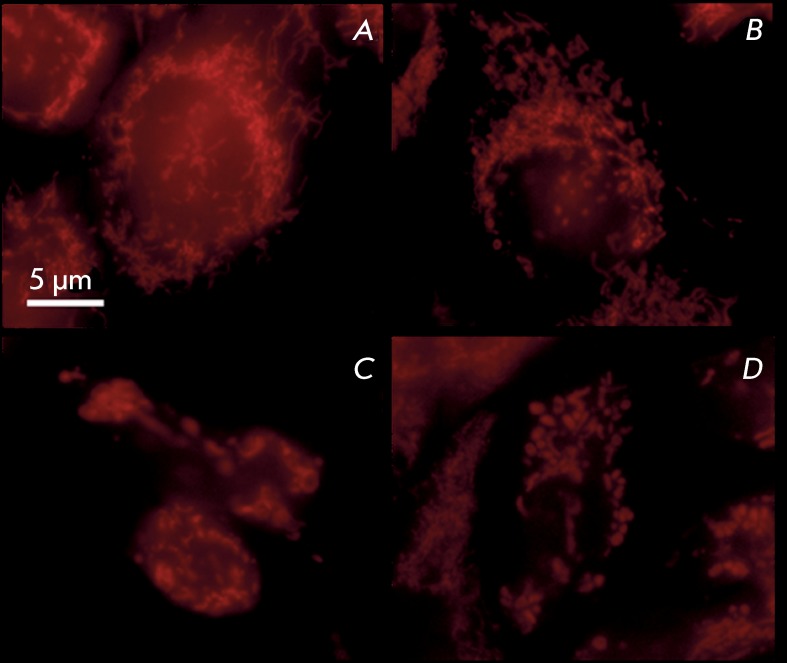
Vitamin E derivatives are known to act as agents exhibiting cytotoxity against tumor cells. The effect of vitamin E succinate on human epidermoid carcinoma cell line A431 was investigated in this study using live imaging, immunocytochemistry, and transmission electron microscopy. α-Tocopheryl succinate-induced apoptotic cell death in A431 cells was shown to be both dose- and time-dependent. The hyperproduction of reactive oxygen species, changes in size, shape and ultrastructural characteristics of mitochondria followed by the release of cytochromecfrom mitochondria to cytosol were observed. These results suggest that α-tocopheryl succinate induces apoptosis that occurs via the mitochondrial pathway. Mitochondria are shown to be crucial targets in α-tocopheryl succinate-induced caspase-dependent cell death in human carcinoma A431 cells.
Keywords: ROS; apoptosis; cytochromec; mitochondrial pathway; α-tocopheryl succinate.
Figures


Similar articles
-
[Proapoptotic action of alpha-tocopheryl succinate in rat thymocytes is conditioned by the inhibition of mitochondrial succinate dehydrogenase].Ukr Biokhim Zh (1999). 2006 Jul-Aug;78(4):104-11. Ukr Biokhim Zh (1999). 2006. PMID: 17236627 Russian.
-
Skin cancer chemopreventive agent, {alpha}-santalol, induces apoptotic death of human epidermoid carcinoma A431 cells via caspase activation together with dissipation of mitochondrial membrane potential and cytochrome c release.Carcinogenesis. 2005 Feb;26(2):369-80. doi: 10.1093/carcin/bgh325. Epub 2004 Nov 4. Carcinogenesis. 2005. PMID: 15528219
-
RRR-alpha-tocopheryl succinate-induced apoptosis of human breast cancer cells involves Bax translocation to mitochondria.Cancer Res. 2003 May 15;63(10):2483-91. Cancer Res. 2003. PMID: 12750270
-
Photodynamic therapy-induced apoptosis in epidermoid carcinoma cells. Reactive oxygen species and mitochondrial inner membrane permeabilization.J Biol Chem. 2001 Dec 14;276(50):47379-86. doi: 10.1074/jbc.M107678200. Epub 2001 Sep 28. J Biol Chem. 2001. PMID: 11579101
-
The role of alpha tocopheryl succinate (α-TOS) as a potential anticancer agent.Nutr Cancer. 2014;66(2):167-76. doi: 10.1080/01635581.2014.863367. Epub 2013 Dec 23. Nutr Cancer. 2014. PMID: 24364743 Review.
References
-
- Prasad K.N., Edwards-Prasad J.. J. Am. Coll. Nutr. 1992;11:487–500. - PubMed
-
- Kelloff G.J., Crowell J.A., Boone C.W., Steele V.E., Lubet R.A., Greenwald P., Alberts D.S., Covey J.M., Doody L.A., Knapp G.G.. J. Cell Biochem. Suppl. 1994;20:282–299. - PubMed
-
- Theriault R.L., Lipton A., Hortobagyi G.N., Leff R., Gluck S., Stewart J.F., Costello S., Seaman J.J.. J. Clin. Oncol. 1999;17:846–854. - PubMed
-
- Fariss M.W., Fortuna M.B., Everett C.K., Smith J.D., Trent D.F., Djuric Z.. Cancer Res. 1994;54:3346–3351. - PubMed
-
- Kline K., Yu W., Sanders B.G.. Mol Carcinog. 1998;22(4):247–257. - PubMed
LinkOut - more resources
Full Text Sources